eBook - ePub
Juvenile Osteology
A Laboratory and Field Manual
- 384 pages
- English
- ePUB (mobile friendly)
- Available on iOS & Android
eBook - ePub
About this book
The need for a laboratory and field manual to assist with the evaluation of juvenile skeletal material is long overdue. This resource is essential for the practising osteoarchaeologist and forensic anthropologist who requires a quick, reliable and easy-to-use reference to aid in the identification, siding and aging of juvenile osseous material. While excellent reference books on juvenile osteology are currently available, no pre-existing source adequately fills this particular niche in the market. This field manual is designed with practicality as its primary directive. Descriptions of each bone contain 1) morphological characteristics useful for identification, 2) other elements with which the bone may be confused, 2) tips for siding, 3) illustrations of varying developmental phases, 4) data useful for ageing, and 5) a summary of developmental timings. Concise, bullet-style descriptions assist with quick retrieval of information.Unique to this manual is the presentation of data collected from a variety of populations, utilizing a range of observational methods, as an alternative to providing one overall aging summary that is derived from a compilation of many individual sources. This manual provides a host of data on a variety of populations to enable the user to select the reference most applicable to their needs. The final chapter combines information from each bone to provide a summary of developmental changes occurring at different life stages to act as an immediate 'ready reckoner' for the knowledgeable practitioner. It also provides forms useful for documenting juvenile material and diagrams to help with the recognition of commingled juvenile remains. The manual is a must for anyone responsible for the evaluation of juvenile osseous material through dry bone assessment, radiographs, sonograms, and or CT scans.
- Identifies every component of the developing skeleton
- Provides detailed analysis of juvenile skeletal remains and the development of bone as a tissue
- Summarizes key morphological stages in the development of every bone
- Provides data on a variety of populations to enable the user to select the reference most applicable to their needs
- Focuses on practicality, with direct, bullet style descriptions
- Provides forms for documenting juvenile material
- Provides diagrams to help with the recognition of commingled juvenile remains
- Final chapter provides summary of developmental changes occurring at different life stages to act as an immediate 'ready reckoner' for the practitioner
Trusted by 375,005 students
Access to over 1.5 million titles for a fair monthly price.
Study more efficiently using our study tools.
Information
Chapter 1. The Head and Neck
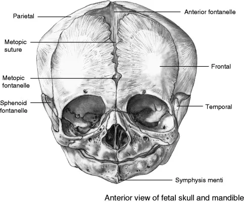
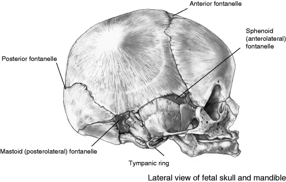
The Fetal Skull
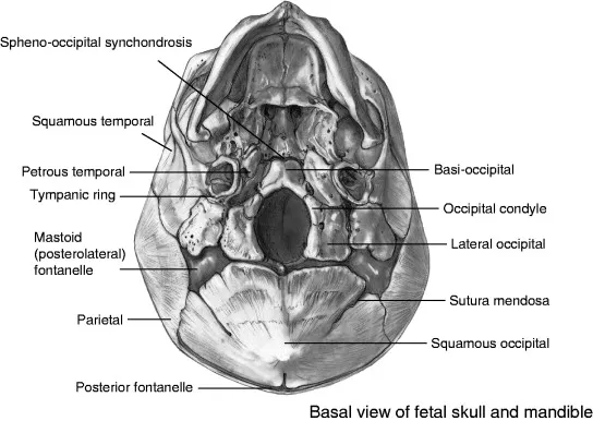
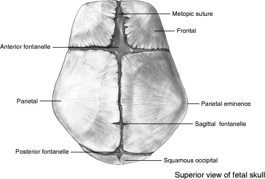
The Occipital
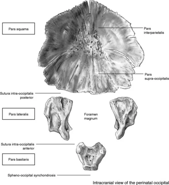
Pars Squama
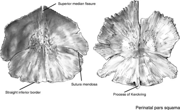
Identification
- Probably indistinguishable from fragments of other vault bones unless a characteristic part, such as the process of Kerckring, is present.
- More robust in the region of the foramen magnum than other vault bones.
Orientation
- Superior border is angled, inferior border is straight.
- Mendosal sutures are obliquely oriented in an inferolateral direction.
Pars Lateralis
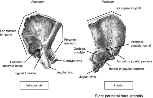
Identification
During perinatal life, the inferior surface resembles that of the scapula (see page 7).
- Within a single skeleton, the scapula is much larger and its blade is more extensive than the body of the pars lateralis.
- Presence of the occipital condyles easily distinguishes the pars lateralis.
Siding
- The condylar and jugular limbs are orientated anteromedially.
- The condylar limb, as identified by the part...
Table of contents
- Cover
- Title
- Brief Table of Contents
- Table of Contents
- Copyright
- Dedication
- Preface
- Chapter 1. The Head and Neck
- Chapter 2. The Dentition
- Chapter 3. The Vertebral Column
- Chapter 4. The Thorax
- Chapter 5. The Pectoral Girdle
- Chapter 6. The Upper Limb
- Chapter 7. The Pelvic Girdle
- Chapter 8. The Lower Limb
- Chapter 9. Summaries, Recording Forms, and Practical Sequencing Information
Frequently asked questions
Yes, you can cancel anytime from the Subscription tab in your account settings on the Perlego website. Your subscription will stay active until the end of your current billing period. Learn how to cancel your subscription
No, books cannot be downloaded as external files, such as PDFs, for use outside of Perlego. However, you can download books within the Perlego app for offline reading on mobile or tablet. Learn how to download books offline
We are an online textbook subscription service, where you can get access to an entire online library for less than the price of a single book per month. With over 1.5 million books across 990+ topics, we’ve got you covered! Learn about our mission
Look out for the read-aloud symbol on your next book to see if you can listen to it. The read-aloud tool reads text aloud for you, highlighting the text as it is being read. You can pause it, speed it up and slow it down. Learn more about Read Aloud
Yes! You can use the Perlego app on both iOS and Android devices to read anytime, anywhere — even offline. Perfect for commutes or when you’re on the go.
Please note we cannot support devices running on iOS 13 and Android 7 or earlier. Learn more about using the app
Please note we cannot support devices running on iOS 13 and Android 7 or earlier. Learn more about using the app
Yes, you can access Juvenile Osteology by Louise Scheuer,Sue Black,Maureen C. Schaefer in PDF and/or ePUB format, as well as other popular books in Law & Forensic Science. We have over 1.5 million books available in our catalogue for you to explore.