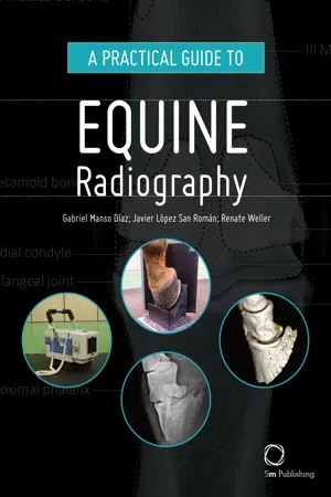
A Practical Guide to Equine Radiography
- English
- ePUB (mobile friendly)
- Available on iOS & Android
A Practical Guide to Equine Radiography
About this book
A Practical Guide to Equine Radiography is designed to accompany the clinical veterinarian either within a hospital setting or out in the field. The book offers an informative step-by-step guide to obtaining high quality radiographs with a focus on image quality, accuracy, consistency and safety. General principles and equipment are covered before working through the anatomy of the horse with separate chapters devoted to each body region, providing a thorough and detailed picture of the skeletal structure of the horse, making the book an ideal reference for professionals involved with horse health and disease.Features provided in the book will guide the veterinarian through the stages of taking and interpreting normal radiographs and include: · Clinical indications of radiographic areas of interest in the horse · Equipment required · Preparation and setup guides, supported by photographs· Projections focusing on radiographic areas of interest, aided by photographs· x-rays presented with detailed labels, providing a close-up view of skeletal structures· Three dimensional images demonstrating normal anatomy A Practical Guide to Equine Radiography is an essential tool for equine practitioners, veterinary students and para-professionals.
Tools to learn more effectively

Saving Books

Keyword Search

Annotating Text

Listen to it instead
Information
Chapter 1
How to get the most from your X-ray system
What determines the success of a diagnostic imaging procedure?
Correct choice of modality: when does taking radiographs make sense?
- confirm a clinically suspected diagnosis
- assess the severity of a disease
- exclude other pathological conditions
- assist in surgery planning
- monitor the progress of disease.
- No imaging technique can replace the clinical examination! Radiographic findings do not indicate pain!
- The clinical findings need to guide radiography, e.g. a wound, swelling or positive regional analgesia need to point to an area.
- Using imaging as a ‘fishing exercise’ is not a good idea, since most changes observed on images may or may not be of clinical significance and their clinical meaning can only be appreciated in conjunction with the clinical findings.
- An exception is pre-purchase and pre-sales radiographs, where an attempt is made to use radiography as a predictor for future soundness and performance.
Basic principles of the underlying physics of radiographs
How are X-rays produced?
- X-rays are electromagnetic waves from the high-energy end of the electromagnetic spectrum (visible light or radio waves are part of the same spectrum, but of lower energy and different wave length).
- X-rays are produced when fast-moving electrons collide with matter. This happens in an X-ray tube that houses a cathode and an anode. By heating up the cathode, a cloud of electrons (negatively charged particles) is produced, which are accelerated by applying a current to the system until they hit the positive anode at high speed.
- The higher the temperature of the cathode, the more electrons are produced; this is related to the mAs (milliampere seconds) settings of the X-ray machine.
- The higher the speed of the electrons, the higher the penetrating power of the resulting X-rays. This is controlled by the kVp settings of the X-ray machine.
- X-rays radiate from the source in straight lines in all directions. For medical purposes, only a small cone of the X-rays, the primary beam, is used.
- The size of the primary beam is set by adjusting the window through which X-rays can leave the housing of the X-ray generator. This is called collimation and is an essential radiation protection mechanism, but also optimizes image quality by reducing scatter.
- The intensity of the X-ray beam is inversely proportional to the square of the distance from the source (‘inverse square law’). This is obviously important for radiation safety consideration and has to be taken into account when adjusting exposure settings. Figure 1.1 illustrates this effect.
How do X-rays interact with matter?
- When X-rays hit matter, they can either penetrate the material or get absorbed by it. The main underlying principles on an atomic level are called the Compton and the photoelectric effect. The Compton effect is less desirable since it is responsible for scatter radiation that degrades image quality.
- The degree of absorption of the X-rays by material is determined by the thickness and the radiodensity of the absorber.
- The radiodensity depends on the physical density and the atomic number of the material, e.g. lead has a very high atomic number which allows complete absorption of X-rays with only a few millimetres of material. This is the reason why, for example, lead is used for shielding purposes, e.g. in protective clothing. The same effect can be achieved with material of lower atomic number by increasing its thickness, e.g. a 20 cm brick wall.
 Figure 1.1 This figure illustrates the effect of distance on X-ray intensity, ‘inverse square law’. The intensity of the X-ray beam is inversely proportional to the square of the distance from the source. This is obviously important for radiation safety consideration and must be taken into account when adjusting exposure settings.
 - The body is composed of materials of different radiodensities and thickness, hence X-rays are absorbed differentially. For example, bone has a higher radiodensity and hence absorbs more X-rays than soft tissue, hence bone appears whiter on the resulting image than soft tissues. This provides the basis for the image contrast that allows differentiation between structures on radiographs.
How are X-rays registered and how is that transformed into an image?
- While the way X-rays are generated has not changed much, the way they are detected has undergone considerable changes in the last few years.
- Conventionally, X-rays are detected using photographic film in combination with intensifying screens. After exposure, the film needs developing in a similar process to film-based photography to produce the final radiograph.
- Computed radiography (CR) systems still require the use of cassettes and a processor. A phosphor-coated plate in the cassette absorbs X-rays and stores them as energy. The stored energy is released as visible light after stimulation of the atoms on the phosphor plate with a laser beam in the processor. The light is registered and converted into a digital signal. After erasing the imaging plate, it can then be reused.
- In digital radiography (DR) systems, the image is displayed directly on a screen without the necessity of processing a plate.
What can radiographs show?
- Radiographs can show changes in tissue density, shape, size, outline and position of structures.
- Radiographs in the horse are primarily used to assess bones but can also provide information about soft tissues.
When do we see changes in bone on radiographs?
- Bone is a dynamic tissue and undergoes constant changes in response to the stress it is put under (Wolff’s law). This results in changes in bone density, size, shape and outline which can be a physiological process but also changes with pathology. A good example of physiological adaptation of bone to increased stress turning into a pathological process is the changes observed in the skeletal system of racehorses, for example in the third carpal bone.
- A 30–50% change in mineralization of a bone is required until it can be visualized on radiographs. This makes radiographs a relatively insensitive tool to detect these changes and they often indicate advanced pathology.
- Once radiographic abnormalities have developed they can persist for a long time without being clinically significant; a good example of this is the presence of osteophytes indicating joint osteoarthritis without any clinical signs.
How does an increase in bone production appear on radiographs?
- Enthesophytes: focal, distinct new bone formation at attachment site of ligaments, tendons and joint capsules, usually associated with chronic strain at this site.
- Osteophytes: periarticular new bone usually associated with osteoarthritis.
- Sclerosis: a term used for localized new bone formation, usually in response to stress (e.g. subchondral bone sclerosis in osteoarthritis) or when the body is walling off areas, e.g. a sequestrum or bone cyst.
- Periosteal new bone: often caused by trauma but can also be caused by infection.
- Endosteal new bone: most commonly associated with trauma, e.g. fractures, but can also be caused by infection or inflammation.
- Cortical thickening: in response to stress.
- Callus formation: fracture repair.
How does a decrease in bone production appear on radiographs?
- Focal lucencies:
- Changes in bone contour, e.g. flattening of trochlear ridges in cases of osteochondrosis.
- Well-defined lucencies within bone, e.g. osseous cyst-like lesions.
- Subchondral bone lucencies, e.g. in osteoarthritis.
- Diffuse lesions:
- Diffuse bone resorption affecting whole bones is, for example, seen with disuse osteopenia and results in a honeycomb appearance of the bone structure. This is often most easily appreciated in the proximal sesamoid bones or the distal phalanx.
- Diffuse heterogenous lesions are a radio-graphic sign of neoplastic processes; however, bone tumours are extremely rare in horses!
What can radiographs tell us about soft tissues?
- Radiographs are not very sensitive when it comes to the assessment of different soft tissue densities: e.g. fluids such as blood or urine have the same radiographic appearance as most other soft tissues (tendons, cartilage, etc.).
- The exception is fat, which appears more radiolucent than other soft tissues, which can, for example, be appreciated in the case of the triangular radiolucency consistent with the patellar fat pad in the stifle or on the dorsal aspect of the carpus. The disappearance of these may indicate pathology of the respective joint.
- Abdominal radiography:
- The sheer size of an adult horse makes it impossible to get detailed radiographs of the adult abdomen. One exception where abdominal radiographs may be useful is to visualize the presence of sand or enteroliths in the gut.
- Abdominal radiographs in foals are commonly performed to evaluate the gastrointestinal tract and render diagnostic results similar to small animals.
- Thoracic radiography:
- This is best performed with a high- output X-ray machine and a grid for scatter reduction, hence is usually only done in hospital settings.
- It is not very sensitive for most thoracic disease and its usefulness should be considered very carefully in each case, especially since it involves high radiation exposures!
Table of contents
- Cover
- Half Title
- Title
- Copyright
- CONTENTS
- List of figures
- About the authors
- 1. How to get the most from your X-ray system
- 2. X-ray equipment and radiation safety in equine practice
- 3. Image quality: how do we assess image quality and what can we do to get the best possible image?
- 4. Foot
- 5. Pastern
- 6. Fetlock
- 7. Metacarpus and metatarsus
- 8. Carpus
- 9. Elbow
- 10. Shoulder
- 11. Tarsus
- 12. Stifle
- 13. Pelvis
- 14. Head
- 15. Cervical spine
- 16. Back
- 17. Thorax
- 18. Abdomen
- Index
Frequently asked questions
- Essential is ideal for learners and professionals who enjoy exploring a wide range of subjects. Access the Essential Library with 800,000+ trusted titles and best-sellers across business, personal growth, and the humanities. Includes unlimited reading time and Standard Read Aloud voice.
- Complete: Perfect for advanced learners and researchers needing full, unrestricted access. Unlock 1.4M+ books across hundreds of subjects, including academic and specialized titles. The Complete Plan also includes advanced features like Premium Read Aloud and Research Assistant.
Please note we cannot support devices running on iOS 13 and Android 7 or earlier. Learn more about using the app