![]()
Chapter I
THE PELVIS
I.1 Physical aspect
Fig. I.1 Caudolateral aspect of the equine pelvis. (A) Bones; (B) superficial aspect.
1- Sacral tuber; 2- Tuber coxae; 3a- Tuber ischiadicum, 3b- point of the croup; 4a- Greater trochanter, 4b- Hip; 5- Third trochanter; 6- Gluteus medius muscle; 7- Gluteus superficialis muscle; 8- Gluteofemoralis muscle; 9- Biceps femoris muscle; 10- Semitendinosus muscle; 11- Semimembranosus muscle; 12- Tensor fascia latae muscle; 13- Sacrum (median sacral crest); 14a- Caudal vertebrae, 14b- Tail.
I.2 Bones
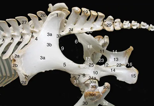
Fig. I.2 Dorsolateral aspect of the equine pelvic bones.
Ilium: 1- Sacral tuber; 2- Tuber coxae, 2a- dorsocranial cuspid, 2b- ventrocranial cuspid, 2c- dorsocaudal cuspid, 2d-ventrocaudal cuspid; 3- Ilium wing, 3a- gluteal face, 3b- accessory gluteal line; 4- Ilium crest, 5- Ilium neck; 6- Psoas minor muscle tubercle; 7- Ilium body; 8- Lateral rectus femoris muscle area; 9- Major sciatic incisura;
Ischium: 10- Ischium body; 11- Lateral (acetabular) ramus; 12- Medial ramus; 13- Ischium table; 14- Minor sciatic incisura; 15- Tuber ischiadicum (ischiatic tuberosity); 16- Ischiatic arch;
Pubis: 17- Cranial ramus; 18- Obturator sulcus;
Sacrum: 19- Median sacral crest (five spinal processes); 20- Lateral sacral crest (fused transverse processes); 21- Dorsal sacral foramen;
Pelvis and connected bones: 22- Pelvic symphysis; 23- Obturator foramen; 24- Acetabulum, 24a- Acetabular margin; 25- Ischiatic spine (sciatic crest); 26- Spinal process of the sixth lumbar vertebra; 27- Fourth lumbar vertebra; 28- Intervertebral foramen; 29- First caudal vertebra; 30- Left femur, 30a- head, 30b- neck, 30c- major trochanter (caudal part); 30d- major trochanter (cranial part); 31- Right femur; 32- Right tibia; 33- Sacroiliac joint.
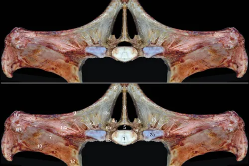
Fig. I.3 Ventral aspect of the equine pelvic bones.
Ilium: 1- Tuber coxae, 1a- dorsocranial cuspid, 1b- ventrocranial cuspid, 1c- ventrocaudal cuspid; 2- Ilium wing, 2a- iliac face, 2b- sacropelvic face (insertion of the interosseous sacroiliac ligament); 3- Ilium crest; 4- Ilium neck; 5- Arch line; 6- Psoas minor muscle tubercle; 7- Ilium body; 8- Medial rectus femoris muscle area; 9- Major sciatic incisura;
Pubis: 10- Pubis body; 11- Cranial ramus; 12- Caudal ramus, 12a- symphysial face; 13- Pubis pecten; 14- Accessory ligament sulcus;
Ischium: 15- Ischium body; 16- Lateral (acetabular) ramus; 17- Ischium table; 18- Medial ramus, 18a- symphysial face; 19- Tuber ischiadicum (ischiatic tuberosity); 20- Ischiatic arch;
Pelvis: 21- Pelvic symphysis; 22- Obturator foramen; 23- Acetabulum, 23a- acetabular margin, 23b- lunar surface, 23c- acetabular fossa, 23d- acetabular notch;
Sacrum: 24- First sacral vertebra; 25- Fourth sacral vertebra; 26- Sacral wing; 27- First ventral (intervertebral) sacral foramen; 28- Third ventral (intervertebral) sacral foramen; 29- Promontory; 30- Sacroiliac joint;
Lumbar spine: 31- Sixth lumbar vertebra (L6); 32- Transverse process of the fifth lumbar vertebra (L5, fused with L6); 33- Lumbar ventral intervertebral foramen; 34- Third lumbar vertebra (L3, ventral crest); 35- Transverse process of L3; 36- Intertransverse lumbosacral joint;37- Intertransverse synostosis between L5 and L6; 38- Intervertebral symphysis between L3 and L4.
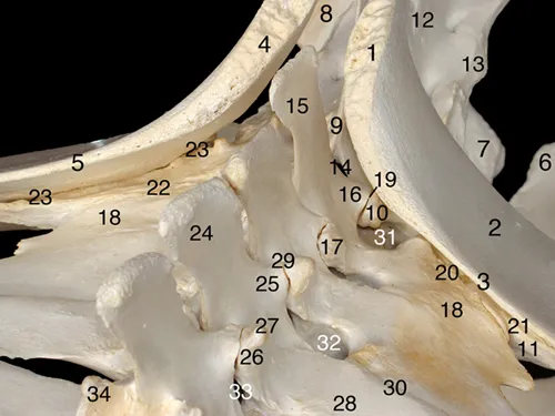
Fig. I.4 Dorsal aspect of the equine pelvic bones.
Ilium: 1- Tuber coxae, 1a- dorsocranial cuspid, 1b- dorsocaudal cuspid, 1c- ventrocaudal cuspid; 2- Sacral tuber; 3- Ilium wing; 4- Ilium crest; 5- Ilium neck; 6- Ilium body; 7- Major sciatic incisura;
Ischium: 8- Ischium body; 9- Lateral (acetabular) ramus; 10- Ischium table; 11- Medial ramus, 11a- symphysial face; 12- Tuber ischiadicum (ischiatic tuberosity); 13- Ischiatic arch;
Pubis: 14- Cranial ramus; 15- Caudal ramus, 15a- symphysial face;
Pelvis: 16- Pelvic symphysis, 16a- cranial (pubic part), 16b- caudal (ischiatic) part; 17- Obturator foramen; 18- Ischiatic spine (sciatic crest); 19- Acetabulum, 19a- acetabular margin;
Sacrum: 20- Median sacral crest; 21- Lateral sacral crest; 22- Transverse process of the first sacral vertebra (sacral wing); 23- Right lumbosacral articular process joint; 24- Sacroiliac joint; 25- Third dorsal (intervertebral) sacral foramen; 26- Sacral canal (caudal opening); 27- First caudal vertebra (vertebral body);
Lumbar spine: 28- Transverse process of the third lumbar vertebra (L3); 29- Transverse process of the fifth lumbar vertebra (L5); 30- Right articular process joint between L3 and L4.
Fig. I.5 Dorsal aspect of the equine sacrum.
1- Spinal process of the first sacral vertebra (S1); 2- Vertebral arch of S1; 3- Left cranial articular process of S1; 4- Transverse process of the first sacral vertebra (sacral wing), 4a- auricular surface (contributing to the sacroiliac joint), 4b- insertion surface of the interosseous sacroiliac ligament; 5- Articular surface of the intertransverse lumbosacral joint seen through the cranial intervertebral incisura; 6- Median sacral crest, 6a- spinal process of the second sacral vertebra, 6b- spinal process of the fifth sacral vertebra; 7- First dorsal (intervertebral) sacral foramen; 8- Second dorsal (intervertebral) sacral foramen; 9- Lateral sacral crest; 10- First caudal vertebra, 10a- vertebral arch, 10b- vertebral body.
Fig. I.6 Craniolaterodorsal aspect of the lumbosacroiliac area.
Coxal bones: 1- Sacral tuber of the left ilium; 2- Ilium wing of the left ilium; 3- Ilium crest of the left ilium; 4- Sacral tuber of the right ilium; 5- Ilium crest of the right ilium; 6- Left ischium (angle between ilium arch and pelvic symphysis); 7- Right ischium (angle between ilium arch and pelvic symphysis); 8- Right tuber ischiadicum;
Sacrum: 9- Spinal process of the first sacral vertebra (S1); 10- (Cranial) articular process of S1; 11- Left transverse process of S1 (sacral wing); 12- Median sacral crest; 13- Lateral sacral crest; 14- Interarcual space between the last lumbar vertebra (L6) and S1;
Lumbar vertebrae and sacroiliac joints: 15- Spinal process of L6; 16- Caudal articular process of L6; 17- Cranial articular process of L6; 18- Transverse process of L6 (fused with the transverse process of the fifth lumbar vertebra (L5)); 19- Left lumbosacral articular process joint; 20- Left lumbosacral intertransverse joint; 21- Left sacroiliac joint; 22- Right lumbosacral intertransverse joint; 23- Right sacroiliac joint; 24- Spinal process of the fourth lumbar vertebra (L4); 25- Caudal articular process of L4; 26- Cranial articular process of L4; 27- Mammillary process; 28- Transverse process of L4; 29- Left articular process joint between L3 and L4; 30- Left intertransverse joint between L4 and L5; 31- Dorsal lumbosacral intertransverse foramen; 32- Dorsal intertransverse foramen between L4 and L5; 33- Left intervertebral foramen between L3 and L4; 34- Right cranial articular process of the third lumbar vertebra.
I.3 Dissected specimen
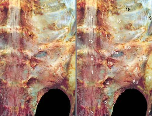
Fig. I.7 Ventral aspect of the roof of the pelvis: bones and ligaments.
1- Body of the first sacral vertebra (S1); 2- Transverse process of S1 (sacral wing); 3- Body of the third sacral vertebra (S3); 4- First ventral (intervertebral) sacral foramen; 5- Second ventral (intervertebral) sacral foramen; 6- Ilium wing; 7- Ilium neck; 8- Sacroiliac joint covered by the ventral sacroiliac ligament, 8a- caudomedial part, 8b- craniolateral part (covered by the iliopsoas muscle); 9- Lumbosacral intervertebral disc (sixth lumbar (L6) disc); 10- Lumbosacral intertransverse joint, 10a- joint space, 10b- lumbosacral intertransverse ligament; 11- Ventral ramus of the sixth lumbar nerve; 12- Body of the sixth lumbar vertebra (L6); 13- Transverse process of L6 (fused with L5 transverse process); 14- Fifth lumbar intervertebral disc; 15- Body of the fifth lumbar vertebra (L5); 16- Transverse process of L5 (fused with L6 transverse process); 17- Ventral intertransverse foramen between L5 and L6; 18- Intertransverse ligament; 19- Iliolumbar ligament.
Fig. I.8 Cranial aspect of the equine pelvis: dorsal part.
1- Vertebral body of the first sacral vertebra (S1); 2- Transverse process of S1 (sacral wing), 2a- articular surface of the intertransverse lumbosacral joint; 3- Spinal process of S1; 4- Left (cranial) articular process of S1, 4a- articular surface of the right artic...