MRI Physics
eBook - ePub

MRI Physics

Tech to Tech Explanations

  1. English
  2. ePUB (mobile friendly)
  3. Available on iOS & Android
eBook - ePub

MRI Physics

Tech to Tech Explanations

About this book

MRI PHYSICS

MRI PHYSICS

TECH TO TECH EXPLANATIONS

Technologists must have a solid understanding of the physics behind Magnetic Resonance Imaging (MRI), including safety, the hows and whys of the quantum physics of the MR phenomenon, and how to competently operate MRI scanners. Generating the highest quality images of the human body involves thorough knowledge of scanner hardware, pulse sequences, image contrast, geometric parameters, and tissue suppression techniques.

MRI Physics: Tech to Tech Explanations is designed to help student MRI technologists and radiotherapists preparing for Advanced MRI certification examinations to better understand difficult concepts and topics in a quick and easy manner.

Written by a highly experienced technologist, this useful guide provides clear and reader-friendly coverage of what every MR Technologist needs to know. Topics include safety considerations associated with the magnetic field and RF, pulse sequences, artifacts, MRI math, the much-feared gradients, and I.V. contrast.

  • Provides basic guidance on safety considerations, protocols options, critical thinking, and image contrast optimization
  • Simplifies the challenging topic of MRI physics using straightforward language and clear explanations
  • Covers content for American Registry of Radiologic Technologists (ARRT) and Continuing Qualifications Requirements (CQR) exams
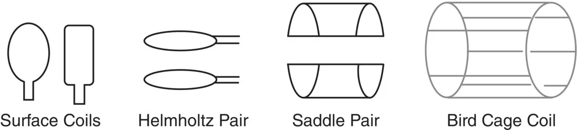
  • Features numerous illustrations and photographs of various MRI concepts, pulse sequence design, artifacts, and the application of concepts in clinical settings

MRI Physics: Tech to Tech Explanations is a must-have resource for the experienced and training MRI technologist, medical students, and radiology residency rotations.

Frequently asked questions

Yes, you can cancel anytime from the Subscription tab in your account settings on the Perlego website. Your subscription will stay active until the end of your current billing period. Learn how to cancel your subscription.
At the moment all of our mobile-responsive ePub books are available to download via the app. Most of our PDFs are also available to download and we're working on making the final remaining ones downloadable now. Learn more here.
Perlego offers two plans: Essential and Complete
  • Essential is ideal for learners and professionals who enjoy exploring a wide range of subjects. Access the Essential Library with 800,000+ trusted titles and best-sellers across business, personal growth, and the humanities. Includes unlimited reading time and Standard Read Aloud voice.
  • Complete: Perfect for advanced learners and researchers needing full, unrestricted access. Unlock 1.4M+ books across hundreds of subjects, including academic and specialized titles. The Complete Plan also includes advanced features like Premium Read Aloud and Research Assistant.
Both plans are available with monthly, semester, or annual billing cycles.
We are an online textbook subscription service, where you can get access to an entire online library for less than the price of a single book per month. With over 1 million books across 1000+ topics, we’ve got you covered! Learn more here.
Look out for the read-aloud symbol on your next book to see if you can listen to it. The read-aloud tool reads text aloud for you, highlighting the text as it is being read. You can pause it, speed it up and slow it down. Learn more here.
Yes! You can use the Perlego app on both iOS or Android devices to read anytime, anywhere — even offline. Perfect for commutes or when you’re on the go.
Please note we cannot support devices running on iOS 13 and Android 7 or earlier. Learn more about using the app.
Yes, you can access MRI Physics by Stephen J. Powers in PDF and/or ePUB format, as well as other popular books in Medicine & Medical Technology & Supplies. We have over one million books available in our catalogue for you to explore.

Information

1
Hardware: Magnet Types and Coils

Magnets

Three types of magnets are used in MRI:
  • Permanent. These are rare to almost nonexistent today. They are made up of brick‐sized magnets that, when placed in the right pattern, combine to make a magnetic field strong enough to image with. These magnets are large, heavy, and have a weak B0 field so signal‐to‐noise ratio (SNR) is at a premium. They cannot however be turned off.
  • Resistive. These consist of a large coil of wire with a lot of electricity passing through it. The amount of electricity circulating is sufficient to make a magnetic field strong enough to image with. These are usually 0.5–0.7 T. They also generate a lot of heat from electrical resistance in the wire.
  • Superconducting. These are resistive magnets but with one major difference. The magnet is cooled with liquid helium, a cryogen. The cryogen decreases electrical resistance in the wires that make up the magnet. The loss of resistance from cooling enables more current to flow so stronger magnetic fields are possible. Typical field strengths are 1–4T. Research magnets can be as strong as 7–9 T. Stronger magnetic fields also come with a larger fringe field compared to the lower‐power fields. Think of the fringe field as being like the “Scatter Radiation” in x‐radiography. The magnetic field is not solely confined to the scanner. Magnetism picks up rapidly as you get closer to the bore.
The field attraction gets stronger the closer you get to isocenter.

Teaching Moment:

Magnetic force of attraction is a cubic exponential function. Halve your distance and the strength of the fringe field you are experiencing goes up by a factor of 8 (23, or 2 × 2 × 2). That is why pens, beepers, and paper clips get pulled out of your pockets and go flying into the bore at about 40 mph.

The Superconducting Magnet

The magnet coil is cooled with liquid helium. Helium is usually a gas, but when it is sufficiently compressed it becomes a liquid which is extremely cold. Liquid helium is a cryogen with a temperature of about 4° Kelvin (very close to absolute zero).
Helium does not want to be a liquid. It wants to be a gas and revert to a lower energy state. If allowed to, it will do so very rapidly. The resulting conversion back to a gas is called a quench. Quenches result in a high conversion ratio of about 750:1, meaning that 1 litre of liquid helium makes about 750 litres of gaseous helium. Superconducting magnets have a large vent pipe leading outside the building so that if there is a quench, the gases vent outside and do not fill the scan room.
Quenching the magnet is only done in a life‐threatening situation such as when someone is pinned between a ferrous object and the magnet. Trust me, if an O2 bottle, IV pole, or stretcher is drawn in to the magnet, you will NOT get it off. A quench will result in the scanner being down for several days.

Coils

There is a plethora of different coils. Some are general purpose; others are highly specific. General purpose or flexible coils are made by multiple manufacturers. They can be used for a shoulder on one patient, a knee on the next, then a soft tissue mass on another.
Coils for more specific use are breast, endo‐rectal, or the long‐lost Temporal‐Mandibular Joint (TMJ) coils. If you still have TMJ coils and they still work on your system, keep them, they are awesome for wrists, orbits, and fingers! Remember, a coil is a coil is a coil. But also know that coils are system specific. You cannot use a 1.5 T coil on a 3 T system and vice versa.
Coils vary in size: small, medium, and large. If you can get it around, under, or over the area to be imaged, it will work. However, you have to choose a coil size that is appropriate to the patient size or coverage needed. You should use the smallest coil possible for best signal to noise. With all scan factors remaining the same, a small coil has better SNR than a large coil. Why? The smaller coil unto itself does not see more signal, it sees less noise. That’s the signal to noise ratio. The same amount of signal with less noise results in a higher SNR.
An imaging coil is just that: loops of wire that when placed next to or around a body part form an integral part of something in MR called Faraday’s law of induction. Faraday’s law of induction states that a moving magnet field passing through a coil of wire will induce current to flow in that wire. Current is signal, and signal is money.
Schematic illustration of the four basic coil types.
Figure 1.1 The four basic coil types. Typical body parts they are used on are:
  • Surface coil: spine
  • Saddle coil: knee, foot/ankle
  • Helmholtz: shoulder (Figure 1.2)
  • Bird cage (quadrature): brain (Figure 1.3).
Figure 1.1 shows the four basic coil types. Remember, t...

Table of contents

  1. Cover
  2. Table of Contents
  3. Title Page
  4. Copyright Page
  5. Dedication Page
  6. About the Author
  7. Preface
  8. Acknowledgements
  9. Introduction
  10. 1 Hardware: Magnet Types and Coils
  11. 2 The Basics
  12. 3 Image Weighting
  13. 4 Introduction to the Basic Pulse Sequences
  14. 5 Multi Echo Spin Echo Sequence
  15. 6 Tissue Suppression
  16. 7 The Gradient Echo Sequence
  17. 8 Gradient Echo Magnetic Resonance Angiography
  18. 9 k‐Space
  19. 10 Echo Planar Sequences
  20. 11 Geometric Parameters
  21. 12 Image Artifacts
  22. 13 Gradients
  23. 14 MRI Math
  24. 15 Parallel Imaging
  25. 16 IV Gadolinium
  26. Glossary
  27. Suggested Reading
  28. Index
  29. End User License Agreement