Emergency Procedures for the Small Animal Veterinarian E-Book
eBook - ePub

Emergency Procedures for the Small Animal Veterinarian E-Book

  1. 688 pages
  2. English
  3. ePUB (mobile friendly)
  4. Available on iOS & Android
eBook - ePub

Emergency Procedures for the Small Animal Veterinarian E-Book

About this book

The new edition of the hugely successful Emergency Procedures for the Small Animal Veterinarian gives you all the information you need to form a diagnosis quick and accurately, establish a prognosis and recommend treatment for a patient suffering and illness, injury or toxic event.Easy-to-read bullet-point text gives quick access to the most essential information needed to treat emergency cases quickly and efficiently. Loads of practical appendices of commonly used drugs and supplements, drugs in special circumstances (e.g. safe drugs in pregnancy, drugs to avoid in renal failure, etc.), clinical chemistry and laboratory data, conversion tables, and many more, are included for easy reference to essential data.With step-by-step coverage of cardiopulmonary emergencies, trauma gastrointestinal emergencies, toxicological events, a greatly expanded chapter on exotic pets, and much more, Emergency Procedures for the Small Animal Veterinarian gives you the facts you need to help you save more lives faster!

  • All you need to know to manage every small animal emergency case you will encounter
  • Many excellent and practical appendices of drugs, poisons, lab data, haematology
  • Takes the diagnosis-prognosis-treatment approach to every emergency situation
  • Easy-to-access format, with concise text and lots of lists
  • Divided into organ systems, making it easy to locate information in a hurry
  • Handy information on what to tell the owner in emergency injury situations
  • Expanded chapter on Emergencies in Exotic Species
  • Text updated throughout
  • Information on drugs is updated
  • New cover design to make the new edition stand out
  • Flexicover to protect the book against heavy usage - this is not a book that will remain on the shelves

Frequently asked questions

Yes, you can cancel anytime from the Subscription tab in your account settings on the Perlego website. Your subscription will stay active until the end of your current billing period. Learn how to cancel your subscription.
At the moment all of our mobile-responsive ePub books are available to download via the app. Most of our PDFs are also available to download and we're working on making the final remaining ones downloadable now. Learn more here.
Perlego offers two plans: Essential and Complete
  • Essential is ideal for learners and professionals who enjoy exploring a wide range of subjects. Access the Essential Library with 800,000+ trusted titles and best-sellers across business, personal growth, and the humanities. Includes unlimited reading time and Standard Read Aloud voice.
  • Complete: Perfect for advanced learners and researchers needing full, unrestricted access. Unlock 1.4M+ books across hundreds of subjects, including academic and specialized titles. The Complete Plan also includes advanced features like Premium Read Aloud and Research Assistant.
Both plans are available with monthly, semester, or annual billing cycles.
We are an online textbook subscription service, where you can get access to an entire online library for less than the price of a single book per month. With over 1 million books across 1000+ topics, we’ve got you covered! Learn more here.
Look out for the read-aloud symbol on your next book to see if you can listen to it. The read-aloud tool reads text aloud for you, highlighting the text as it is being read. You can pause it, speed it up and slow it down. Learn more here.
Yes! You can use the Perlego app on both iOS or Android devices to read anytime, anywhere — even offline. Perfect for commutes or when you’re on the go.
Please note we cannot support devices running on iOS 13 and Android 7 or earlier. Learn more about using the app.
Yes, you can access Emergency Procedures for the Small Animal Veterinarian E-Book by Signe J. Plunkett in PDF and/or ePUB format, as well as other popular books in Medicine & Veterinary Medicine. We have over one million books available in our catalogue for you to explore.

Information

Publisher
Saunders Ltd.
Year
2012
eBook ISBN
9780702054303
Edition
3
1

Supportive therapy

Acid–base disturbances

Evaluation of the blood pH, bicarbonate (HCO3−),
image
and base excess may be very helpful in the assessment of a critically ill or injured animal. Arterial blood samples are necessary to evaluate the respiratory status and are the preferred sample, but venous blood samples are helpful to evaluate the status of the patient at the cellular level.
The normal arterial blood gas reference ranges for dogs and cats inspiring room air are listed in Table 1.1.
Table 1.1
Typical reference ranges for normal arterial blood gas values for dogs and cats inspiring room air
DogCat
pH7.41 (7.35–7.46)7.39 (7.31–7.46)
image
(mm Hg)
37 (31–43)31 (25–37)
[HCO3−] (mEq/L)22 (19–26)18 (14–22)
image
(mm Hg)
92 (81–103)107 (95–118)
Evaluation of blood gas results is a process involving many steps.
1. Determine whether the pH is normal.
2. Evaluate the partial pressure of carbon dioxide (PCO2) – the respiratory component.
3. Evaluate the bicarbonate concentration [HCO3−] – the nonrespiratory component.
4. What is the primary disorder? The direction of the expected changes are shown in Table 1.2.
Table 1.2
The changes in simple primary acid–base disorders
DisorderpH
image
[HCO3−]
Respiratory acidosis↓↑↑ or normal
Respiratory alkalosis↑↓↓ or normal
Nonrespiratory (Metabolic) acidosis↓↓↓
Nonrespiratory (Metabolic) alkalosis↑↑↑
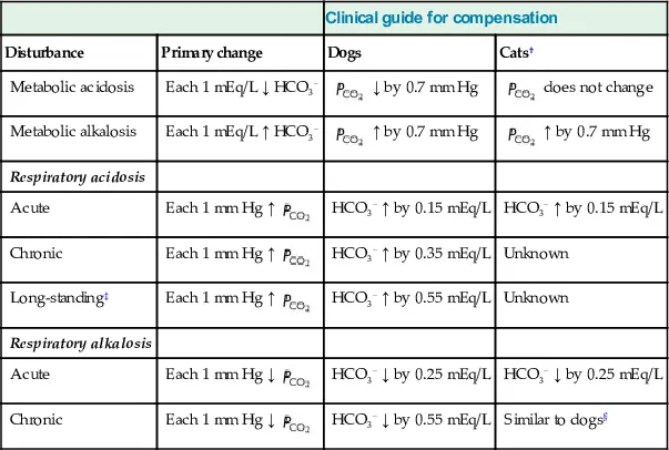
5. Is the secondary or adaptive response as expected? The expected responses are shown in Table 1.3.
Table 1.3
Expected compensatory response in simple acid–base disturbances in dogs and cats*
Clinical guide for compensation
DisturbancePrimary changeDogsCats†
Metabolic acidosisEach 1 mEq/L ↓ HCO3−
image
↓ by 0.7 mm Hg
image
does not change
Metabolic alkalosisEach 1 mEq/L ↑ HCO3−
image
↑ by 0.7 mm Hg
image
↑ by 0.7 mm Hg
Respiratory acidosis
AcuteEach 1 mm Hg ↑
image
HCO3− ↑ by 0.15 mEq/LHCO3− ↑ by 0.15 mEq/L
ChronicEach 1 mm Hg ↑
image
HCO3− ↑ by 0.35 mEq/LUnknown
Long-standing‡Each 1 mm Hg ↑
image
HCO3− ↑ by 0.55 mEq/LUnknown
Respiratory alkalosis
AcuteEach 1 mm Hg ↓
image
HCO3− ↓ by 0.25 mEq/LHCO3− ↓ by 0.25 mEq/L
ChronicEach 1 mm Hg ↓
image
HCO3− ↓ by 0.55 mEq/LSimilar to dogs§
*From DiBartola SP (ed), Fluid, Electrolyte, and Acid-Base Disorders in Small Animal Practice, 3rd edn, St Louis, Elsevier, 2006, p 298, reprinted with permission from Elsevier Ltd. Data in dogs from de Morais & DiBartola (1991). See DiBartola (2006) for reference in cats.
†Data from cats are derived from a very limited number of cats.
‡More than 30 days.
§Exact degree of compensation has not been determined, but in cats with chronic respiratory alkalosis maintain normal arterial pH.
6. Which disease process(es) is(are) responsible for the acid–base disorder?

Respiratory acidosis

Respiratory acidosis or primary hypercapnia indicates hypoventilation and hypoxemia. The blood pH is decreased, the
image
is increased and, with compensation, the HCO3− increases.
Sympathetic activation, increased cardiac output and possibly tachyarrhythmias may occur in the presence of moderately elevated
image
. As the
image
increases, intracranial pressure and cerebral blood flow increase. Disorientation, narcosis, and coma may occur at extremely high
image
levels (60–70 mm Hg).
Causes of respiratory acidosis include:
• Depression of the respiratory center
â—‹ Associated with medications (inhalant anesthetics, opioids, barbiturates)
â—‹ Neurologic disease
– Cervical spinal cord lesion
– Brainstem lesion
• Neuromuscular disease
â—‹ Myasthenia gravis
â—‹ Botulism
â—‹ Tetanus
â—‹ Tick paralysis
â—‹ Severe hypokalemia
â—‹ Associated with medications or chemicals (organophosphates, aminoglycosides)
• Obstruction of large airways
â—‹ Aspiration
â—‹ Kinked or plugged endotracheal tube
â—‹ Tracheal co...

Table of contents

  1. Cover image
  2. Title Page
  3. Table of Contents
  4. Dedication
  5. Copyright
  6. Preface
  7. Acknowledgments
  8. List of contributors
  9. 1 Supportive therapy
  10. 2 Shock
  11. 3 Cardiovascular emergencies
  12. 4 Respiratory emergencies
  13. 5 Traumatic emergencies
  14. 6 Environmental emergencies
  15. 7 Dermatologic emergencies
  16. 8 Hematologic emergencies
  17. 9 Gastrointestinal emergencies
  18. 10 Metabolic and endocrine emergencies
  19. 11 Urinary emergencies and electrolyte disorders
  20. 12 Reproductive emergencies
  21. 13 Neurologic and ocular emergencies
  22. 14 Toxicologic emergencies
  23. 15 Emergencies in exotic species
  24. Appendices
  25. Index