"This is a 'go-to' reference text for a serious cytologist. " Reviewed by: Kathleen Tennant on behalf of Veterinary Record, November 2015- Comprehensive coverage of all body systems and body fluids — and the pathological changes associated with various infectious agents — emphasizes areas in which the application of cytology has the greatest diagnostic value.- Exceptional-quality, full-color photomicrographs show both normal and abnormal tissue and also include detailed legends.- Discussions of clinical, differential, and cytological diagnosis accompany the illustrations of lesions and conditions in each chapter.- Helpful hints for improving specimen quality are provided in discussions of common errors and problems encountered in the preparation of cytological specimens.- Coverage of histology in organ system chapters demonstrates the histological or histopathologic corollary of cytologic findings.- Clear, concise descriptions include sampling techniques, slide preparation and examination, and guidelines for interpretation, leading to accurate in-house and commercial laboratory diagnosis.- Easy-to-use, well-organized format includes many tables, algorithms, boxes, and Key Point callouts for at-a-glance reference.- NEW! Chapter on Fecal Cytology- Highlighted boxes featuring Key Points provide helpful tips for best conceptual understanding and diagnostic effectiveness- Photomicrographs now include more comparative histology- Discussions of broader uses of stains and immunocytochemistry for differential cytologic characterization- Expanded chapter on Advanced Diagnostic Techniques includes more methodology and application of current tools, representing advances in both aspiration and exfoliative cytology.
eBook - ePub
Canine and Feline Cytology - E-Book
Canine and Feline Cytology - E-Book
- 544 pages
- English
- ePUB (mobile friendly)
- Available on iOS & Android
eBook - ePub
About this book
Trusted by 375,005 students
Access to over 1.5 million titles for a fair monthly price.
Study more efficiently using our study tools.
Information
Topic
MedicineSubtopic
Veterinary MedicineChapter 1
The Acquisition and Management of Cytology Specimens
Denny J. Meyer
The classification of events that depend on the accuracy of observation is limited by the ability of the observer to describe and of the interpreter to decipher.
—Michael Podell, M.Sc., D.V.M.
For the microscopic examination of tissue, one important factor that affects the accuracy of observation is specimen management. The successful use of aspiration cytology depends on several interrelated procedures: acquisition of a representative specimen, proper application to a glass side, adequate staining, and examination with a high-quality microscope. A deficiency in one or more of these steps will adversely affect the yield of diagnostic information. The objective of this chapter is to provide general recommendations for managing samples in order to ensure accurate diagnosis.
General Sampling Guidelines
Before executing any sampling procedure, a cytology kit should be prepared and dedicated for that purpose. An inexpensive plastic tool caddie works well. Suggested contents are listed in Box 1-1. Six or more slides are placed on a firm, flat surface such as a surgical tray immediately before initiating the sampling procedure. The surface of the glass slide should be routinely wiped with a paper towel, or at least on a shirtsleeve, to remove “invisible” glass particles that interfere with the spreading procedure.
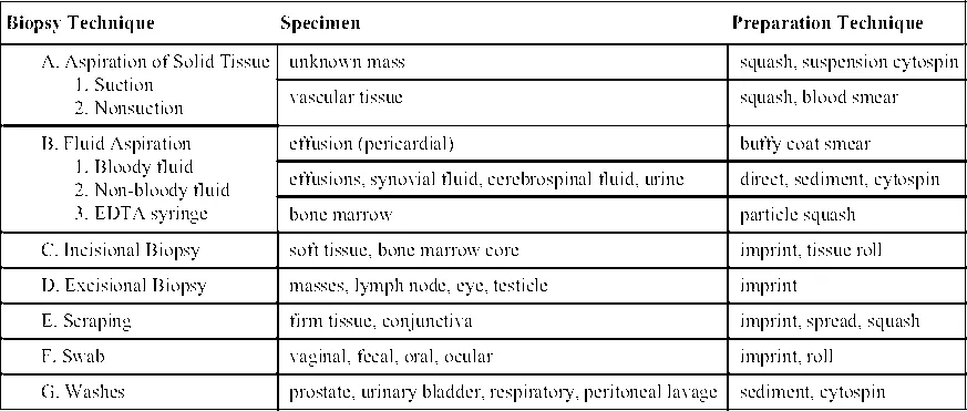
Table 1-1 lists biopsy techniques, example specimens, and suggested cytologic preparation techniques. The collection of specimens for cytologic evaluation from cutaneous and subcutaneous tissues and abdominal organs and masses in smaller animals is generally accomplished with a 20- or 22-gauge, 1- to 1½-inch needle firmly attached to a 6- or 12-mL syringe. For internal organs that are more difficult to reach, a 2½- to 3½-inch spinal needle is used. The added length amplifies the area for cell collection and enhances the diagnostic yield—cores of hepatic tissue can be obtained with a longer needle. The stylet can be left in place as the cavity is entered to avoid contamination during the “searching” process of locating the tissue of sampling interest. Coating the needle and syringe hub with sterile 4% disodium ethylenediaminetetraacetic acid (EDTA) before aspiration biopsy sampling of vascular tissues, notably the bone marrow, reduces the risk of clot formation that will compromise the quality of the cytologic specimen. For the relatively inexperienced, this may be a practice to consider routinely when sampling any tissue. Clotted specimens are a frequent cause of cytologic preps of poor quality.
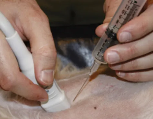
The general steps for obtaining a cytologic specimen are illustrated in Fig. 1-1A-E. Following appropriate cleansing and disinfectant application, the tip of the needle is inserted into the tissue of interest, the plunger retracted slightly (0.5 to 1 mL of vacuum), the needle advanced and retracted in several different directions, the plunger released, the needle withdrawn, and the specimen placed on a glass slide or in an EDTA (purple-topped) tube as appropriate. Commercial aspiration guns (Fig. 1-1B) are available that can be loaded with various size syringes (Fig. 1-1B). The syringe plunger sits within the trigger, which allows for easier and more stable retraction. If fluid is obtained from a mass lesion, the site is completely drained, the needle withdrawn, the fluid placed in an EDTA tube, and the procedure repeated with a new needle directed at firm tissue. Both specimens are examined microscopically. To enhance operator flexibility, a butterfly needle can be used to attach the needle and syringe. Positioning and redirection of the needle is easier and accommodates patient movement (Fig. 1-1C).
Aspiration is not a prerequisite for obtaining a cytologic specimen. A technique based on the principle of capillarity, referred to as fine-needle capillary sampling, can be performed by placing a needle into the lesion with or without a syringe attached (Mair et al., 1989; Yue and Zheng, 1989). The technique has diagnostic sensitivity similar to that of aspiration biopsy when used to sample a variety of tissues. Its major advantage is to reduce blood contamination from vascular tissues such as liver, spleen, kidney, and thyroid. Cells are displaced into the cylinder of the needle by capillary action as the needle is incompletely retracted and redirected into the tissue three to six times. Personal preference is justified when deciding between aspiration and nonaspiration sampling for collection of the specimen. Through trial and error, the operator may determine that each has value for sampling different tissues.
Table 1-1
Biopsy Techniques, Associated Specimens, and Cytologic Preparation Techniques
| Biopsy Technique | Specimen | Preparation Technique |
A. Aspiration of Solid Tissue 1. Suction 2. Nonsuction | ||
| unknown mass | squash, suspension cytospin | |
| vascular tissue | squash, blood smear | |
B. Fluid Aspiration 1. Bloody fluid 2. Non-bloody fluid 3. EDTA syringe | ||
| effusion (pericardial) | buffy coat smear | |
| effusions, synovial fluid, cerebrospinal fluid, urine | direct, sediment, cytospin | |
| bone marrow | particle squash | |
C. Incisional Biopsy | soft tissue, bone marrow core | imprint, tissue roll |
D. Excisional Biopsy | masses, lymph node, eye, testicle | imprint |
E. Scraping | firm tissue, conjunctiva | imprint, spread, squash |
F. Swab | vaginal, fecal, oral, ocular | imprint, roll |
G. Washes | prostate, urinary bladder, respiratory, peritoneal lavage | sediment, cytospin |
Diagnostic Imaging-Guided Sample Collection
Cytology sample collection can be performed under the guidance of fluoroscopy, ultrasound, and computed tomography. Ultrasound guidance is the preferred method because of its widespread availability and portability. In addition, ultrasound provides real-time monitoring of precise needle placement. The technique and indications are detailed elsewhere (Nyland et al., 2002a). Ultrasound-guided fine-needle aspiration biopsy (FNAB) is indicated for cytologic evaluation of nodules and masses detected on ultrasound and to evaluate organomegaly when a diffuse cellular infiltrate such as lymphoma and mast cell tumor is suspected. Most sarcomas exfoliate sparsely or not at all. A surgical or ultrasound-guided cutting needle biopsy is recommended if the FNAB sample is not conclusive. Ultrasound-guided FNAB can be performed in most patients without chemical restraint or local anesthesia. If chemical restraint is needed, agents that promote panting should be avoided because this will lead to excessive movement and gas ingestion (Nyland et al., 2002a).
Biopsy Guidance
Ultrasound-guided FNAB can be performed by freehand technique or with the aid of a biopsy guide fastened to the transducer. Freehand technique consists of holding the transducer in one hand and inserting the needle with the other at an oblique angle to the long axis of the transducer but still within the scan plane (Fig. 1-1D). This technique requires more skill but allows for greater flexibility. If the needle can...
Table of contents
- Cover image
- Title page
- Table of Contents
- Copyright
- Contributors
- Dedication
- Preface
- Acknowledgments
- Chapter 1. The Acquisition and Management of Cytology Specimens
- Chapter 2. General Categories of Cytologic Interpretation
- Chapter 3. Skin and Subcutaneous Tissues
- Chapter 4. Hemolymphatic System
- Chapter 5. Respiratory Tract
- Chapter 6. Body Cavity Fluids
- Chapter 7. Oral Cavity, Gastrointestinal Tract, and Associated Structures
- Chapter 8. Dry-Mount Fecal Cytology
- Chapter 9. The Liver
- Chapter 10. Urinary Tract
- Chapter 11. Microscopic Examination of the Urinary Sediment
- Chapter 12. Reproductive System
- Chapter 13. Musculoskeletal System
- Chapter 14. The Central Nervous System
- Chapter 15. Eyes and Adnexa
- Chapter 16. Endocrine/Neuroendocrine System
- Chapter 17. Advanced Diagnostic Techniques
- Appendix 1. Microscope Basics and Telecytology
- Appendix 2. Selected Cytologic Stains and Techniques
- Appendix 3. Interference and Polarizing Substances
- Appendix 4. Chromatin Patterns
- Appendix 5. Advanced Collection and Preparation Techniques
- Appendix 6. Immunocytochemistry Staining Protocol
- Appendix 7. List of Specialized Diagnostic Testing Sites
- Appendix 8. Quality Assurance and Diagnostic Test Reporting
- Index
Frequently asked questions
Yes, you can cancel anytime from the Subscription tab in your account settings on the Perlego website. Your subscription will stay active until the end of your current billing period. Learn how to cancel your subscription
No, books cannot be downloaded as external files, such as PDFs, for use outside of Perlego. However, you can download books within the Perlego app for offline reading on mobile or tablet. Learn how to download books offline
We are an online textbook subscription service, where you can get access to an entire online library for less than the price of a single book per month. With over 1.5 million books across 990+ topics, we’ve got you covered! Learn about our mission
Look out for the read-aloud symbol on your next book to see if you can listen to it. The read-aloud tool reads text aloud for you, highlighting the text as it is being read. You can pause it, speed it up and slow it down. Learn more about Read Aloud
Yes! You can use the Perlego app on both iOS and Android devices to read anytime, anywhere — even offline. Perfect for commutes or when you’re on the go.
Please note we cannot support devices running on iOS 13 and Android 7 or earlier. Learn more about using the app
Please note we cannot support devices running on iOS 13 and Android 7 or earlier. Learn more about using the app
Yes, you can access Canine and Feline Cytology - E-Book by Rose E. Raskin,Denny Meyer in PDF and/or ePUB format, as well as other popular books in Medicine & Veterinary Medicine. We have over 1.5 million books available in our catalogue for you to explore.