- NEW! Advances in joint surgeries, specifically the knee, shoulder, and elbow, keep practitioners abreast of the latest technology and best practices.- NEW! Coverage of minimally invasive surgery has been added to the many chapters.- NEW! Advances in imaging (MRI, CT, and radiographs) are included to keep practitioners up to date on the latest technology.- Updates on new fixation technologies include angle stable interlocking nails and locking plate fracture fixation systems.- Updates on common surgeries include triple pelvic osteotomy and total hip replacement- NEW! High-definition clinical photographs have been added to give readers a closer view of various fractures and repair techniques.
eBook - ePub
Brinker, Piermattei and Flo's Handbook of Small Animal Orthopedics and Fracture Repair
- 880 pages
- English
- ePUB (mobile friendly)
- Available on iOS & Android
eBook - ePub
Brinker, Piermattei and Flo's Handbook of Small Animal Orthopedics and Fracture Repair
About this book
Trusted by 375,005 students
Access to over 1.5 million titles for a fair monthly price.
Study more efficiently using our study tools.
Information
Topic
MedicineSubtopic
Veterinary MedicinePart I
Diagnosis and treatment of fractures, lameness, and joint disease
Outline
1. Orthopedic examination and diagnostic tools
2. Fractures: Classification, diagnosis, and treatment
3. Bone grafting
4. Delayed union and nonunion
5. Treatment of acute and chronic bone infections
6. Arthrology
7. Principles of joint surgery
8. Arthroscopy in joint surgery
Chapter 1
Orthopedic examination and diagnostic tools
General examination
An orthopedic examination must begin with an adequate history and general physical examination. A systemic approach to the examination ensures that multiple problems are discovered. The animal’s general health should be ascertained before focusing on the orthopedic complaint. The entire examination varies with case complexity, a history of recent trauma, the intended use of the animal (e.g., breeding, showing, racing, hunting), and economics dictated by owners. Severely traumatized animals with hemorrhaging wounds and unstable fractures that could become open fractures obviously need different immediate steps; these animals are not discussed in this chapter. This chapter focuses on the examination for orthopedic problems (Table 1-1) and presents some of the diagnostic tools available.
TABLE 1-1.
Causes of Lameness in the Dog (Excluding Fractures and Minor Soft Tissue Injuries)
| Pelvic Limb | Forelimb |
| Growing Dog | |
| 1. Hip dysplasia 2. Avascular necrosis (Legg-Calvé-Perthes) 3. Avulsion of long digital extensor 4. OCD—stifle 5. OCD—hock 6. Luxating patella complex 7. Genu valgum 8. Panosteitis Medium to large breeds = 1, 3-8 Toy to small breeds =1, 2, 6 Chondrodystrophied breeds = 1, 2, 6, 8 | 1. OCD—shoulder 2. Luxation/subluxation shoulder—congenital 3. Avulsion supraglenoid tubercle 4. OCD—elbow 5. UAP 6. FCP 7. UME 8. Elbow incongruity a. Congenital b. Physeal injury 9. Radius curvus 10. Retained cartilaginous cores (ulna) 11. Panosteitis 12. HOD Medium to large breeds = 1, 4-7, 8b, 9-12 Toy to small breeds = 2, 8, 9 Chondrodystrophied breeds = 5, 8a, 8b, 9, 11, 13 |
| Adult Dog | |
| A. Arthritis (or continuum): 1-7 B. Luxating patella complex C. Panosteitis D. Cruciate/meniscal syndrome E. Inflammatory joint disease F. Neoplasia Medium to large breeds = A1, A 3-7, B, D, F Toy to small breeds = A2, B, D-F Chondrodystrophied breeds = A1, B, D-F | A. Arthritis (or continuum): 1-6, 8, 9 B. UME C. Panosteitis D. Bicipital tenosynovitis/biceps rupture E. Calcification of supraspinatus tendon F. Contracture of infraspinatus or supraspinatus G. Bone/soft tissue neoplasia H. Luxation/subluxation—shoulder I. Inflammatory joint disease J. HO Medium to giant breeds = A, 7, 11, I, J Toy to small breeds = 2, G, H, I, J Chondrodystrophied breeds = A-5, A-8, A-9, C, H, I, J |
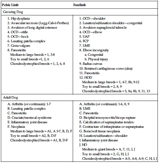
OCD, Osteochondritis dissecans; UAP, ununited anconeal process; FCP, fragmented coronoid process; UME, ununited medial epicondyle; HOD, hypertrophic osteodystrophy; HO, hypertrophic osteopathy
History
Specific historical information is useful for ruling out categories of orthopedic problems. This information includes breed, age, gender, occurrence of trauma, owner identification of limb(s) involved, description of the lameness or gait abnormality, chronological progression of the problem, efficacy of treatments tried, and variability with weather, exercise, and arising from recumbency. Other features such as fever, inappetance, lethargy, and weight loss may indicate some systemic problem, such as inflammatory joint conditions or internal injury from trauma.
Certain historical facts and deviation from the “normal” presentation of certain orthopedic conditions alert the clinician to investigate further by asking appropriate questions or performing additional tests or procedures. For example, a 10-year-old dog that falls down only two stairs and sustains a fractured radius and ulna should be carefully scrutinized for pathological fracture. Normally, chronic luxating patellas usually do not suddenly cause a carrying-leg lameness, and cruciate ligament rupture may have become the more recent problem. Chronic osteoarthritic conditions usually do not cause severe pain. In older animals with severe progressive pain, neoplasia must always be considered. With pelvic fractures, trauma to the chest, abdomen, or spine often occurs. Answers to specific questions help assess concurrent problems. For example, knowing whether the recumbent animal has been eating, voiding large pools of urine, or moving the legs spontaneously is helpful. A good appetite probably does not occur with significant internal injuries. “Urinating” or dribbling small amounts of urine does not mean the bladder is intact, and voluntary leg movement usually means serious thoracolumbar spinal injury has not occurred.
Distant observation and gait evaluation
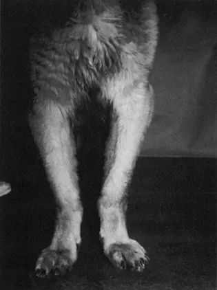
The animal should be observed for general thriftiness and relative weight status. Patient disposition and potential lack of animal or owner cooperation should be noted. Sedation should not be used if possible, or at least until the area of involvement is known, because tranquilizers may mask detection of painful regions. The animal should be observed for body conformation, decreased weight bearing, trembling, asymmetrical joint or soft tissue swellings, muscle atrophy, and digit and joint alignment. Dogs with tarsocrural osteochondritis dissecans (OCD) tend to be very straight legged in the pelvic limb, whereas dogs with elbow problems tend to have curvature of the forelimbs (Figure 1-1).
Gait
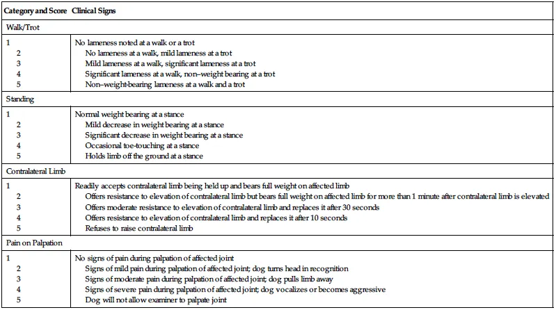
Observing the lameness is helpful before examining the limb. It helps confirm or contradict owner complaints. Often in an examination room environment, however, a mild chronic lameness disappears. The gait is observed at a walk and if necessary a trot. Covert lameness may become apparent during tight circles or stair climbing. Abnormalities include a shortened stride, dragging of the toenails, “toeing-in” or “toeing-out,” limb circumduction, hypermetria, stumbling, generalized weakness, ataxia, crisscrossing of the legs, abnormal sounds (e.g., clicks, snaps), and a head “bob,” which is a bobbing motion of the head that occurs with foreleg lameness. The head elevates as the painful leg strikes the ground. It is important to record specific observations of lameness for a patient, but there is also value in describing lameness using a known subjective grading system or scale. The use of a lameness scale for standing, walking, and trotting may improve consistency of recorded observations in medical records over time. Numerous subjective lameness scales have been used, and one example of a study of cruciate ligament disease in dogs is presented here (Table 1-2).1, 2
TABLE 1-2.
Modified Lameness Grading System (after Vasseur et al)1 Used to Assess Response to Treatment in Dogs After Cranial Cruciate Surgery (Horstman)2
| Category and Score | Clinical Signs |
| Walk/Trot | |
| 1 2 3 4 5 | No lameness noted at a walk or a trot No lameness at a walk, mild lameness at a trot Mild lameness at a walk, significant lameness at a trot Significant lameness at a walk, non–weight bearing at a trot Non–weight-bearing lameness at a walk and a trot |
| Standing | |
| 1 2 3 4 5 | Normal weight bearing at a stance Mild decrease in weight bearing at a stance Significant decrease in weight bearing at a stance Occasional toe-touching at a stance Holds limb off the ground at a stance |
| Contralateral Limb | |
| 1 2 3 4 5 | Readily accepts contralateral limb being held up and bears full weight on affected limb Offers resistance to elevation of contralateral limb but bears full weight on affected limb for more than 1 minute after contralateral limb is elevated Offers moderate resistance to elevation of contralateral limb and replaces it after 30 seconds Offers resistance to elevation of contralateral limb and replaces it after 10 seconds Refuses to raise contralateral limb |
| Pain on Palpation | |
| 1 2 3 4 5 | No signs of pain during palpation of affected joint Signs of mild pain during palpation of affected joint; dog turns head in recognition Signs of moderate pain during palpation of affected joint; dog pulls limb away Signs of severe pain during palpation of affected joint; dog vocalizes or becomes aggressive Dog will not allow examiner to palpate joint |
Standing observation and palpation
With the animal standing as symmetrically as possible, both hands exam...
Table of contents
- Cover image
- Title page
- Table of Contents
- Copyright
- Dedication
- Preface
- 1. Diagnosis and treatment of fractures, lameness, and joint disease
- 2. Fractures and orthopedic conditions of the forelimb
- 3. Fractures and orthopedic conditions of the hindlimb
- 4. Other fractures and reconstruction of bone deformity
- 5. Miscellaneous conditions of the musculoskeletal system
- Index
Frequently asked questions
Yes, you can cancel anytime from the Subscription tab in your account settings on the Perlego website. Your subscription will stay active until the end of your current billing period. Learn how to cancel your subscription
No, books cannot be downloaded as external files, such as PDFs, for use outside of Perlego. However, you can download books within the Perlego app for offline reading on mobile or tablet. Learn how to download books offline
Perlego offers two plans: Essential and Complete
- Essential is ideal for learners and professionals who enjoy exploring a wide range of subjects. Access the Essential Library with 800,000+ trusted titles and best-sellers across business, personal growth, and the humanities. Includes unlimited reading time and Standard Read Aloud voice.
- Complete: Perfect for advanced learners and researchers needing full, unrestricted access. Unlock 1.5M+ books across hundreds of subjects, including academic and specialized titles. The Complete Plan also includes advanced features like Premium Read Aloud and Research Assistant.
We are an online textbook subscription service, where you can get access to an entire online library for less than the price of a single book per month. With over 1.5 million books across 990+ topics, we’ve got you covered! Learn about our mission
Look out for the read-aloud symbol on your next book to see if you can listen to it. The read-aloud tool reads text aloud for you, highlighting the text as it is being read. You can pause it, speed it up and slow it down. Learn more about Read Aloud
Yes! You can use the Perlego app on both iOS and Android devices to read anytime, anywhere — even offline. Perfect for commutes or when you’re on the go.
Please note we cannot support devices running on iOS 13 and Android 7 or earlier. Learn more about using the app
Please note we cannot support devices running on iOS 13 and Android 7 or earlier. Learn more about using the app
Yes, you can access Brinker, Piermattei and Flo's Handbook of Small Animal Orthopedics and Fracture Repair by Charles E. DeCamp,Spencer A. Johnston,Loïc M. Dejardin,Susan Schaefer in PDF and/or ePUB format, as well as other popular books in Medicine & Veterinary Medicine. We have over 1.5 million books available in our catalogue for you to explore.