**Selected for Doody's Core Titles® 2024 with "Essential Purchase" designation in Veterinary Medicine** Learn to recognize, diagnose, and manage a wide range of common ocular conditions with Slatter's Fundamentals of Veterinary Ophthalmology, 6th Edition. This thoroughly updated text provides the latest, most practical information on structure and function of the eye, the ophthalmic examination and diagnostic techniques, medical and surgical management of ocular disease, and management of ocular emergencies. Enhanced and logically organized coverage includes dogs, cats, horses, livestock, birds, and exotic pets. In addition, over 1, 000 color photos and illustrations accurately depict ocular conditions encountered in practice and demonstrate diagnostic and surgical techniques. Edited by three of the most revered authorities in the field of veterinary ophthalmology, this reference is an essential aid to successful veterinary practice and education.- Clinical Tips boxes such as "The Controversy Remains", "Did You Know?", "Look Again", and "Note" offer helpful practice advice and facts.- UPDATED Additional species added to the exotics chapter include birds, small mammals, and others.- A team of internationally respected veterinary ophthalmologists provide comprehensive, clinical expertise in all areas needed to evaluate, diagnose, manage, and monitor a patient with ophthalmic disease.- Practical, clinically focused coverage provides a one-stop diagnostic guide to ophthalmic disease in small and large animals including dogs, cats, horses, livestock (cows, sheep, goats), birds, and exotic pets.- Chapters on equine, livestock, and exotic pet ophthalmology written by specialists in these fields for the most clinically relevant coverage.- NEW! Chapter on ophthalmic surgical techniques describes instrument and suture choice, patient positioning, surgical preparation, and general techniques.- NEW! Additional drawings depict ophthalmic surgeries.- NEW! In-depth equine and livestock ophthalmology content- NEW! Suggested reading lists included at the end of each chapter.
eBook - ePub
Slatter's Fundamentals of Veterinary Ophthalmology E-Book
Slatter's Fundamentals of Veterinary Ophthalmology E-Book
- 584 pages
- English
- ePUB (mobile friendly)
- Available on iOS & Android
eBook - ePub
Slatter's Fundamentals of Veterinary Ophthalmology E-Book
Slatter's Fundamentals of Veterinary Ophthalmology E-Book
About this book
Trusted by 375,005 students
Access to over 1.5 million titles for a fair monthly price.
Study more efficiently using our study tools.
Information
1
The Eye and Vision
Paul E. Miller
Evolution of the Eye
Vision is a fundamental sensory phenomenon that affords a distinct survival advantage to most animals in existence today. The range of the types of eyes is breathtaking, as exemplified in the phylum Mollusca, which has members with eyes ranging from simple clusters of light-sensitive cells to complex mammalian-like structures with a cornea, lens, and retina that can control the depth of focus and the amount of light entering the eye (Figure 1-1). Light-sensitive visual pigments appear to have a common ancestry across a diverse range of species, as all seven subfamilies of light-detecting proteins (opsins) were already present in the last common ancestor of all animals. This commonality suggests that light-sensing pigments evolved from one common ancestral protein.
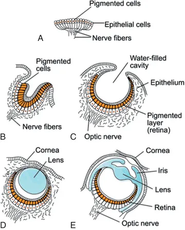
FIGURE 1-1 Varying levels of complexity in the visual organs of living mollusk species. A, A simple, flat patch of pigmented cells connected to nerve fibers as may be found in jellyfish. B, A slightly more complex pigment cup as found in the limpet Patella, which does not form an image but provides information about the direction of incoming light. C, A pinhole camera–type eye filled with water as found in Nautilus; this allows for an image to be focused on the retina. D, A camera-type eye with a basic lens and cornea as found in the marine snail Murex. E, A complex camera-type eye with a cornea, lens, iris, and retina as in a cuttlefish, squid, or octopus. Muscles moving the lens allow for control of depth of focus and the iris controls the amount of light entering the eye. This shows visual organs as they occur in modern species—none is ancestral to another, and this does not necessarily reflect a historical series of steps in the evolution of complex eyes. (Image courtesy Wikipedia public domain.)
In contrast, as reviewed by Schwab, the anatomic and physiologic diversity of complex image-forming eyes suggests these structures may have evolved on 50 to 100 separate occasions, affirming the considerable survival advantage that vision affords. As there is intense evolutionary pressure for each species to optimize its visual capabilities to exploit a specific ecological niche and not to simply evolve toward human visual capabilities, it is inaccurate to describe one species as having vision that is “superior” or “inferior” to another.
For example, although humans have an additional color-detecting photopigment and greater visual acuity than most mammals, most domestic mammals have a much larger visual field and greater ability to see in dim light than do humans. Additionally, it must be remembered that vision did not evolve in isolation from the other senses. When one considers that a dog's sense of smell is estimated to be 1000 times more acute than humans' and that their sense of hearing is approximately four times more acute than humans', it can easily be argued that dogs are at least as “advanced” as humans in terms of their sensory capabilities. Therefore it is more appropriate to evaluate a species based on how well adapted it is to exploit its environmental niche rather than on specific sensory capabilities.
Development of the Eye
The development of the mammalian eye mimics many of the stages observed in the evolution of the eye (Figure 1-2). As the neural tube closes, fluid within it pressurizes, causing the evagination of the optic stalks and bulbous optic vesicles. As described by Aguirre et al, this begins on day 15 of gestation in the dog. As the optic vesicles enlarge, the overlying surface ectoderm forms the lens placode and contact between the vesicle and placode induces the optic vesicle to invaginate, forming a double-layered optic cup. The anterior rim of the cup forms the anterior uvea (iris and ciliary body), and the posterior part forms the retina. The double epithelium of this structure forms the two epithelial layers of the uvea and retina. The invagination of the vesicle is initially not completed on the ventral side of the optic cup, forming an embryonic optic fissure that allows the hyaloid vascular system to enter the eye and nourish the inner layer of the optic cup and developing lens. Failure of the optic fissure to completely close later in development results in a coloboma, a condition in which a portion of the eye is usually lacking.
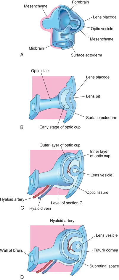
FIGURE 1-2 Early eye development. A, The optic vesicles extend from the neural tube forming the forebrain and approach the future lens (lens placode). B, Contact between the optic vesicle and the lens placode results in invagination of the lens vesicle forming a double-layered optic cup. C, The lens invaginates into the optic cup and is nourished by hyaloid vessels that enter the eye through the optic fissure. D, With continued development the lens separates from the overlying tissue that will become the cornea
Simultaneously, the lens placode thickens and forms the lens vesicle, which becomes encircled by the optic cup and nourished by the branches of the hyaloid artery called the posterior and lateral tunica vasculosa lentis. This process results in the lens basement membrane (which eventually becomes the lens capsule) completely surrounding the lens epithelial cells and immunologically isolating much of the lens proteins from the developing immune system. Therefore leakage of large amounts of lens proteins through the lens capsule after birth can result in immune-mediated intraocular inflammation.
The anterior lens is nourished by the anterior tunica vasculosa lentis, which is a network of blood vessels originating at the future iris collarette region that, when infiltrated by secondary mesenchymal cells, forms the pupillary membrane that covers the future pupil. Failure of these fetal blood vessels to completely regress results in certain pathologic conditions after birth, including persistent tunica vasculosa lentis or persistent pupillary membranes. Lens epithelial cells eventually fill in the lens vesicle, and the ectoderm covering the lens becomes transparent, forming the cornea. Once organogenesis is complete, the various tissues of the eye differentiate into their adult form. In many domestic mammals, development of some structures such as the retina is not complete until several months after birth.
Basic Anatomy of the Eye
The fundamental anatomy of the eye and its associated tissues (adnexa) is described in Figures 1-3, 1-4, and 1-5. The eye can be functionally divided into three main layers (tunics):
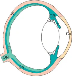
FIGURE 1-3 The three layers of the eye. The cornea and sclera form the fibrous or outer layer (tan), the uvea forms the vascular or middle layer (dark green), and the retina and optic nerve form the neuroectodermal or inner layer (light green). CB, Ciliary body; Ch, choroid; Co, cornea; ON, optic nerve; R, retina; S, sclera.
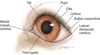
FIGURE 1-4 Frontal view of the external structures of the canine eye
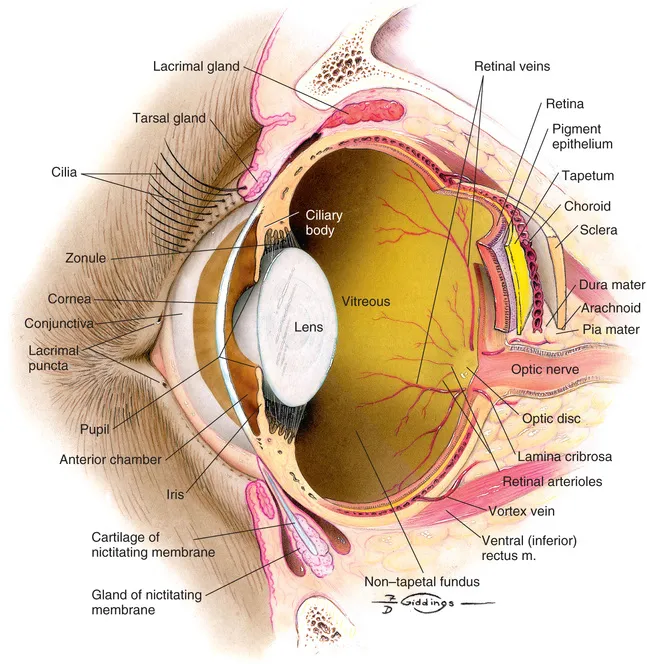
FIGURE 1-5 Internal structures of the canine eye.
1. The fibrous tunic (tunica fibrosa oculi), which is principally collagenous in nature and composed of the cornea and sclera. These tissues protect the eye's internal structures and give the eye its shape. The regular arrangement of the collagen bundles in the cornea results in a transparent structure, whereas the absence of such an arrangement causes the sclera to appear white and opaque. The limbus represents the junction between the cornea and sclera.
2. The highly vascular and often heavily pigmented uveal tract (tunica vasculosa oculi), which is composed of the iris, ciliary body, and choroid. These tissues nourish the interior of the eye, control the amount of light reaching the retina, adjust the focal power of the lens, and, in pigmented animals, create a darkened interior to the globe that limits light scattering inside of the eye. When the eye is examined, the iris is usually the first structure that is appreciated due to the transparency of the cornea, and the pupil appears black because at most angles no light is reflected out of the interior of the globe.
3. The retina/optic nerve (tunica nervosa oculi) that form the neuroectodermal or inner layer. The retina contains light-sensitive cells (rod and cone photoreceptors) that converge through a series of retinal layers eventually culminating in the retinal ganglion cells (the axons of which form the optic nerve). In addition to rods and cone photoreceptors, a small portion of the retina...
Table of contents
- Cover image
- Title Page
- Table of Contents
- Copyright
- Contributors
- Dedication
- Preface and Acknowledgments
- 1 The Eye and Vision
- 2 The Ophthalmic Examination and Diagnostic Testing
- 3 Ophthalmic Medications and Therapeutics
- 4 Principles of Ophthalmic Surgery
- 5 Diseases of the Orbit
- 6 Diseases of the Eyelids
- 7 Diseases of the Conjunctiva
- 8 Diseases of the Third Eyelid
- 9 Diseases of the Lacrimal System
- 10 Diseases of the Cornea and Sclera
- 11 Diseases of the Uvea
- 12 The Glaucomas
- 13 Diseases of the Lens
- 14 Diseases of the Vitreous
- 15 Diseases of the Retina
- 16 Neuroophthalmic Diseases
- 17 Ophthalmic Emergencies
- 18 Equine Ophthalmology
- 19 Livestock Ophthalmology
- 20 Exotic Pet and Avian Ophthalmology
- Appendix Breed Predisposition to Eye Disorders
- Glossary
- Index
Frequently asked questions
Yes, you can cancel anytime from the Subscription tab in your account settings on the Perlego website. Your subscription will stay active until the end of your current billing period. Learn how to cancel your subscription
No, books cannot be downloaded as external files, such as PDFs, for use outside of Perlego. However, you can download books within the Perlego app for offline reading on mobile or tablet. Learn how to download books offline
Perlego offers two plans: Essential and Complete
- Essential is ideal for learners and professionals who enjoy exploring a wide range of subjects. Access the Essential Library with 800,000+ trusted titles and best-sellers across business, personal growth, and the humanities. Includes unlimited reading time and Standard Read Aloud voice.
- Complete: Perfect for advanced learners and researchers needing full, unrestricted access. Unlock 1.5M+ books across hundreds of subjects, including academic and specialized titles. The Complete Plan also includes advanced features like Premium Read Aloud and Research Assistant.
We are an online textbook subscription service, where you can get access to an entire online library for less than the price of a single book per month. With over 1.5 million books across 990+ topics, we’ve got you covered! Learn about our mission
Look out for the read-aloud symbol on your next book to see if you can listen to it. The read-aloud tool reads text aloud for you, highlighting the text as it is being read. You can pause it, speed it up and slow it down. Learn more about Read Aloud
Yes! You can use the Perlego app on both iOS and Android devices to read anytime, anywhere — even offline. Perfect for commutes or when you’re on the go.
Please note we cannot support devices running on iOS 13 and Android 7 or earlier. Learn more about using the app
Please note we cannot support devices running on iOS 13 and Android 7 or earlier. Learn more about using the app
Yes, you can access Slatter's Fundamentals of Veterinary Ophthalmology E-Book by David J. Maggs,Paul E. Miller,Ron Ofri,David Maggs,Paul Miller in PDF and/or ePUB format, as well as other popular books in Medicine & Veterinary Medicine. We have over 1.5 million books available in our catalogue for you to explore.