Veterinary Neuroanatomy: A Clinical Approach is written by veterinary neurologists for anyone with an interest in the functional, applied anatomy and clinical dysfunction of the nervous system in animals, especially when of veterinary significance. It offers a user-friendly approach, providing the principal elements that students and clinicians need to understand and interpret the results of the neurological examination. Clinical cases are used to illustrate key concepts throughout. The book begins with an overview of the anatomical arrangement of the nervous system, basic embryological development, microscopic anatomy and physiology. These introductory chapters are followed by an innovative, hierarchical approach to understanding the overall function of the nervous system. The applied anatomy of posture and movement, including the vestibular system and cerebellum, is comprehensively described and illustrated by examples of both function and dysfunction. The cranial nerves and elimination systems as well as behaviour, arousal and emotion are discussed. The final chapter addresses how to perform and interpret the neurological examination.Veterinary Neuroanatomy: A Clinical Approach has been prepared by experienced educators with 35 years of combined teaching experience in neuroanatomy. Throughout the book great care is taken to explain key concepts in the most transparent and memorable way whilst minimising jargon. Detailed information for those readers with specific interests in clinical neuroanatomy is included in the text and appendix. As such, it is suitable for veterinary students, practitioners and also readers with a special interest in clinical neuroanatomy.- Contains nearly 200 clear, conceptual and anatomically precise drawings, photographs of clinical cases and gross anatomical specimens- Keeps to simple language and focuses on the key concepts- Unique 'NeuroMaps' outline the location of the functional systems within the nervous system and provide simple, visual aids to understanding and interpreting the results of the clinical neurological examination- The anatomical appendix provides 33 high-resolution gross images of the intact and sliced dog brain and detailed histological images of the sectioned sheep brainstem.- An extensive glossary explains more than 200 neuroanatomical structures and their function.
eBook - ePub
Veterinary Neuroanatomy
A Clinical Approach
- 208 pages
- English
- ePUB (mobile friendly)
- Available on iOS & Android
eBook - ePub
About this book
Trusted by 375,005 students
Access to over 1.5 million titles for a fair monthly price.
Study more efficiently using our study tools.
Information
Topic
MedicineSubtopic
Veterinary MedicineChapter 1 Regional neuroanatomy
Basic systems arrangement of the nervous system
Introduction to regions
Peripheral nervous system
Spinal nerves
Cranial nerves
Autonomic nervous system
Central nervous system
Spinal cord
Brain – forebrain, brainstem, cerebellum
Functional systems: introduction to the neurological examination
Neuroanatomy and lesion localisation
Basic systems arrangement of the nervous system
Nerves comprise a cell body, or soma (plural = somata), and cell processes/neurites. Dendrites are the processes that receive information, whereas axons/nerve fibres convey efferent information as action potentials/nerve impulses. The axons may be unmyelinated or myelinated with a lipid-rich, insulating myelin sheath that both speeds up nerve conduction and protects the axon. Ganglia are collections of nerve cell bodies outside the CNS. Nuclei are collections of nerve cell bodies with a similar function, inside the CNS.
Nerve fibres are of three basic types:
1. Afferent/sensory fibres bring information into the central nervous system (CNS) from the periphery and convey that information to higher centres for processing.
2. Efferent/motor fibres take information from motor planning centres through the CNS to connect with other motor fibres that take information into the periphery.
3. Integrating fibres that may connect afferent fibres with storage centres, other processing centres or efferent fibres. They process, organise and/or store information.
Introduction to regions
The anatomical descriptions are based primarily on canine anatomy but they are also relevant to most domestic animals.
There are three main components to the nervous system: the central nervous system (CNS), the peripheral nervous system (PNS) and the autonomic nervous system (ANS).
Peripheral nervous system
Key points

The PNS consists of the nerves and ganglia located outside the brain and the spinal cord and principally functions to connect the central nervous system (CNS) to the head, body, limbs and viscera. With respect to nomenclature, a ‘nerve(s)’ is by definition, in the PNS, making the word ‘peripheral’ (as in ‘peripheral nerve’) redundant; it is myelinated by Schwann cells. Unlike the CNS, the PNS is not protected by bone, leaving it more vulnerable to mechanical injury. Schwann cells form the insulating myelin sheaths surrounding peripheral axons, whereas in the CNS that task is performed by oligodendrocytes. The change from oligodendrocytes to Schwann cells occurs where the dura mater surrounding the spinal cord abuts the perineurium at the origin of the spinal nerves (Fig. 1.1). Afferent and efferent axons of the PNS form the spinal and cranial nerves (CNN).
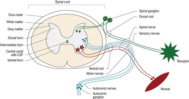
Fig. 1.1 Transverse section of thoracic spinal cord showing the spinal nerve and its components.
Spinal nerves
Spinal nerves arise as roots from the spinal cord. A dorsal and a ventral root attach on each side of the spinal cord, and define each spinal cord segment. For example, the third cervical spinal cord segment has two dorsal roots and two ventral roots attaching to it. The dorsal roots convey primarily sensory nerve fibres into the spinal cord. Each dorsal root contains a spinal ganglion (old name ‘dorsal root ganglion’), housing the nerve cell bodies of these sensory fibres (Fig. 1.1). The ventral roots convey motor nerve fibres away from the spinal cord. Motor fibres may be somatic and innervate striated muscle, or autonomic and innervate smooth or cardiac muscle. The dorsal and ventral roots fuse at the level of the intervertebral foramen to form a spinal nerve. Distal to the intervertebral foramen, the mixed sensory and motor spinal nerve usually splits into a dorsal and ventral branch. The dorsal branch supplies the epaxial muscles and skin, while the ventral branch supplies the hypaxial muscles and skin. A third branch, carrying autonomic fibres, may also arise and pass ventrally towards the midline to supply the viscera.
Spinal nerves may remain as single, discrete nerves all the way out into the periphery, in which case they are named for the number of the spinal cord segment from which they ...
Table of contents
- Cover image
- Title page
- Table of Contents
- Copyright
- Preface
- Acknowledgements
- Terminology, glossary and abbreviations
- Chapter 1: Regional neuroanatomy
- Chapter 2: Neuroembryology
- Chapter 3: Neurohistology, physiology and supporting structures
- Chapter 4: Hierarchical organisation in the nervous system
- Chapter 5: Reflexes and motor systems
- Chapter 6: Ascending somatic sensory tracts and conscious sensory systems
- Chapter 7: The cerebellum
- Chapter 8: Vestibular system
- Chapter 9: Posture and movement in quadrupeds
- Chapter 10: Cranial nerves
- Chapter 11: Behaviour, emotion and arousal
- Chapter 12: The autonomic nervous system
- Chapter 13: The neurological examination and lesion localisation
- Appendix
- Index
Frequently asked questions
Yes, you can cancel anytime from the Subscription tab in your account settings on the Perlego website. Your subscription will stay active until the end of your current billing period. Learn how to cancel your subscription
No, books cannot be downloaded as external files, such as PDFs, for use outside of Perlego. However, you can download books within the Perlego app for offline reading on mobile or tablet. Learn how to download books offline
Perlego offers two plans: Essential and Complete
- Essential is ideal for learners and professionals who enjoy exploring a wide range of subjects. Access the Essential Library with 800,000+ trusted titles and best-sellers across business, personal growth, and the humanities. Includes unlimited reading time and Standard Read Aloud voice.
- Complete: Perfect for advanced learners and researchers needing full, unrestricted access. Unlock 1.5M+ books across hundreds of subjects, including academic and specialized titles. The Complete Plan also includes advanced features like Premium Read Aloud and Research Assistant.
We are an online textbook subscription service, where you can get access to an entire online library for less than the price of a single book per month. With over 1.5 million books across 990+ topics, we’ve got you covered! Learn about our mission
Look out for the read-aloud symbol on your next book to see if you can listen to it. The read-aloud tool reads text aloud for you, highlighting the text as it is being read. You can pause it, speed it up and slow it down. Learn more about Read Aloud
Yes! You can use the Perlego app on both iOS and Android devices to read anytime, anywhere — even offline. Perfect for commutes or when you’re on the go.
Please note we cannot support devices running on iOS 13 and Android 7 or earlier. Learn more about using the app
Please note we cannot support devices running on iOS 13 and Android 7 or earlier. Learn more about using the app
Yes, you can access Veterinary Neuroanatomy by Christine E Thomson,Caroline Hahn,Christine Thomson in PDF and/or ePUB format, as well as other popular books in Medicine & Veterinary Medicine. We have over 1.5 million books available in our catalogue for you to explore.