Fundamentals of Canine Neuroanatomy and Neurophysiology introduces the fundamentals of veterinary neuroanatomy and neurophysiology, demonstrating structure and function as it relates to clinical applications with a highly visual approach.
Offers a straightforward yet comprehensive introduction to structure and function of the nervous system
Demonstrates the relevance of the basic principles to the clinical setting
Illustrates concepts using line drawings, photographs, micrographs, and MRIs
Includes access to a companion website with review questions and answers and the figures from the book at www.wiley.com/go/uemura/neuroanatomy
Trusted by 375,005 students
Access to over 1.5 million titles for a fair monthly price.
The brain and spinal cord are organized through a series of developmental events. They start as a thickened neural plate and then transform into a simple tubular structure, the neural tube. The cranial end of the tubular structure enlarges to become the brain, whereas the remaining neural tube develops into the spinal cord. As precursor cells of the neural tube proliferate and differentiate into neurons and neuroglia, they migrate to appropriate target locations. This process of proliferation, differentiation, and migration is crucial for further developmental events, including outgrowth of axons and formation of synapses. Any interference with such developmental processes risks congenital malformations, perinatal mortality, and postnatal morbidity.
Formation of the neural tube
What embryonic germ layer becomes the nervous tissue?
Explain how the neural ectoderm forms the neural tube.
The development of the nervous system, like all other organ systems, starts at fertilization. An oocyte swept from the ovary is transported in the uterine tube to be fertilized. A fertilized ovum undergoes repeated cell division (also called cleavage). The first cleavage of the ovum results in two blastomeres, and successive cell divisions of blastomeres produce a spherical ball of cells. The blastomeres start to rearrange and a fluid-filled cavity, the blastocele, is formed. The wall of the blastocyst is only a single cell in thickness, except the area where a cluster of cells, the inner cell mass, appears. The inner cell mass is destined to be concerned primarily with the formation of the embryonic body. Within the inner cell mass a cavity starts to develop, separating the amnion from the embryo-formative cells, the embryonic disc. The embryonic disc gives rise to the three germ layers (endoderm, mesoderm, ectoderm) that form all the tissue and organs of the embryo.
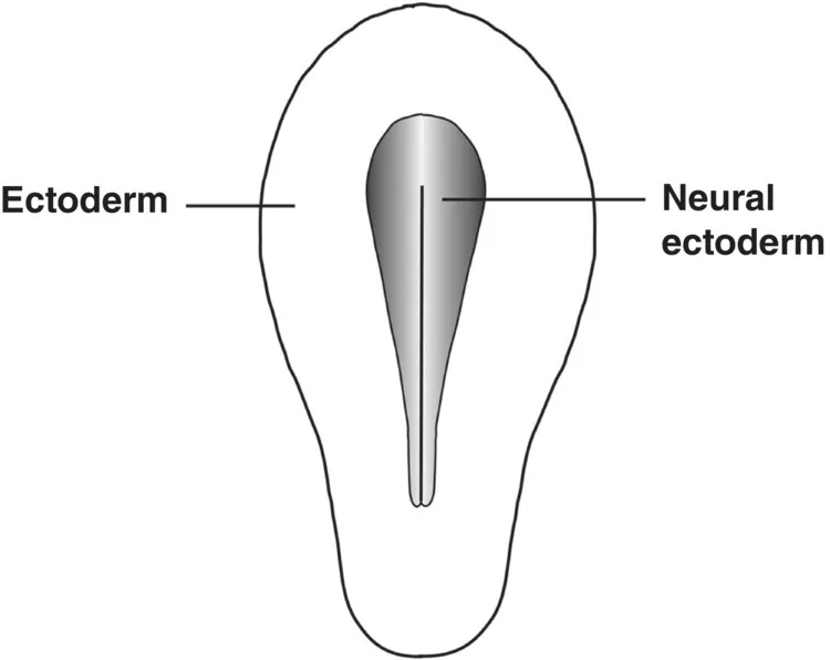
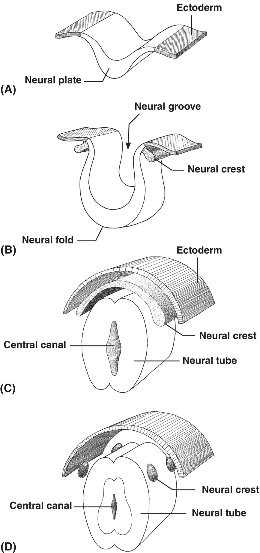
The dorsomedial area of the ectoderm differentiates to become the neural ectoderm (Fig. 1.1). As the embryo develops, the neural ectoderm separates from the remaining ectoderm. Initially, the neural ectoderm is a flat area made of a single cell layer. There are three developmental stages (neural plate, neural fold, and neural tube) that form a tubular primordium of the central nervous system (CNS) (Fig. 1.2). The neural ectoderm thickens to become the neural plate, which folds into a neural groove. As the neural fold continues to become thicker, the groove becomes narrower and the dorsal edges of the fold fuse and the neural fold becomes the neural tube. The rostral end of the neural tube becomes the brain and the remaining neural tube develops into the spinal cord.
Fig. 1.1 The neural plate develops from the neural ectoderm that occupies the central portion of the ectoderm.
Fig. 1.2 Development of the neural tube and neural crest from the ectoderm. (A) Transverse section of the embryonic disc. The neural plate is the thickened area of the ectoderm. (B) Invagination of the neural plate to form the neural fold. The neural crest appears at the dorsolateral edge of the neural fold. (C) Fusion of the dorsal edges of the neural fold forms the neural tube. (D) The neural crest gives rise to small aggregates of cells that start to migrate ventrally to become ganglia.
Neural plate
The neural ectoderm thickens to form a neural plate (Figs 1.1 and 1.2A). The first step in the formation of the brain and spinal cord is its transformation from a thickened neural plate into a tubular mass of cells.
Neural fold
The neural groove appears as a result of differential growth of the neural plate along the longitudinal axis of the embryo (Fig. 1.2B). The groove deepens and the elevated lateral margins of the neural plate form the neural fold. The neural folds, as they become more elevated, grow toward each other. The neural fold at the rostral end (also referred to as the cephalic end because the rostral neural fold develops into the brain) is much greater in size than it is further caudally. This results in the differentiation of the rostral neural fold into the brain and the remaining caudal portion into the spinal cord. At about the time the neural groove deepens, a cluster of cells appears and forms the neural crest at the area where the neural fold borders on the ectoderm. The neural crest detaches from the ectoderm to become ganglia of the cranial and spinal nerves. A cluster of cells associated with the cephalic end differentiates into the ganglia of the cranial nerves. Those cells associated with the remaining neural fold differentiate into the dorsal root ganglia and ganglia of the autonomic nervous system (ANS).
Neural tube
The neural tube results from fusion of the dorsal edges of the neural fold (Fig. 1.2C and D). Prior to the closure of the neural groove, the neural plate is continuous laterally with the ectoderm. When the two neural folds fuse with each other, the ectoderm also fuses to overlie the newly formed neural tube. The closure of the neural groove begins in the middle of the embryo and proceeds toward the two ends. However, progression of closure is more rapid towards the cephalic end than towards the caudal end (Fig. 1.3). As a result, three stages of neural development (i.e., the neural plate, fold, and tube) coexist simultaneously in different regions of the embryo. The cephalic end of the neural t...
Table of contents
Cover
Title Page
Table of Contents
Preface
Abbreviations
About the Companion Website
1 Developmental Anatomy
2 Structure and Function of Neurons and Neuroglia
3 Anatomy of the Canine Brain
4 Meninges and Ventricular System
5 Spinal Cord
6 Spinal Reflexes
7 Somatosensory System
8 Viscerosensory System
9 Brain Stem
10 Midbrain
11 Pons
12 Medulla Oblongata
13 Reticular Formation
14 Thalamus
15 Cerebrum
16 Motor System
17 Cerebellum
18 Vestibular System
19 Auditory System
20 Visual System
21 Hypothalamus
22 Autonomic Nervous System
Self-Evaluation Answers
Index
End User License Agreement
Frequently asked questions
Yes, you can cancel anytime from the Subscription tab in your account settings on the Perlego website. Your subscription will stay active until the end of your current billing period. Learn how to cancel your subscription
No, books cannot be downloaded as external files, such as PDFs, for use outside of Perlego. However, you can download books within the Perlego app for offline reading on mobile or tablet. Learn how to download books offline
Perlego offers two plans: Essential and Complete
Essential is ideal for learners and professionals who enjoy exploring a wide range of subjects. Access the Essential Library with 800,000+ trusted titles and best-sellers across business, personal growth, and the humanities. Includes unlimited reading time and Standard Read Aloud voice.
Complete: Perfect for advanced learners and researchers needing full, unrestricted access. Unlock 1.5M+ books across hundreds of subjects, including academic and specialized titles. The Complete Plan also includes advanced features like Premium Read Aloud and Research Assistant.
Both plans are available with monthly, semester, or annual billing cycles.
We are an online textbook subscription service, where you can get access to an entire online library for less than the price of a single book per month. With over 1.5 million books across 990+ topics, we’ve got you covered! Learn about our mission
Look out for the read-aloud symbol on your next book to see if you can listen to it. The read-aloud tool reads text aloud for you, highlighting the text as it is being read. You can pause it, speed it up and slow it down. Learn more about Read Aloud
Yes! You can use the Perlego app on both iOS and Android devices to read anytime, anywhere — even offline. Perfect for commutes or when you’re on the go. Please note we cannot support devices running on iOS 13 and Android 7 or earlier. Learn more about using the app
Yes, you can access Fundamentals of Canine Neuroanatomy and Neurophysiology by Etsuro E. Uemura in PDF and/or ePUB format, as well as other popular books in Medicine & Veterinary Medicine. We have over 1.5 million books available in our catalogue for you to explore.