Mass Spectrometry in Food Analysis
  1. 442 pages
  2. English
  3. ePUB (mobile friendly)
  4. Available on iOS & Android
eBook - ePub

About this book

The quality and safety of food are crucial for human nutrition. However, evaluating the chemical composition of food is challenging for the analyst and requires powerful methods. Chromatography and mass spectrometry (MS) is the gold standard for analyzing complex food samples, including raw materials and intermediate and finished products.

Mass Spectrometry in Food Analysis covers the MS-based analysis of different aspects of food quality, which include nutritional value, profile of macronutrients (proteins, lipids, and carbohydrates), micronutrients (vitamins), and nutraceutical active compounds. Additionally, sensory quality, flavor, food pigments, safety, and detection of pesticides, contact materials, veterinary drugs and pharmaceuticals, organic pollutants, and pathogens are covered.

Key Features:


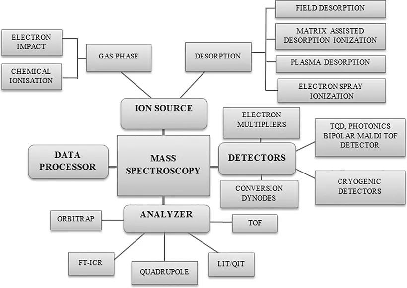
  • Contains the basics of mass spectrometry and experimental strategies


  • Explores determination of macro- and micronutrients


  • Analyzes sensory and nutraceutical food quality


  • Discusses detection of contaminants and proof of authenticity


  • Presents emerging methods for food analysis

This book contains an introductory section that explains the basics of MS and the difference between targeted and untargeted strategies for beginners. Further, it points out new analytical challenges, such as monitoring contaminants of emerging concern, and presents innovative techniques (e.g., ambient ionization MS and data mining).

Also available in the Food Analysis & Properties Series:

Nanoemulsions in Food Technology: Development, Characterization, and Applications, edited by Javed Ahmad and Leo M.L. Nollet (ISBN: 978-0-367-61492-8)

Sequencing Technologies in Microbial Food Safety and Quality, edited by Devarajan Thangadurai, Leo M.L. Nollet, Saher Islam, and Jeyabalan Sangeetha (ISBN: 978-0-367-35118-2)

Chiral Organic Pollutants: Monitoring and Characterization in Food and the Environment, edited by Edmond Sanganyado, Basil K. Munjanja, and Leo M.L. Nollet (ISBN: 978-0-367-42923-2)

For a complete list of books in this series, please visit our website at:

www.crcpress.com/Food-Analysis--Properties/book-series/CRCFOODANPRO

Frequently asked questions

Yes, you can cancel anytime from the Subscription tab in your account settings on the Perlego website. Your subscription will stay active until the end of your current billing period. Learn how to cancel your subscription.
No, books cannot be downloaded as external files, such as PDFs, for use outside of Perlego. However, you can download books within the Perlego app for offline reading on mobile or tablet. Learn more here.
Perlego offers two plans: Essential and Complete
  • Essential is ideal for learners and professionals who enjoy exploring a wide range of subjects. Access the Essential Library with 800,000+ trusted titles and best-sellers across business, personal growth, and the humanities. Includes unlimited reading time and Standard Read Aloud voice.
  • Complete: Perfect for advanced learners and researchers needing full, unrestricted access. Unlock 1.4M+ books across hundreds of subjects, including academic and specialized titles. The Complete Plan also includes advanced features like Premium Read Aloud and Research Assistant.
Both plans are available with monthly, semester, or annual billing cycles.
We are an online textbook subscription service, where you can get access to an entire online library for less than the price of a single book per month. With over 1 million books across 1000+ topics, we’ve got you covered! Learn more here.
Look out for the read-aloud symbol on your next book to see if you can listen to it. The read-aloud tool reads text aloud for you, highlighting the text as it is being read. You can pause it, speed it up and slow it down. Learn more here.
Yes! You can use the Perlego app on both iOS or Android devices to read anytime, anywhere — even offline. Perfect for commutes or when you’re on the go.
Please note we cannot support devices running on iOS 13 and Android 7 or earlier. Learn more about using the app.
Yes, you can access Mass Spectrometry in Food Analysis by Leo Nollet, Robert Winkler, Leo Nollet,Robert Winkler,Leo M. L. Nollet, Leo M. L. Nollet, Robert Winkler in PDF and/or ePUB format, as well as other popular books in Technology & Engineering & Food Science. We have over one million books available in our catalogue for you to explore.

Information

Section 1 Mass Spectrometry – Theoretics

1 Basic Principles and Fundamental Aspects of Mass Spectrometry

Javed Ahamad
Tishk International University
Faraat Ali
Botswana Medicines Regulatory Authority
Manjoor Ahmad Sayed
Mettu University
Javed Ahmad
Najran University
Leo M.L. Nollet
University College Ghent
DOI: 10.1201/9781003091226-2

Contents

  1. 1.1 Introduction
  2. 1.2 Principle of Mass Spectrometry
  3. 1.3 Instrumentation of Mass Spectrometry
    1. 1.3.1 Ionization Techniques
      1. 1.3.1.1 Gas Phase
      2. 1.3.1.2 Desorption Phase
    2. 1.3.2 Mass Analyzers
      1. 1.3.2.1 Time-of-Flight
      2. 1.3.2.2 Quadrupole
      3. 1.3.2.3 Orbitrap Mass Analyzer
      4. 1.3.2.4 Fourier Transform–Ion Cyclotron Resistance
      5. 1.3.2.5 Tandem Mass Analyzer
    3. 1.3.3 Mass Detectors
      1. 1.3.3.1 Electron Multiplier
      2. 1.3.3.2 Faraday Cup
      3. 1.3.3.3 Photomultipliers
  4. 1.4 Interpretation of Mass Spectra
    1. 1.4.1 Types of Peaks in Mass Spectroscopy
    2. 1.4.2 Fragmentation Rules
  5. 1.5 Application of Mass Spectrometry
    1. 1.5.1 Qualitative Applications
      1. 1.5.1.1 Determination of Molecular Weight
      2. 1.5.1.2 Determination of the Molecular Formula
      3. 1.5.1.3 Determination of the Partial Molecular Formula
      4. 1.5.1.4 Determination of the Compound Structure
    2. 1.5.2 Quantitative Applications
      1. 1.5.2.1 Isotope Abundance Assessment
      2. 1.5.2.2 Determination of Isotope Ratio
      3. 1.5.2.3 Differentiation between cis-Isomers and trans-Isomers
      4. 1.5.2.4 Mass Spectrometry in Thermodynamics
      5. 1.5.2.5 Measurement of Ionization Potential
      6. 1.5.2.6 Determination of Ion–Molecule Reactions
      7. 1.5.2.7 Detection and Identification of Impurity
      8. 1.5.2.8 Identification of Unknown Compounds
    3. 1.5.3 Both Qualitative and Quantitative Applications
      1. 1.5.3.1 Phytochemical Analysis
      2. 1.5.3.2 Structural Elucidation of Unknown Phytochemicals
      3. 1.5.3.3 Drug Metabolism Studies
      4. 1.5.3.4 Clinical Studies
      5. 1.5.3.5 Forensic Applications
  6. 1.6 Conclusion
  7. References

1.1 Introduction

Over the past decades, mass spectrometry (MS) has developed exponentially, with its applications ranging from each discipline across the health and life disciplines (Henderson and McIndoe, 2005; Banoub et al., 2005). Depending on their mass-to-charge (m/z) ratio, MS relies on the creation of gas-phase ions (negatively or positively charged) that can be electrically (or magnetically) separated (Korfmacher, 2005). The x-axis reflects m/z values in an MS spectrum, while the y-axis shows complete ion counts. Mass spectrometers, technically speaking, should indeed be called m/z spectrometers (Grayson, 2002). The mass spectrometric study, including their composition, potency, and purity, may provide considerable details about the analytes (Kitson et al., 1996; Herbert and Johnstone, 2003).
The use of advanced mass spectrometric methods has been restricted to volatile compounds with a low-weight spectrum of molecules (>1,000 Da) (Beckey, 1969; Torgerson et al., 1974). Furthermore, a traditional ionization technique, namely electron impact (EI), is extreme and can contribute to the degradation of complex organic compounds (e.g., enzymes, nucleic acids, and carbohydrates). In particular, MS was restricted to the gas chromatography (GC)-MS instrument separating volatile compounds within biology laboratories (Barber et al., 1981). Under such circumstances, without substantial destruction and deterioration, highly complex compounds, such as proteins, cannot be transmitted to the gas phase (Tanaka et al., 1988). Only after soft ionization techniques were introduced, MS was used for genomics studies (Hillenkamp and Karas, 1990). Many separation methods, such as high-pressure liquid chromatography (HPLC-MS) and capillary electrophoresis (CE), can now be combined with MS (Pramanik et al., 2002). MS has emerged as a powerful technique for ensuring the quality and safety of food products such as honey, cereals, meat products, wine, and milk and dairy products (Ahamad et al., 2018; 2020a,b). MS analysis of purified materials or synthetic conjugates without using a separation technique is also popular (Baldwin, 2005). This chapter comprehensively summarizes the principles, recent advancements in ionization and detectors, and MS applications.

1.2 Principle of Mass Spectrometry

MS measures the specific fragments or ions that occur from organic molecules’ breakdown (Griffiths et al., 2001). The fundamental concept requires the bombardment of organic matter. A compound is attacked with a beam of electrons to generate positively charged ions (Emmett and Caprioli, 1994; Gale and Smith, 1993). A mass spectrum represents each ion in the form of peak strength. Ion deflection is based on charge, mass, and velocity; ion separation is based on the m/z ratio; and detection is proportional to the abundance of ions (Karas et al., 2000; Banoub et al., 2003; Banoub et al., 2004).

1.3 Instrumentation of Mass Spectrometry

In 1912, the first mass spectroscope was developed and used by J.J. Thompson to analyze the atomic weight of elements and to track the abundance of elemental isotopes. With the advent of techniques to vaporize organic compounds in the 1950s, it became possible to study bioactive compounds via a mass spectroscope. MS consists of an ionization source, analyzer, detector, and data processor. The analyzer and detector are kept in a vacuum so that air molecules do not collide with the ions created, and the trajectories of the ions are held at a certain speed. The sample is ionized in the ionization chamber after it is pumped into the instrument through the inlet. In the analyzer, ionized species (cations/anions) are then separated, resolving the ions based on their m/z ratio. Finally, these ions are detected by detectors, and the relative abundance is recorded in the form of mass spectra for each resolved ionic species (Silverstein and Webster, 1998). Figure 1.1 shows the schematic diagram of different parts of MS.
FIGURE 1.1 Schematic diagram representing different parts of mass spectrometry.

1.3.1 Ionization Techniques

Depending on the types of organic compounds, different types of ionization techniques are used. Volatile samples are subjected to either electron or chemical ionization (CI), and fast-atom bombardment (FAB), matrix-assisted laser desorption ionization (MALDI), and electrospray ionization (ESI) methods are used to ionize non-volatile samples (Silverstein and Webster, 1998). In Table 1.1, the characteristics of various methods of ionization used in MS are summarized.
TABLE 1.1 Summary of the Features of Different Ionization Methods Used in Mass Spectrometry
Ionization Mode Nature Sample Range of Mass Description
Electron impact (EI) Volatile: thermally stable GC solid or liquid probe <1,100 Da Hard method; mainly fragmented ions
Chemical ionization (CI) Volatile: thermally stable GC solid or liquid probe <1,000 Da Soft method; molecular ion
Fast-atom bombardment (FAB) Organometallic compounds LC direct injection <5,200 Da; optimal range 250–2,000 Soft method; require matrix
Matrix-assisted laser desorption ionization (MALDI) Proteomics, genomics Sample is...

Table of contents

  1. Cover
  2. Half Title Page
  3. Series Page
  4. Title Page
  5. Copyright Page
  6. Table of Contents
  7. Series Preface
  8. Preface
  9. Editors
  10. Contributors
  11. Section 1 Mass Spectrometry – Theoretics
  12. Section 2 MS Analysis of Food Components
  13. Section 3 MS Analysis of Residues
  14. Section 4 MS Analysis in Food Authentication
  15. Section 5 Emerging Fields
  16. Index