eBook - ePub
The Brain Atlas
A Visual Guide to the Human Central Nervous System
- English
- ePUB (mobile friendly)
- Available on iOS & Android
eBook - ePub
The Brain Atlas
A Visual Guide to the Human Central Nervous System
About this book
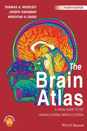
The Brain Atlas: A Visual Guide to the Human Central Nervous System integrates modern neuroscience with clinical practice and is now significantly revised and updated for a Fourth Edition. The book's five sections cover: Background Information, The Brain and Its Blood Vessels, Brain Slices, Histological Sections, and Pathways. These are depicted in over 350 high quality intricate figures making it the best available visual guide to human neuroanatomy.
Frequently asked questions
Yes, you can cancel anytime from the Subscription tab in your account settings on the Perlego website. Your subscription will stay active until the end of your current billing period. Learn how to cancel your subscription.
No, books cannot be downloaded as external files, such as PDFs, for use outside of Perlego. However, you can download books within the Perlego app for offline reading on mobile or tablet. Learn more here.
Perlego offers two plans: Essential and Complete
- Essential is ideal for learners and professionals who enjoy exploring a wide range of subjects. Access the Essential Library with 800,000+ trusted titles and best-sellers across business, personal growth, and the humanities. Includes unlimited reading time and Standard Read Aloud voice.
- Complete: Perfect for advanced learners and researchers needing full, unrestricted access. Unlock 1.4M+ books across hundreds of subjects, including academic and specialized titles. The Complete Plan also includes advanced features like Premium Read Aloud and Research Assistant.
We are an online textbook subscription service, where you can get access to an entire online library for less than the price of a single book per month. With over 1 million books across 1000+ topics, we’ve got you covered! Learn more here.
Look out for the read-aloud symbol on your next book to see if you can listen to it. The read-aloud tool reads text aloud for you, highlighting the text as it is being read. You can pause it, speed it up and slow it down. Learn more here.
Yes! You can use the Perlego app on both iOS or Android devices to read anytime, anywhere — even offline. Perfect for commutes or when you’re on the go.
Please note we cannot support devices running on iOS 13 and Android 7 or earlier. Learn more about using the app.
Please note we cannot support devices running on iOS 13 and Android 7 or earlier. Learn more about using the app.
Yes, you can access The Brain Atlas by Thomas A. Woolsey,Joseph Hanaway,Mokhtar H. Gado in PDF and/or ePUB format, as well as other popular books in Biological Sciences & Neurology. We have over one million books available in our catalogue for you to explore.
Information
PART I
PART I is an illustrated overview of The Brain Atlas. The main features of the central nervous system and the organization of this book are described. Sources of the specimens and the methods by which they were prepared and photographed are detailed. Key aspects of the various radiological techniques for the images included are outlined. Selected references are listed at the end.
Introduction
Overview
The human nervous system is complex and sophisticated. It is the most remarkable system in biology. A major challenge for neuroscience, psychology, medicine, and, indeed, for civilization is to understand the nervous system at the same fundamental levels at which we now understand other organ systems. Early in the 21st century, only 50 years after the discovery of the genetic “alphabet,” the complete human genome has been mapped. Likewise, new knowledge about the brain and diseases that afflict the nervous system is exploding. One goal for future work on the human brain is to reach a level of detailed understanding similar to that now possible for the genome.
An anchor in this quest is information about the structure and organization of the central nervous system (CNS). The Brain Atlas: A Visual Guide to the Human Central Nervous System was prepared to help students and professionals understand the normal human brain and guide interpretation of clinical and experimental work.
Clear charts and maps of biological structures have been teaching aids from the earliest times. In the biological sciences, the first detailed and illustrated text based on direct observation was the De Fabrica Humani Corporis (1543) and its synoptic Epitome (1543) by Andreas Vesalius (1514–1564). Those books have been said to “mark the beginning of modern science.” Publication of the Fabrica also was a major landmark in book publishing. The highly popular Epitome was intended as a primer but served very much as a modern day atlas. Such works have evolved and today are used in the same way maps are used to plan travel and understand geographic relationships.
In the mid to late 19th century, instructional programs in universities and medical schools were developed to teach students to make accurate observations from specimens. This skill enables students to generate and retain mental conceptualizations of complex three-dimensional (3D) structures in the body. In part, this was to prepare students to interpret observations that could be made only at the surfaces of living organisms. Experience with these teaching aids was so positive that, even today, instruction at nearly every level now uses charts and atlases to aid the study of gross anatomy, embryology, histology, and neuroscience. Atlases have been developed for a wide range of other related disciplines, such as pathology, radiology, and surgery. These books support varied and flexible learning plans, styles, and objectives. At their best, such works are ready references for efficient recall and lifelong study—rapidly accessible sources of information. The Brain Atlas is intended to be such a work: a reference serving different needs for students learning about the human brain and a resource for rapid clarification in self-directed study, in the classroom, in the laboratory, and in the clinic.
Because of recent stunning advances in imaging, the information included in The Brain Altas is more crucial than ever for medical practice, human and animal brain research, and certain branches of psychology. For example, strokes (brain attacks) resulting from insufficient blood supply to parts of the CNS are the leading cause of disability in adults and the third leading cause of death in the United States. Intense efforts are now directed at reducing risk and improving therapy for this disease. The quick access to information on the brain and its blood supply in The Brain Atlas is crucial for such efforts. Other forms of “brain disease,” such as mental illness, dementia, substance abuse, and a host of genetic syndromes, can be investigated and understood only by reference to the detailed organization of the human brain. Alterations in brain function, such as learning difficulties or speech problems, have also now been directly linked to altered brain structures. In the future, access to basic information about brain structure will be even more essential for evaluating patients at risk for specific diseases and for monitoring and assessing the effects of therapeutic interventions.
New imaging and other innovative techniques have spurred a revolution in the study of the way in which the brain works. Functional imaging of healthy individuals at all ages provides a wide range of new and compelling information on how the brain executes different tasks, from speaking different languages to reacting to pain. Such imaging promises to reveal, for example, the ways in which the brains of individuals with special talents may differ. The human brain is no longer a “black box” from which one only rarely and fortuitously records activity. Instead, the precise locations in the brain associated with many uniquely human tasks can be specified. Therefore, ready anatomical reference works are crucial for cognitive psychologists and research scientists.
The Brain Atlas is divided into five parts, with key features summarized at the beginning of each. This introduction (Part I) summarizes several general aspects of the brain to help the novice get started or refresh the knowledge of advanced students and practicing professionals. The balance of this overview outlines terminology used in this volume, as well as special features designed to assist in identification, study, and navigation. Information on the sources and preparation of the anatomical images that appear in this book is provided. The main parts of the volume (Parts II–V) are designed to flow logically and progressively, from overall surface anatomy of the CNS (Part II), through cross-section-al gross anatomy (Part III) and selected regional histology (Part IV), and ending with diagrams of the major neuronal systems that are responsible for the brain’s magnificent array of functions (Part V). Because each part illustrates a different aspect of the structure and organization of the CNS, the book is arranged so that users can navigate easily between topics fo...
Table of contents
- Cover
- Title Page
- Table of Contents
- Preface
- Acknowledgements
- About the Digital Companion
- PART I:
- PART II:
- PART III:
- PART IV
- PART V:
- Index
- End User License Agreement