Part I General Post-Mortem Processes: Degradation of Soft Tissue, Bone and Associated Materials
Chapter 1 Gross Post-Mortem Changes in the Human Body
Stuart J. Hamilton and Michael A. Green
1.1 Introduction
All organisms die and the natural consequence of this is decomposition. The rate of decomposition is determined by numerous factors, both intrinsic to the body such as body habitus, and environmental factors such as predators, ambient temperature and humidity. Decomposition may broadly be divided into putrefaction and arrested decay, although these processes are not mutually exclusive and may be seen in the same body.
Post-mortem changes produce both artefacts that may mislead the unwary and obscure findings that point to the events immediately before, and indeed leading directly to, the death of the person. Recommended literature regarding forensic pathology is DiMaio and DiMaio (2001) and Knight and Saukko (2004). In this chapter the processes that occur in the body after death are discussed, along with the ways in which human and other environmental factors can affect these processes. The potential pitfalls faced in examination of the deceased are highlighted. In particular, some post-mortem changes may be mistaken for injuries sustained shortly before death.
1.2 The Immediate Post-Mortem Period
While most people know what death is, it is remarkably difficult to define scientifically and is best considered a process during which the cells and tissues that make up an organism cease to function. This is not the same as the ‘time of death’ recorded on the death certification, which is usually the time at which life is pronounced extinct by a suitably qualified person. In the UK, to diagnose brain death, a series of appropriate tests are performed twice at different times and the time of death is considered to be the time of the first set of tests. The tests include shining a torch into both eyes to see if they react to the light, stroking the cornea, pinching the nose, inserting ice-cold water in each ear, trying to provoke gagging or coughing by placing a plastic tube down the trachea, amongst other tests (National Health Service 2015).
One of the first notable features is that thermoregulation stops and the body temperature will naturally move towards ambient temperature. In most cases, this means the body will cool, but in particularly warm climates the body may actually increase in temperature.
The cooling of the body has long been of interest to the forensic community, particularly with respect to estimating the post-mortem interval (PMI). A historical review on the early work of estimating time since death is described by Knight and Madea (2016). A common ‘rule of thumb’ is that a body tends to cool at a rate of one degree Celsius per hour. A slightly more refined version takes into account the sigmoid nature of the cooling curve and assumes that the body does not begin to cool for around three hours after death, but cools at around one degree Celsius per hour thereafter, called the ‘initial temperature plateau’ (Al-Alousi et al. 2002). The authors politely submit that such calculations are so ‘rough and ready’ as to be of no real value in criminal investigations. By way of example, the case of R v Lattimore and Leighton (1974) involved a body recovered from a fire scene. Because of the possibility of sexual activity, a rectal temperature was not taken at the scene and later excessive reliance on post-mortem phenomena to estimate the time of death (particularly given that the body had been exposed to an unusually high ambient temperature) became an important aspect of the case with respect to the defendants' alibis. This ultimately led to a successful appeal and the case initiated research funded by the UK Home Office. Unfortunately, this extensive and expensive study only confirmed the unreliabilities, which had been known for years.
A more refined calculation is the Henssge equation and nomogram that take into account numerous factors including body weight, gender, body coverings, whether the body is wet or dry and ambient temperature (Henssge and Madea 2004; Henssge et al. 2002; Madea and Henssge 2016). The nomogram is useful, but as with any hand-drawn graphical method there is potential for user error. While the equation may appear daunting, there are now apps available for smartphones that can perform the calculation with relative ease. It should also be noted that any estimate of the PMI is based on a ‘normal’ initial body temperature, but particularly in cases of overwhelming infection or strenuous exercise, the initial temperature may be higher, and indeed in cases of sepsis, a transient post-mortem rise in temperature may be seen. Equally, hypothermia will cause a low core body temperature in life, also affecting estimates of the PMI. Even without such extreme changes, it is known that the human core temperature may vary under normal circumstances (the changes associated with the menstrual cycle are so well recognised that they can be used for family planning) and so one must bear in mind that there is an inherent potential error of up to two hours in any PMI calculation, and even the Henssge calculation includes a range of ±2.8 hours as a minimum range, rising to as much as ±7 hours.
Furthermore, the so-called ‘normal temperature’ in any individual may vary by as much as 1°C from the assumed 37.4, so all calculations start with a built-in potential error of up to 2 hours. It is also the case that the ambient temperature is based on a single measurement, and as this temperature can fluctuate over time, both outdoors and indoors with automatic central heating, its measurement may not accurately reflect the average temperature to which the body has been exposed throughout the post-mortem period.
Concurrently with cooling, rigor mortis (post-mortem stiffening) will develop as the muscles become depleted of adenosine triphosphate (ATP) and the muscle fibres permanently cross-link. As the reader may recall from his or her studies of basic physiology, muscle fibres are composed of actin and myosin, with the myosin heads binding to actin during muscle contraction and hydrolysing ATP to adenosine diphosphate (ADP) to allow the myosin to release the actin. Once ATP is depleted then cross-linking of the actin/myosin becomes permanent until decomposition causes breakdown of the molecules, or mechanical force is applied as discussed below. A logical corollary of this is that individuals who die shortly after exercise may have already depleted their stores of energy and ATP and therefore may develop rigor mortis more rapidly than an identical person who died at rest.
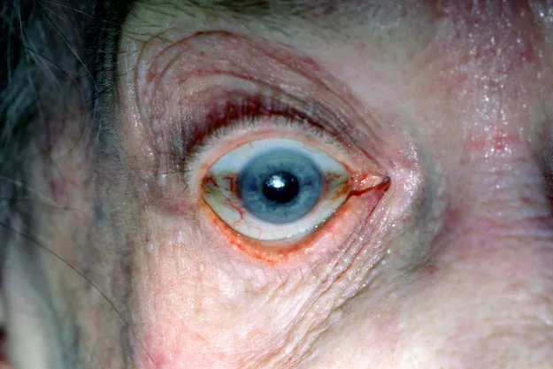
Rigor mortis is first observed in the small muscle groups such as the fingers, eyelids and lower jaw, which has produced the age-old practices of ‘pennies on the eyelids’, and the timely application of a jaw bandage or prop. The appearance of a slack-jawed, half-staring body can be highly distressing to relatives. Furthermore, if the eyes remain open, a dark discoloration occurs in the exposed conjunctiva, the so-called tâche noire de la sclérotique (Figure 1.1). In the early stages of its development, this may be mistaken for petechial haemorrhage (pinpoint haemorrhages resulting from ruptured venules, a common finding in strangulation), which the inexperienced may interpret as a sign of asphyxial death.
Figure 1.1 The tâche noire de la sclérotique, a post-mortem artefact.
Rigor mortis then gradually becomes apparent in larger muscle groups until the body is essentially stiff. As the muscle decomposes, rigor mortis will pass. The rate at which rigor appears and disappears is extremely variable, as is its intensity. Individuals with large muscle bulk can develop very strong degrees of rigor, while particularly cachexic elderly people with very low muscle mass may show only weak rigor, or in extreme cases no recognisable rigor mortis at all. The ‘strength’ of rigor mortis is not a matter of forensic relevance. However, there are two aspects of rigor mortis that are worthy of consideration by the forensic pathologist. First, rigor in the heart can make it appear contracted and hypertrophic; therefore any assessment of cardiac hypertrophy should also include weight as well as wall thickness. Second, rigor developing in the muscles of the iris may cause dilation or contraction after death and therefore clini...
Table of contents
Cover
Title Page
Copyright
Table of Contents
List of Contributors
Notes on Contributors
Foreword
Acknowledgements
Introduction
Part I: General Post-Mortem Processes: Degradation of Soft Tissue, Bone and Associated Materials
Part II: The Depositional Environment
Part III: Anti-, Peri- and Post-Mortem Modifications to the Body
Part IV: Case Studies
Part V: Past, Present and Future Considerations
Index
End User License Agreement
Frequently asked questions
Yes, you can cancel anytime from the Subscription tab in your account settings on the Perlego website. Your subscription will stay active until the end of your current billing period. Learn how to cancel your subscription
No, books cannot be downloaded as external files, such as PDFs, for use outside of Perlego. However, you can download books within the Perlego app for offline reading on mobile or tablet. Learn how to download books offline
Perlego offers two plans: Essential and Complete
Essential is ideal for learners and professionals who enjoy exploring a wide range of subjects. Access the Essential Library with 800,000+ trusted titles and best-sellers across business, personal growth, and the humanities. Includes unlimited reading time and Standard Read Aloud voice.
Complete: Perfect for advanced learners and researchers needing full, unrestricted access. Unlock 1.5M+ books across hundreds of subjects, including academic and specialized titles. The Complete Plan also includes advanced features like Premium Read Aloud and Research Assistant.
Both plans are available with monthly, semester, or annual billing cycles.
We are an online textbook subscription service, where you can get access to an entire online library for less than the price of a single book per month. With over 1.5 million books across 990+ topics, we’ve got you covered! Learn about our mission
Look out for the read-aloud symbol on your next book to see if you can listen to it. The read-aloud tool reads text aloud for you, highlighting the text as it is being read. You can pause it, speed it up and slow it down. Learn more about Read Aloud
Yes! You can use the Perlego app on both iOS and Android devices to read anytime, anywhere — even offline. Perfect for commutes or when you’re on the go. Please note we cannot support devices running on iOS 13 and Android 7 or earlier. Learn more about using the app
Yes, you can access Taphonomy of Human Remains by Eline M. J. Schotsmans, Nicholas Márquez-Grant, Shari L. Forbes, Eline M. J. Schotsmans,Nicholas Márquez-Grant,Shari L. Forbes in PDF and/or ePUB format, as well as other popular books in Medicine & Forensic Medicine. We have over 1.5 million books available in our catalogue for you to explore.