The Renal System at a Glance
eBook - ePub

The Renal System at a Glance

Christopher O'Callaghan

Share book
  1. English
  2. ePUB (mobile friendly)
  3. Available on iOS & Android
eBook - ePub

The Renal System at a Glance

Christopher O'Callaghan

Book details
Book preview
Table of contents
Citations

About This Book

Highly Commended in Internal medicine in the 2017 BMA Medical Book Awards 
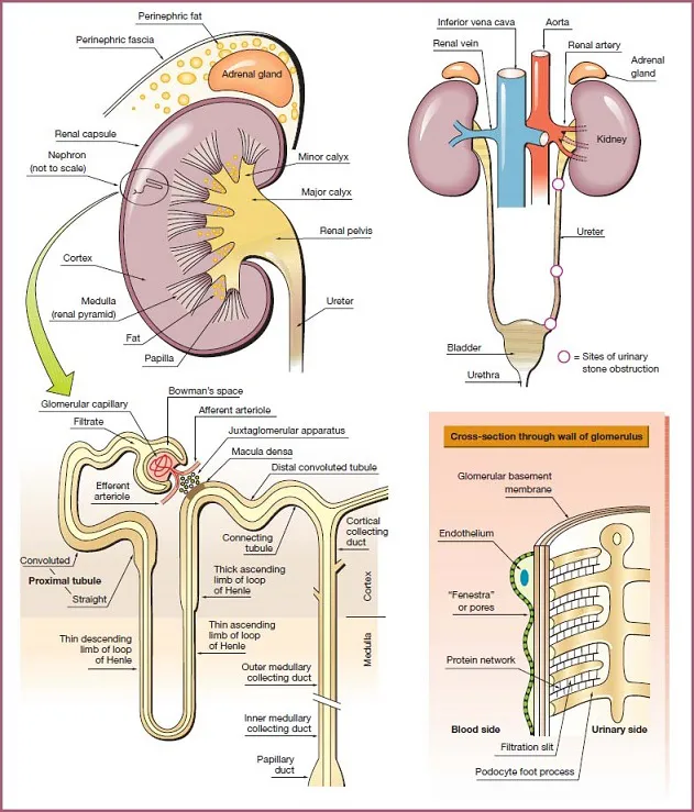
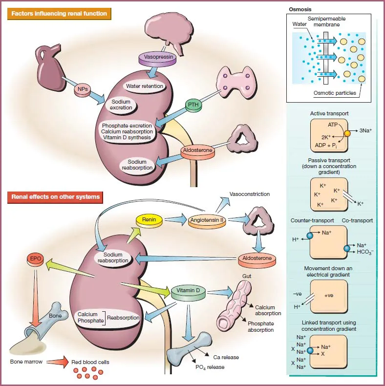
The Renal System at a Glance is a highly illustrated and practical guide to the structure and function of the kidney, renal, and urinary system. It also covers related disorders and abnormalities and their treatment.

Fully updated to reflect the many exciting new developments in the understanding of nephrology, this new edition has been restructured to better integrate basic science and clinical examples to the medical school curricula. New chapters on glomerular filtration and global kidney medicine are included, while the latest guidance and approaches to acute kidney injury, chronic kidney diseases, and renal replacement therapy have also been incorporated.

The Renal System at a Glance:

  • Offers clear explanations on tricky topics such as electrolytes, fluid balance and acid-base handling
  • Features new sections on glomerular filtration, and a new chapter on the global differences in kidney problems
  • Includes cross-referencing between basic science and related clinical content
  • Focuses on clinical disorders and investigations – ideal for those embarking on medicine rotations
  • Illustrates each topic in a double page spread, complete with charts, graphs, and photographs
  • An updated companion website is available at www.ataglanceseries.com/renalsystem featuring animations and MCQs

This new edition is the perfect guide for medical students, junior doctors, and allied health professionals, including specialist nurses, who wish to learn, or refresh their knowledge, on the kidney and renal system in health and disease.

Frequently asked questions

How do I cancel my subscription?
Simply head over to the account section in settings and click on “Cancel Subscription” - it’s as simple as that. After you cancel, your membership will stay active for the remainder of the time you’ve paid for. Learn more here.
Can/how do I download books?
At the moment all of our mobile-responsive ePub books are available to download via the app. Most of our PDFs are also available to download and we're working on making the final remaining ones downloadable now. Learn more here.
What is the difference between the pricing plans?
Both plans give you full access to the library and all of Perlego’s features. The only differences are the price and subscription period: With the annual plan you’ll save around 30% compared to 12 months on the monthly plan.
What is Perlego?
We are an online textbook subscription service, where you can get access to an entire online library for less than the price of a single book per month. With over 1 million books across 1000+ topics, we’ve got you covered! Learn more here.
Do you support text-to-speech?
Look out for the read-aloud symbol on your next book to see if you can listen to it. The read-aloud tool reads text aloud for you, highlighting the text as it is being read. You can pause it, speed it up and slow it down. Learn more here.
Is The Renal System at a Glance an online PDF/ePUB?
Yes, you can access The Renal System at a Glance by Christopher O'Callaghan in PDF and/or ePUB format, as well as other popular books in Medicine & Urology. We have over one million books available in our catalogue for you to explore.

Information

Year
2016
ISBN
9781118393864
Edition
4
Subtopic
Urology

Part 1
Introduction

Chapters

  1. 1 The kidney: structural overview
  2. 2 The kidney: functional overview
  3. 3 Development of the renal system
  4. 4 Clinical features of kidney disease
  5. 5 The kidney: laboratory investigations and diagnostic imaging

Don’t forget to visit the companion website for this book
www.ataglanceseries.com/renalsystem

1 The kidney: structural overview

Diagram of kidney has nephron which is a tubular structure having bowman’s space, et cetera. Three sites of urinary stone obstruction on ureter. Cross-section of glomerulus below.

Gross anatomy

The kidney

The kidneys lie behind the peritoneum at the back of the abdominal cavity, extending from the twelfth thoracic vertebra (T12) to the third lumbar vertebra (L3). The right kidney is lower than the left because of the presence of the liver. During inspiration, both kidneys are pushed down as the diaphragm contracts. Each kidney is covered by a fibrous capsule. This is further surrounded by perinephric fat and then by the perinephric (perirenal) fascia, which also enclose the adrenal gland. The renal cortex is the outer zone of the kidney and the renal medulla is the inner zone made up of the renal pyramids. The cortex contains all the glomeruli, and the medulla contains the loops of Henle, the vasa recta, and the final portions of the collecting ducts.

Vessels and nerves

Blood vessels and the ureter connect with the kidney at the renal hilus. The renal artery arises from the aorta and usually divides into three branches. Two pass in front of the ureter and one goes behind it. Five or six small veins come together to form the renal vein, which leaves the kidney in front of the anterior branch of the renal artery and enters the inferior vena cava. The position of the lymphatics and the renal sympathetic nerves is variable. The lymphatics drain to the lateral aortic lymph nodes. Sympathetic nerves supply the renal vasculature and juxtaglomerular apparatus, and to a lesser extent the rest of the nephron. Afferent fibers enter the spinal cord at T10, T11, and T12.

The draining system for urine

Within the kidney, the pelvis of the ureter divides into two or three major calyces, each of which subdivides into two or three minor calyces. Each minor calyx contains a renal papilla, which is the apex of a medullary pyramid. The ureter passes out of the kidney behind the peritoneum on the psoas muscle and then enters the pelvis in front of the sacroiliac joint. It moves down the lateral pelvic wall toward the ischial spine and then turns forward and medially to enter the bladder. It passes through the bladder wall for 2 cm before opening into the bladder. Urine passes along the ureter by peristalsis. The ureter has three constrictions where kidney stones can become lodged (see Chapter 51). Afferent nerves from the ureter enter the spinal cord at T11, T12, L1, and L2. The bladder is innervated by S3, S4, and S5.

Microanatomy

The nephron

The nephron is the basic unit of the kidney. Each kidney has 400,000–800,000 nephrons, although this number falls with age. A nephron consists of the glomerulus and the associated tubule that leads to the collecting duct. Urine is formed by filtration in the glomerulus; it is then modified in the tubules by the reabsorption and secretion of substances. Cortical nephrons occur throughout the renal cortex and have short loops of Henle; juxtamedullary nephrons begin near the corticomedullary junction and have long loops of Henle, which descend deep into the medulla and enable them to concentrate urine effectively. Cortical nephrons outnumber juxtamedullary nephrons by 7 : 1.

Interstitial cells in the kidney

The cortex contains two types of interstitial cell: phagocytic and fibroblast-like cells. Erythropoietin is made in the fibroblast-like cells. Three types of medullary interstitial cells have been identified. One type contains lipid droplets, which may provide precursors for synthesis of prostaglandins in the kidneys.

The glomerulus as a filtration barrier (see Chapter 7)

The glomerulus is a ball of capillaries surrounded by the Bowman’s capsule, a hollow capsule of the tubular epithelium into which urine is filtered. The space into which the urine is filtered is known as Bowman’s space. The glomerulus also contains mesangial cells, which provide a scaffold to support capillary loops and have contractile and phagocytic properties. Blood enters the glomerular capillaries from an afferent arteriole and leaves through an efferent arteriole, rather than a venule. Vasoconstriction of this efferent arteriole creates a high hydrostatic pressure in the glomerular capillary, forcing water, ions, and small molecules through the filtration barrier into Bowman’s space. Whether a substance is filtered depends on both its molecular size and charge. The filtration barrier has three layers, all of which have a negative charge:
1 Endothelial cells. The endothelial cells of the glomerular capillary wall are thin, with numerous 70-nm pores filled with negatively charged glycoprotein, mostly podocalyxin.
2 Glomerular basement membrane. This specialized capillary basement membrane has three layers and contains negatively charged glycoproteins attached to a three-dimensional framework of collagen and other proteins.
3 Epithelial cells of Bowman’s capsule. The epithelial cells or podocytes have long projections from which foot processes arise and attach to the urinary side of the glomerular basement membrane. Foot processes from different podocytes interdigitate, leaving filtration slits of 25–65 nm between them. Across these slits, a protein network rich in nephrin protein forms “slit pores.” The pores are the key selectivity barrier in the filtration process and prevent the passage of larger molecules such as albumin.

2 The kidney: functional overview

Diagram of renal function: heart, brain, et cetera lead to kidney; renal effects on systems: kidney to bone, gut, et cetera; osmosis: active transport, et cetera of osmotic particles.
The kidney maintains a stable extracellular environment, which supports the function of all body cells. It controls water and ionic balance by regulating the excretion of water, sodium, potassium, chloride, calcium, magnesium, phosphate, and many other substances, and by managing acid-base status.

Tubular function

The urinary filtrate is formed in the glomerulus and flows into the tubules where its volume and content are altered by reabsorption or secretion. Most solute reabsorption occurs in the proximal tubules, and fine adjustments to urine composition are then made in the distal tubule and collecting ducts. The loop of Henle serves to concentrate urine.
The tubular epithelium is only one cell thick. Tubular cells have tight junctions at their apical or luminal edges which separate tubular fluid from peritubular plasma, allowing transport processes to establish concentration gradients across the tubular epithelium. The movement of molecules through these tight junctions is termed “paracellular movement” and is controlled by the properties of proteins called claudins, which form the main barrier t...

Table of contents