The Respiratory System at a Glance has been thoroughly updated in line with current practice guidelines and new techniques to provide a highly illustrated and comprehensive guide to normal lung structure and function, as well as associated pathophysiology. Each topic has been fully revised and is accompanied by clear diagrams to encapsulate essential knowledge.
Reflecting changes to the content, teaching and assessment methods used in medical education, this new edition now includes more information on acid base and its clinical ramifications, further detail on defence mechanisms and immunology, and also features online access to clinical cases and flashcards.
The Respiratory System at a Glance:
• Integrates basic and clinical science – ideal for integrated and systems-based courses
• Includes both the pathophysiology and clinical aspects of the respiratory system
• Is fully revised and updated to reflect current practice guidelines and new therapies
• Provides online clinical cases, brand new flashcards, and MCQs
• Includes a companion website at www.ataglanceseries.com/respiratory featuring interactive multiple choice questions and digital flashcards
- English
- ePUB (mobile friendly)
- Available on iOS & Android
eBook - ePub
The Respiratory System at a Glance
About this book
Trusted by 375,005 students
Access to over 1.5 million titles for a fair monthly price.
Study more efficiently using our study tools.
Information
Part 1
Structure and function
Chapters
- 1 Structure of the respiratory system: lungs, airways and dead space
- 2 The thoracic cage and respiratory muscles
- 3 Pressures and volumes during normal breathing
- 4 Gas laws
- 5 Diffusion
- 6 Lung mechanics: elastic forces
- 7 Lung mechanics: airway resistance
- 8 Carriage of oxygen
- 9 Carriage of carbon dioxide
- 10 Acid–base balance
- 11 Acid–base disorders
- 12 Control of breathing I: chemical mechanisms
- 13 Control of breathing II: neural mechanisms
- 14 Pulmonary circulation and anatomical right-to-left shunts
- 15 Ventilation–perfusion mismatching
- 16 Exercise, altitude and diving
- 17 Development of the respiratory system and birth
- 18 Complications of development and congenital disease
- 19 Lung defence mechanisms
- 20 Immunology of the lung
1
Structure of the respiratory system: lungs, airways and dead space
Lungs
The respiratory system consists of a pair of lungs within the thoracic cage (Chapter 2). Its main function is gas exchange, but other roles include speech, filtration of microthrombi arriving from systemic veins and metabolic activities such as conversion of angiotensin I to angiotensin II and removal or deactivation of serotonin, bradykinin, norepinephrine, acetylcholine and drugs such as propranolol and chlorpromazine. The right lung is divided by transverse and oblique fissures into three lobes: upper, middle and lower. The left lung has an oblique fissure and two lobes (Fig. 1a). Vessels, nerves and lymphatics enter the lungs on their medial surfaces at the lung root or hilum. Each lobe is divided into a number of wedge-shaped bronchopulmonary segments with their apices at the hilum and bases at the lung surface. Each bronchopulmonary segment is supplied by its own segmental bronchus, artery and vein and can be removed surgically with little bleeding or air leakage from the remaining lung.
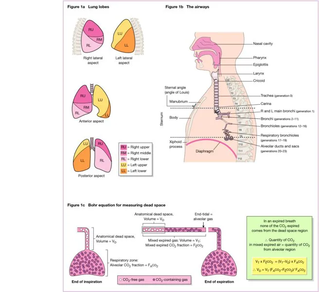
Figure 1
The pulmonary nerve plexus lies behind each hilum, receiving fibres from both vagi and the second to fourth thoracic ganglia of the sympathetic trunk. Each vagus contains sensory afferents from lungs and airways, parasympathetic bronchoconstrictor and secretomotor efferents, and non-adrenergic, non-cholinergic (NANC). Sympathetic noradrenergic fibres supplying airway smooth muscle are sparse in humans, and the β2-adrenergic receptors are stimulated by circulating catecholamines from the adrenal glands (Chapter 7).
Each lung is lined by a thin membrane, the visceral pleura, which is continuous with the parietal pleura, lining the chest wall, diaphragm, pericardium and mediastinum. The space between the parietal and visceral layers is tiny in health and lubricated with pleural fluid. The right and left pleural cavities are separate and each extends as the costodiaphragmatic recess below the lungs even during full inspiration. The parietal pleura is segmentally innervated by intercostal nerves and by the phrenic nerve, and so pain from pleural inflammation (pleurisy) is often referred to the chest wall or shoulder tip. The visceral pleura lacks sensory innervation.
Lymph channels are absent in alveolar walls, but accompany small blood vessels conveying lymph towards the hilar bronchopulmonary nodes and from there to tracheobronchial nodes at the tracheal bifurcation. Some lymph from the lower lobe drains to the posterior mediastinal nodes.
The upper respiratory tract consists of the nose, pharynx and larynx. The lower respiratory tract (Fig. 1b) starts with the trachea at the lower border of the cricoid cartilage, level with the sixth cervical vertebra (C6). It bifurcates into right and left main bronchi at the level of the sternal angle and T4/5 (lower when upright and in inspiration). The right main bronchus is wider, shorter and more vertical than the left, so inhaled foreign bodies enter it more easily.
Airways
The airways divide repeatedly, with each successive generation approximately doubling in number. The trachea and main bronchi have U-shaped cartilage, linked posteriorly by smooth muscle. Lobar bronchi supply the three right and two left lung lobes and divide to give segmental bronchi (generations 3 and 4). The total cross-sectional area of each generation is minimum here, after which it rises rapidly, as increased numbers more than make up for their reduced size. Generations 5–11 are small bronchi, the smallest being 1 mm in diameter. The lobar, segmental and small bronchi are supported by irregular plates of cartilage, with bronchial smooth muscle forming helical bands. Bronchioles start at about generation 12 and from this point onwards cartilage is absent. These airways are embedded in lung tissue, which holds them open like tent guy ropes. The terminal bronchioles (generation 16) lead to respiratory bronchioles, the first generation to have alveoli (Chapter 5) in their walls. These lead to alveolar...
Table of contents
- Cover
- Series
- Titlepage
- Copyright
- Preface to fourth edition
- Units and symbols
- List of abbreviations
- About the companion website
- Part 1: Structure and function
- Part 2: History, examination and investigation
- Part 3: Diseases and treatment
- Index
- EULA
Frequently asked questions
Yes, you can cancel anytime from the Subscription tab in your account settings on the Perlego website. Your subscription will stay active until the end of your current billing period. Learn how to cancel your subscription
No, books cannot be downloaded as external files, such as PDFs, for use outside of Perlego. However, you can download books within the Perlego app for offline reading on mobile or tablet. Learn how to download books offline
Perlego offers two plans: Essential and Complete
- Essential is ideal for learners and professionals who enjoy exploring a wide range of subjects. Access the Essential Library with 800,000+ trusted titles and best-sellers across business, personal growth, and the humanities. Includes unlimited reading time and Standard Read Aloud voice.
- Complete: Perfect for advanced learners and researchers needing full, unrestricted access. Unlock 1.5M+ books across hundreds of subjects, including academic and specialized titles. The Complete Plan also includes advanced features like Premium Read Aloud and Research Assistant.
We are an online textbook subscription service, where you can get access to an entire online library for less than the price of a single book per month. With over 1.5 million books across 990+ topics, we’ve got you covered! Learn about our mission
Look out for the read-aloud symbol on your next book to see if you can listen to it. The read-aloud tool reads text aloud for you, highlighting the text as it is being read. You can pause it, speed it up and slow it down. Learn more about Read Aloud
Yes! You can use the Perlego app on both iOS and Android devices to read anytime, anywhere — even offline. Perfect for commutes or when you’re on the go.
Please note we cannot support devices running on iOS 13 and Android 7 or earlier. Learn more about using the app
Please note we cannot support devices running on iOS 13 and Android 7 or earlier. Learn more about using the app
Yes, you can access The Respiratory System at a Glance by Jeremy P. T. Ward,Jane Ward,Richard M. Leach in PDF and/or ePUB format, as well as other popular books in Medicine & Pulmonary & Thoracic Medicine. We have over 1.5 million books available in our catalogue for you to explore.