Small Animal ECGs
eBook - ePub

Small Animal ECGs

An Introductory Guide

  1. English
  2. ePUB (mobile friendly)
  3. Available on iOS & Android
eBook - ePub

Small Animal ECGs

An Introductory Guide

About this book

Small Animal ECGs: An Introductory Guide provides all the information veterinarians need when using electrocardiography techniques for the first time.

  • An ideal introduction to veterinary electrocardiography written in a very easy to understand way, for what can be a daunting subject
  • The author is RCVS Recognised Specialist in Veterinary Cardiology and regularly speaks on this subject
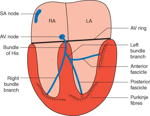
  • Full of examples of colour ECG tracings, as well as colour illustrations to explain arrhythmias
  • Covers techniques that can readily be used in first opinion small animal practice
  • Includes new chapters on mechanisms of supraventricular arrhythmias, accelerated idioventricular rhythm and use of Holters

Frequently asked questions

Yes, you can cancel anytime from the Subscription tab in your account settings on the Perlego website. Your subscription will stay active until the end of your current billing period. Learn how to cancel your subscription.
At the moment all of our mobile-responsive ePub books are available to download via the app. Most of our PDFs are also available to download and we're working on making the final remaining ones downloadable now. Learn more here.
Perlego offers two plans: Essential and Complete
  • Essential is ideal for learners and professionals who enjoy exploring a wide range of subjects. Access the Essential Library with 800,000+ trusted titles and best-sellers across business, personal growth, and the humanities. Includes unlimited reading time and Standard Read Aloud voice.
  • Complete: Perfect for advanced learners and researchers needing full, unrestricted access. Unlock 1.4M+ books across hundreds of subjects, including academic and specialized titles. The Complete Plan also includes advanced features like Premium Read Aloud and Research Assistant.
Both plans are available with monthly, semester, or annual billing cycles.
We are an online textbook subscription service, where you can get access to an entire online library for less than the price of a single book per month. With over 1 million books across 1000+ topics, we’ve got you covered! Learn more here.
Look out for the read-aloud symbol on your next book to see if you can listen to it. The read-aloud tool reads text aloud for you, highlighting the text as it is being read. You can pause it, speed it up and slow it down. Learn more here.
Yes! You can use the Perlego app on both iOS or Android devices to read anytime, anywhere — even offline. Perfect for commutes or when you’re on the go.
Please note we cannot support devices running on iOS 13 and Android 7 or earlier. Learn more about using the app.
Yes, you can access Small Animal ECGs by Mike Martin,Mike Martin in PDF and/or ePUB format, as well as other popular books in Medicine & Veterinary Medicine. We have over one million books available in our catalogue for you to explore.

Information

Year
2015
Print ISBN
9781118409732
eBook ISBN
9781118409725
Edition
3

Part 1
Understanding the electricity of the heart and how it produces an ECG complex

  1. Chapter 1: What is an ECG?
  2. Chapter 2: The electricity of the heart
  3. Chapter 3: Rhythms of sinus origin

Chapter 1
What is an ECG?

An electrocardiograph (ECG), in its simplest form, is a voltmeter (or galvanometer) that records the changing electrical activity in the heart by means of positive (+ve) and negative (−ve) electrodes (Fig. 1.1). Electrocardiography is the process of recording these changing potential differences.
c01f001
Figure 1.1 An ECG records the ‘electricity of the heart’.
A +ve and a −ve electrode can be placed almost anywhere on, or in, the body to record electrical changes. One of the most common and simplest methods is to place these electrodes on the limbs of animals – referred to as a body surface limb ECG recording. However, if monitoring an ECG during anaesthesia or echocardiography, for example, it is sufficient to place the –ve electrode anywhere cranial to the heart (e.g. the forelimb or neck) and the +ve electrode caudal to the heart (e.g. the hind limb, abdomen or flank). Electrodes can also be placed on the chest (precordial chest ECG recording – commonly used in humans) or inside the cardiac chambers (used in electrophysiological studies). This book focuses on the conventional limb ECG recording, which is the method most commonly used in veterinary medicine for clinical diagnosis of arrhythmias.

Chapter 2
The electricity of the heart

Electrical coordination of atrioventricular contraction

For the heart to function efficiently as a ‘circulatory pump’, it must have a coordinated contraction: the two atria contracting and passing blood into the two ventricles, followed by contraction of the ventricles, pumping blood into the aorta and pulmonary artery; that is, there must be a coordinated atrioventricular (AV) contraction. In order for the cardiac muscle cells to contract, they must first receive an electrical stimulus. It is this electrical activity that is detected by an ECG.
The electrical stimulus must first depolarise the two atria. Then, after an appropriate time interval, it must depolarise the two ventricles. The heart must then repolarise (and ‘refill’) in time for the next stimulus and contraction. Additionally, it must repeatedly do so, increasing in rate with an increase in demand and conversely slowing at rest.

Formation of the normal P–QRS–T complex

All cells within the heart have the potential to generate their own electrical activity; however, the sinoatrial (SA) node is the fastest part of the electrical circuit to do so and is therefore the ‘rate controller’, termed the pacemaker. The sinus node rate is, therefore, the dominant pacemaker (over the other cells in the heart) by being the fastest and by a mechanism termed overdrive suppression. The rate of the SA node is influenced by the balance in the autonomic tone, that is, the sympathetic (increases rate) and parasympathetic (decreases rate) systems.
The electrical discharge for each cardiac cycle (Fig. 2.1) starts in the SA node. Depolarisation spreads through the atrial muscle cells. The depolarisation wave then spreads through the AV node; however, it does so at a relatively slower rate, creating a delay. Conduction passes through the AV ring (from the atria into the ventricles) through a narrow pathway called the bundle of His. This then divides in the ventricular septum into left and right bundle branches (going to the left and right ventricles). The left bundle branch divides further into anterior and posterior fascicles. The conduction tissue spreads into the myocardium as very fine branches called Purkinje fibres.
c02f001
Figure 2.1 Illustration of the heart's electrical circuit. SA – sinoatrial; AV – atrioventricular; RA – right atrium; LA – left atrium.

Formation of the P wave

The SA node is therefore the start of the electrical depolarisation wave. This depolarisation wave spreads t...

Table of contents

  1. Cover
  2. Title Page
  3. Dedication
  4. Copyright
  5. Table of Contents
  6. Preface to third edition
  7. About the author
  8. Acknowledgements
  9. Abbreviations
  10. Part 1: Understanding the electricity of the heart and how it produces an ECG complex
  11. Part 2: Abnormal electricity of the heart
  12. Part 3: More advanced electrocardiography
  13. Part 4: Management, clinical significance and treatment of arrhythmias
  14. Part 5: Recording and interpreting ECGs
  15. Further reading
  16. Index
  17. End User License Agreement