Pocket Guide to Echocardiography
eBook - ePub

Pocket Guide to Echocardiography

Andro G. Kacharava, Alexander T. Gedevanishvili, Guram G. Imnadze, Dimitri M. Tsverava, Craig M. Brodsky, Andro G. Kacharava, Alexander T. Gedevanishvili, Guram G. Imnadze, Dimitri M. Tsverava, Craig M. Brodsky

Buch teilen
  1. English
  2. ePUB (handyfreundlich)
  3. Über iOS und Android verfügbar
eBook - ePub

Pocket Guide to Echocardiography

Andro G. Kacharava, Alexander T. Gedevanishvili, Guram G. Imnadze, Dimitri M. Tsverava, Craig M. Brodsky, Andro G. Kacharava, Alexander T. Gedevanishvili, Guram G. Imnadze, Dimitri M. Tsverava, Craig M. Brodsky

Angaben zum Buch
Buchvorschau
Inhaltsverzeichnis
Quellenangaben

Über dieses Buch

With its easy accessibility, low cost, and ability to deliver, essential bedside information about the cardiac structure and function, echocardiography has become one of the most relied-upon diagnostic tools in clinical medicine. As a result, more clinicians than ever before must be able to accurately interpret echocardiographic information in order to administer appropriate treatment.

Based on the authors' experience teaching echocardiography in busy clinical settings, this new pocketbook provides reliable guidance on everyday clinical cardiac ultrasound and the interpretation of echocardiographic images. It has been designed to help readers develop a stepwise approach to the interpretation of a standard transthoracic echocardiographic study and teach how to methodically gather and assemble the most important information from each of the standard echocardiographic views in order to generate a complete final report of the study performed.

What's included:

•A summary of TTE examination protocol and a comprehensive listing of useful formulas and normal values

•Atrial and ventricular dimensions, LV and RV systolic function, LV diastolic patterns

•Echocardiographic findings in the most commonly encountered cardiac diseases and disorders, including various cardiomyopathies, cardiac tamponade, constrictive pericarditis, valvular heart disease, pulmonary hypertension, infective endocarditis, and congenital heart disease

•Companion website with video clips and over 70 self-assessment questions


Packed with essential information and designed for quick look-up, this pocketbook will be of great assistance for anyone who works in busy clinical settings and who needs a ready and reliable guide to interpreting echocardiographic information to help deliver optimal patient care.

Häufig gestellte Fragen

Wie kann ich mein Abo kündigen?
Gehe einfach zum Kontobereich in den Einstellungen und klicke auf „Abo kündigen“ – ganz einfach. Nachdem du gekündigt hast, bleibt deine Mitgliedschaft für den verbleibenden Abozeitraum, den du bereits bezahlt hast, aktiv. Mehr Informationen hier.
(Wie) Kann ich Bücher herunterladen?
Derzeit stehen all unsere auf Mobilgeräte reagierenden ePub-Bücher zum Download über die App zur Verfügung. Die meisten unserer PDFs stehen ebenfalls zum Download bereit; wir arbeiten daran, auch die übrigen PDFs zum Download anzubieten, bei denen dies aktuell noch nicht möglich ist. Weitere Informationen hier.
Welcher Unterschied besteht bei den Preisen zwischen den Aboplänen?
Mit beiden Aboplänen erhältst du vollen Zugang zur Bibliothek und allen Funktionen von Perlego. Die einzigen Unterschiede bestehen im Preis und dem Abozeitraum: Mit dem Jahresabo sparst du auf 12 Monate gerechnet im Vergleich zum Monatsabo rund 30 %.
Was ist Perlego?
Wir sind ein Online-Abodienst für Lehrbücher, bei dem du für weniger als den Preis eines einzelnen Buches pro Monat Zugang zu einer ganzen Online-Bibliothek erhältst. Mit über 1 Million Büchern zu über 1.000 verschiedenen Themen haben wir bestimmt alles, was du brauchst! Weitere Informationen hier.
Unterstützt Perlego Text-zu-Sprache?
Achte auf das Symbol zum Vorlesen in deinem nächsten Buch, um zu sehen, ob du es dir auch anhören kannst. Bei diesem Tool wird dir Text laut vorgelesen, wobei der Text beim Vorlesen auch grafisch hervorgehoben wird. Du kannst das Vorlesen jederzeit anhalten, beschleunigen und verlangsamen. Weitere Informationen hier.
Ist Pocket Guide to Echocardiography als Online-PDF/ePub verfügbar?
Ja, du hast Zugang zu Pocket Guide to Echocardiography von Andro G. Kacharava, Alexander T. Gedevanishvili, Guram G. Imnadze, Dimitri M. Tsverava, Craig M. Brodsky, Andro G. Kacharava, Alexander T. Gedevanishvili, Guram G. Imnadze, Dimitri M. Tsverava, Craig M. Brodsky im PDF- und/oder ePub-Format sowie zu anderen beliebten Büchern aus Medicine & Cardiology. Aus unserem Katalog stehen dir über 1 Million Bücher zur Verfügung.

Information

Jahr
2012
ISBN
9781118480038
Auflage
1
Chapter 1
Comprehensive Transthoracic Echocardiographic Examination Protocol

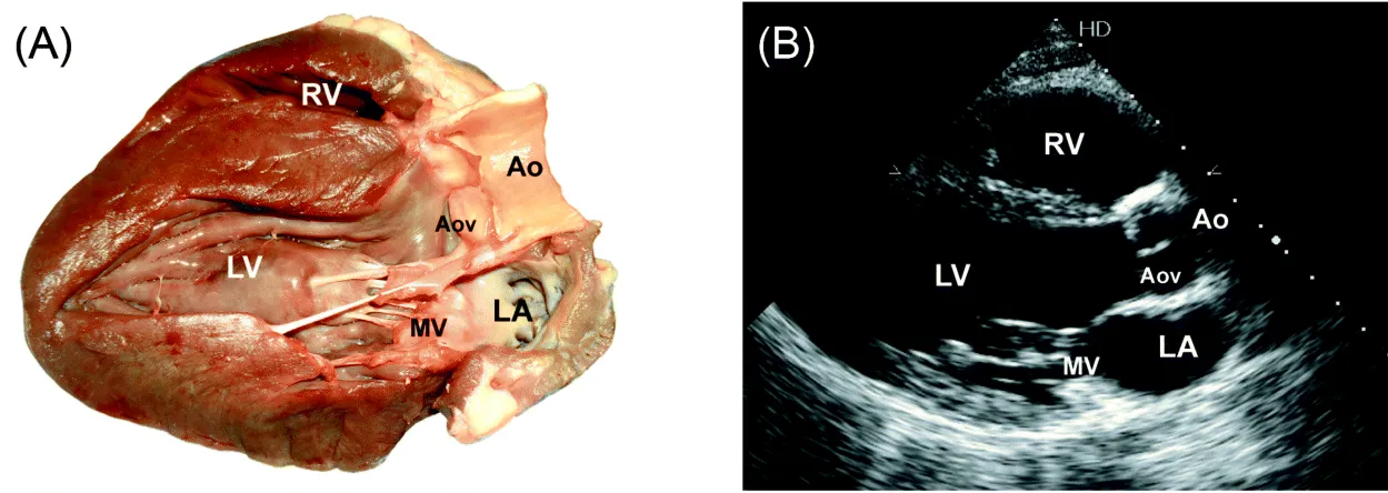
Parasternal Long-axis view (Fig. 1)

1. 2D image (4 beats) (measure LVOT diameter).
2. Color Doppler through MV and AoV (4 beats).
3. M-mode through Aortic root (measure root and LA diameter, and aortic cusp separation).
4. Color Doppler M-mode through the aortic root (4 beats).
5. M-mode through MV (± Valsalva test to check for MV prolapse and SAM; measure “E” to “S” separation distance).
6. Color Doppler M-mode through MV (4 beats).
7. M-mode through mid LV (4 beats) (measure septal and inferolateral wall thickness LVEDD and LVESD).
Figure 1 (A and B) Parasternal long-axis view.
img

RA/RV view

From parasternal LAX view tilt transducer to point it to right hip:
1. 2D image (4 beats);
2. Color Doppler through TV (4 beats);
3. CW Doppler through TV to measure max TR velocity if TR jet present.

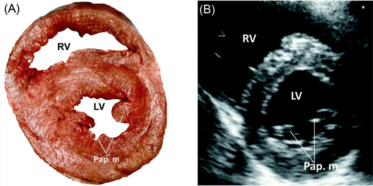
Parasternal Short-axis view (Fig. 2 and Fig. 3)
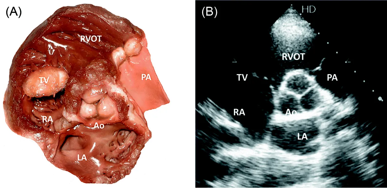
1. 2D through AoV (4 beats) (to assess structure and mobility; use zoom).
2. Color Doppler through AoV (4 beats).
3. 2D through PV (4beats).
4. Color Doppler through PV (4 beats).
5. PW Doppler at the tips of PV to measure PAT (4 beats).
6. CW Doppler through PV to measure PR velocity if present, and maximum outflow velocity through PV.
7. 2D through TV (4 beats).
8. Color Doppler through TV (4 beats).
9. PW Doppler at the tips of TV leaflets to assess inflow pattern (4 beats).
10. If inflow jet max velocity is >1.5 m/sec, trace the diastolic flow to measure mean transvalvular gradient.
11. CW Doppler through TV to measure max TR velocity if TR jet present.
12. Serial 2D short axis images through the LV from base toward apex.
Figure 2 (A and B) Parasternal short-axis view.
img
Figure 3 (A and B) Parasternal short-axis view.
img

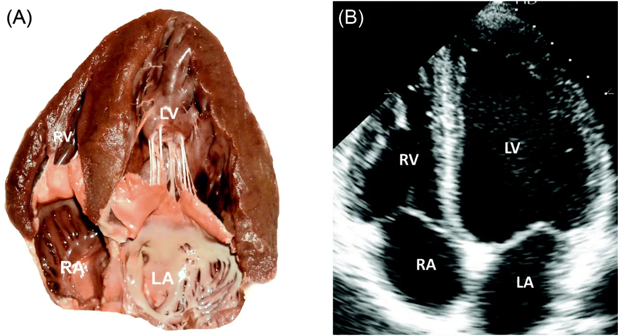
Apical 4-chambers view (Fig. 4)

1. 2D image (4 beats).
2. Color Doppler through the MV (4 beats).
3. Color Doppler M-mode through the MV at the end expiration (4 beats).
4. PW Doppler at the tips of MV leaflets to assess inflow pattern and velocity (4 beats); if pseudonormal or restrictive inflow pattern observed, decrease the preload and reassess the inflow pattern; in impaired relaxation act opposite.
5. PW Doppler at the right/left upper PV to assess inflow pattern (4 beats).
6. PW tissue Doppler at the basal and mid septal and lateral walls (4 beats).
7. If inflow jet max velocity is > 1.9 m/sec, trace the diastolic flow to measure mean transvalvular gradient, then measure PHT of the jet in CW mode.
8. CW Doppler through MV to measure max MR velocity if MR jet present. (Obtain simultaneous SBP measurement to calculate mean LAP.)
9. PW tissue Doppler at basal lateral walls of the RV to assess TEI index and systolic velocity.
10. M-mode through the RV basal lateral wall to measure TAPSE.
Figure 4 (A and B) Apical 4-chambers view.
img

Apical 5-chambers view

From apical 4-chambers view tilt transducer slightly anteriorly; examiner's hand moves downward toward the patient's bed:
1. 2D image (4 beats).
2. Color Doppler through AoV and MV (4 beats).
3. PW Doppler through LVOT and trace the flow (4 beats).
4. CW Doppler through AoV and if outflow max velocity is >1.9 m/sec, trace...

Inhaltsverzeichnis