![]()
Development of the Pulpodentin Complex
Rena D’Souza, DDS, MS, PhD
Chunlin Qin, DDS, MS, PhD
Dentin is a unique, avascular mineralized connective tissue that forms the bulk of the tooth. It underlies enamel in the crown and cementum in the roots, providing these tissues structural support and the tooth its resilience. In a mature tooth, dentin encloses a richly innervated and highly vascularized soft connective tissue, the dental pulp. Dentin and pulp are derived from the dental papilla, whose cells migrate to the first branchial arch from within the ectomesenchyme of the cranial neural crest. The tissues remain closely associated during development and throughout the life of an adult tooth and are hence most commonly referred to as the pulpodentin complex. It is this biologic intimacy that dictates the response of the pulpodentin complex to physiologic and pathologic stimuli.
Because the practice of endodontics involves manipulation of both the dentin and pulp, learning about the mechanisms that lead to their formation is crucial and will create a better understanding of the response to and treatment of pulpal injuries. The purpose of this chapter is to provide a background for the succeeding chapters that discuss the biology of the mature pulpodentin complex during health and disease and in aging. The chapter has two goals: The first goal is to review classic and current knowledge of the events in tooth development that lead to odontoblast differentiation and to convey the excitement of this flourishing field. Attention is focused on the common themes that have emerged and what is known about the influence of tooth-signaling molecules and transcription factors on the development and homeostasis of the pulpodentin complex. The second goal of this chapter is to describe the general principles of dentin matrix formation, particularly the synthesis and secretion of extracellular matrix molecules and their postulated roles in the biomineralization of dentin. Examples of how basic information about the normal biology of the pulpodentin complex can be applied toward solving clinical problems are integrated throughout the chapter. The overarching goal is to emphasize how fundamental theories about development and homeostasis of differentiated and undifferentiated or stem cell populations can be translated to regenerative approaches targeted at restoring the integrity of the adult pulpodentin complex.
Tooth Development (Odontogenesis)
For several decades, the developing tooth organ has served as a valuable paradigm to study the fundamental processes involved in organogenesis. These processes are (1) the determination of position, through which the precise site of tooth initiation is established; (2) the determination of form or morphogenesis, through which the size and shape of the tooth organ are set; and (3) cell differentiation, through which organ-specific tissues are formed by defined cell populations, each with unique properties. The dental literature is enriched with excellent reviews on tooth development, and the reader is encouraged to study the topic in further detail.1–4
General features
Although the tooth is a unique organ, the principles that guide its development are shared in common with other organs such as the lung, kidney, heart, mammary glands, and hair follicles.5,6 The most important among developmental events are those guiding epithelial-mesenchymal interactions, which involve a molecular crosstalk between the ectoderm and mesenchyme, two tissues that have different origins. Although only vertebrates have teeth, their development involves genetic pathways that are also active in invertebrates. This conservation of a “molecular toolbox” for organogenesis throughout evolution proves that certain master regulatory molecules are critical to all tissue interactions during development. New studies have also shown that tooth-signaling molecules are repeatedly used at various stages of development. Both tooth morphogenesis and cell differentiation occur as a result of sequential interactions. Hence, it is not one biologic event involving a single molecule but a series of interactions involving several molecules that leads to the development of the pulpodentin complex.3,4
Importantly, signaling is reciprocal, whereby an exchange of information occurs in both directions, from dental epithelium to mesenchyme and from dental mesenchyme to epithelium. For example, in experiments where dental epithelium was separated from mesenchyme, cusp patterning failed to occur. Similarly, in the absence of dental epithelium, odontoblasts are unable to differentiate from dental mesenchyme.7–9
It is only logical to apply these basic developmental principles to the current understanding of how the adult or mature pulpodentin complex responds to injury and repair. The latter clearly involves a series of molecules that operate in concert to dictate the outcome of pulpal disease and therapies. In the adult situation, whether certain cells and molecules can mimic the inductive influence of dental epithelium during development has yet to be definitively proven and remains a subject of interest in pulpal biology research.
Stages of tooth development
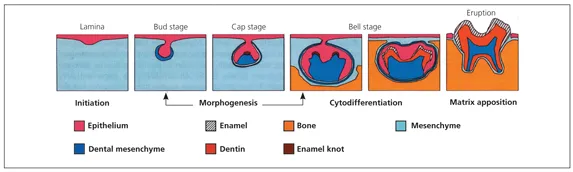
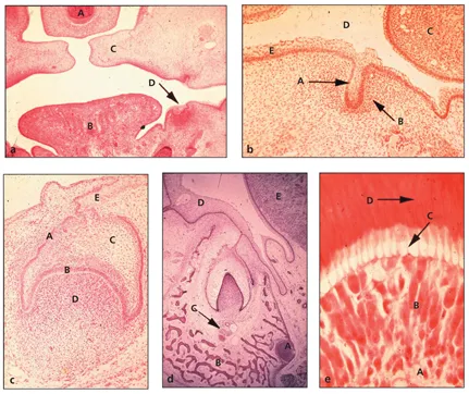
Teeth develop in distinct stages that are easily recognizable at the microscopic level. These stages in odontogenesis, described by the histologic appearance of the tooth organ, are termed, from early to late, the lamina, bud, cap, and bell (early and late) stages of tooth development.10–12 Although the following descriptions use these common terms, the modern literature uses functional terminology to describe odontogenesis in four phases: (1) initiation, (2) morphogenesis, (3) cell differentiation or cytodifferentiation, and (4) matrix apposition (Fig 1-1). The photomicrographs in Fig 1-2 depict the morphologic stages of tooth development.
Fig 1-1 Stages of tooth development. Note the sequential transformation from the dental lamina to a distinctly shaped dental organ. The transient appearance of the enamel knot in the region of the forming cusp tips precedes the terminal differentiation of cells and the formation of specialized matrices. (Reprinted from Thesleff and Sharpe12 with permission.)
Fig 1-2 Histologic survey of odontogenesis in a pig embryo. (Courtesy of the University of Texas Health Science Center at Houston, Dental Branch.) (a) Lamina stage: A, nasal septum; B, tongue; C, palatal shelves; D, dental lamina (hematoxylin-eosin [H&E] stain; original magnification ×4). (b) Bud stage: A, ectodermal outgrowth; B, dental mesenchyme; C, tongue; D, oral cavity space; E, oral ectoderm (H&E stain; original magnification ×10). (c) Cap stage or transition to early bell stage: A, outer dental epithelium; B, internal dental epithelium; C, stellate reticulum; D, dental papilla ectomesenchyme; E, dental lamina (H&E stain; original magnification ×10). (d) Late bell stage: A, nerve bundle; B, alveolar bone; C, vas-culature; D, oral ectoderm; E, tongue. Note the extension of the dental lamina on the right aspect of the dental organ that will form the succeda-neous incisor (H&E stain; original magnification ×10). (e) Onset of dentinogenesis: A, dental pulp; B, cluster of odontoblasts that appear crowded at the tip; C, odontoblast process; D, dentin (H&E stain; original magnification ×20).
Lamina stage
The dental lamina is the first morphologic sign of tooth development and is visible around 5 weeks of human development and at embryonic day 11 (E11) in mouse gestation. This thickening of the oral epithelium lining the frontonasal, maxillary, and mandibular arches occurs only at sites where tooth organs will develop. At the lamina stage, cells in the dental epithelium and underlying ectomesenchyme are dividing at different rates, more rapidly in the latter. As explained later, ...