![]()
Chapter 1
Introduction and Pipelines
1.1 Introduction
1.2 Pipelines and Five Topics
1.3 Challenges and Advances
An introduction is provided to explain the pipelines from imaging to meshing and simulation. The pipelines involve five main topics: imaging, image processing, geometric modeling and computer graphics, mesh generation and finite element simulations. These five topics can be applied to many applications in computational biology, medicine, material sciences and engineering.
1.1 Introduction
Over recent years, finite element method (FEM) and scanning technology have been developed and advanced rapidly, especially in cyberinfrastructure-revolutionized research areas such as computational science and engineering. They have very broad applications in computational biology, medicine, materials sciences and engineering. There is an emerging need for quality mesh generation of the spatially realistic domains to enable the analysis, understanding and prediction of complex physical or biological phenomena. In images obtained from computer tomography (CT), magnetic resonance imaging (MRI) or microscopy scanning, the domain of focus often possesses homogeneous, heterogeneous materials and/or functionally different properties. For each of the partitioned materials, high-fidelity (correct topology and accurate geometry) geometric models and quality meshes are needed, with meshes conforming at the manifold or non-manifold boundaries. However, quality meshes for such complex geometries are typically generated manually and thus usually take months to be created. It is well known that FEM is currently well developed and efficient, but mesh generation for complex geometries (e.g., the human body) still takes 80% of the total analysis time and is the major obstacle to reduce the total computation time. High-fidelity geometric modeling for such sophisticated domains is critical for a lot of cyber-enabled innovative applications. As a new emerging interdisciplinary research area, “geometric modeling and mesh generation from scanned images” integrates image processing, geometric modeling and mesh generation with FEM to solve problems in computational biology, medicine, material sciences and engineering.
Image-based mesh generation is a relatively new field. Normally researchers first extract boundary surfaces using isocontouring [189, 256] which usually involves manual interaction, and then construct tetrahedral or hexahedral (hex) meshes. The research on mesh generation is dominated by tetrahedral meshing algorithms, which can be grouped into Delaunay triangulation [109, 396], advancing front [251, 252], or grid-based methods [338, 377]. Fewer algorithms exist for automatic all-hex mesh generation due to its intrinsic complexities, and all these existing methods have limitations. For example, the frequently used, easy to implement block-structured method [71, 280] produces non-conforming boundaries and a large number of elements. The grid-based method [325, 326], which puts structured grids inside the volume while adding elements at the boundaries afterwards, cannot be extended to all-hex mesh generation for heterogeneous domains with conformal non-manifold boundaries. Today, the key barriers scientists face are:
• A lack of automatic and robust meshing techniques for multiscale modeling and heterogeneous domains;
• Robust unstructured all-hex mesh generation with sharp feature preservation for complicated geometry and topology is still a challenge;
• The inability of existing methods to effectively improve the quality of non-manifold meshes with feature preservation and topology validation; and
• A lack of volumetric parameterization (e.g., NURBS and T-spline) techniques for complicated domains to support a new development of FEM called isogeometric analysis.
Many simulations cannot hereby be effectively carried out due to the lack of analysis-suitable finite element meshes. In this book, we introduce the fundamentals of imaging, image processing, computational geometry, mesh generation, visualization, finite element analysis as well as their novel and advanced applications in computational biology, medicine, material sciences and other engineering fields.
1.2 Pipelines and Five Topics
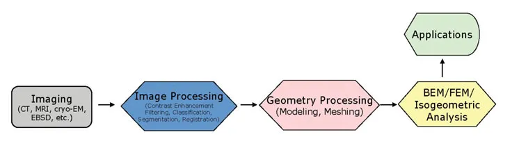
As a newly emerging interdisciplinary research area, “geometric modeling and mesh generation from scanned images” integrates mechanical engineering, biomedical engineering, computer science, and mathematics together. From image scanning to meshing and then to simulation applications, the entire procedure forms comprehensive image-mesh-simulation pipelines [426]. As shown in Figure 1.1, there are mainly five modules in the : imaging, image processing, geometry processing, finite element methods, and application.
FIGURE 1.1: Image-mesh-simulation pipeline with five basic topics.
There are many techniques and processes developed to create images, such as CT, MRI, nuclear medicine, ultrasound, and fluorescence for biomedical imaging. CT utilizes X-rays while MRI utilizes radio wave and magnetic field to scan objects. For both CT and MRI, the resulting images provide anatomy structure. Unlike CT and MRI, Nuclear Medicine injects radioactive source into the body, and then measures the radiation emitted from the body. The resulting images provide physiological functions of the object. Ultrasound utilizes high frequency sound waves in real time, generally 3~10 megahertz. Fluoroscopy uses a constant of X-rays, and it is also in real time. Cryo-EM (cryo-electron microscopy) is a popular scanning technique in structural biology to study small-scale objects like viruses at the level of Angstroms. In addition, people also scan metal materials. For example, electron backscatter diffraction (EBSD) is a microstructural-crystallographic technique used to examine the crystallographic orientation and elucidate texture or preferred orientation of polycrystalline materials. Instead of destroying the metal material like EBSD, high-energy X-ray is a nondestructive scanning technique to study polycrystalline materials. These various scanning techniques produce images for different types of objects. In some research areas like computer-aided design (CAD), people also use computational ways like signed distance function to compute volumetric imaging data. For biomolecules or proteins, people use the atomic resolution information to build electron density map and solve partial differential equations to obtain electron static potential distribution on regular grids.
The scanned image data V is a scalar field over sampled rectilinear grids, V = {F(i,j,k)| i,j,k are indices of x,y,z coordinates in a rectilinear grid}. Due to the limitation of the scanning resolution, object motion and other issues, the obtained images may have low contrast, noises and unclear boundary for materials of interest. Therefore, people develop different kinds of image processing techniques to improve the quality of images and try to extract useful information to enable a better understanding. More specifically, people use computational methods to enhance the contrast, remove noises, classify and segment materials or regions of interest, or register images from difference time phases or modalities. After that, the processed images are fed into the third module in the pipeline to perform geometry processing. It includes geometric modeling, mesh generation and quality improvement. Various quality finite element meshes can be created in this module, which are ready for the following finite element simulations.
The fourth module includes various mechanics simulation techniques, such as boundary element methods, finite element methods and isogeometric analysis [179]. The last module is for applications, it can be very broad ranging from mechanical engineering, biomedical engineering, material sciences, petroleum engineering, electrical engineering to civil engineering and architecture. Generally, various applications have different research aims and they need different techniques in the first four modules to handle the entire process. For example, in cardiovascular blood flow simulation and brain biomechanics applications, image segmentation and quality piecewise-linear elements like tetrahedral/hexahedral meshes or solid NURBS (non-uniform rational B-splines)/T-spline models [332, 334] are required. In dynamic lung modeling, image registration techniques are needed to track the motion of tumor in the lung. In cardiac mechanical property study, high order elements like tricubic Hermite models are preferable. These five topics provide various fundamental technologies, based on which users can develop their own detailed pipelines.
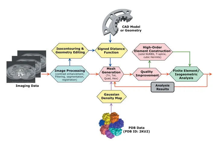
Figure 1.2 shows a comprehensive image-mesh-simulation computational framework that includes several pipelines. Following the red arrows, we start with scanned images, use image processing techniques to extract and segment the region of interest, and then we generate piecewise-linear elements (triangle, quadrilateral, tetrahedra or hexahedra) and improve the mesh quality. The resulting meshes can be used in finite element analysis and the analysis results are then fed to the meshing process to fine tune the finite element meshes. Following the green arrows, the obtained piecewise-linear meshes can be used as control meshes to build high-order elements like solid NURBS, T-splines or cubic Hermite models, which are then used in isogeometric analysis. Another pipeline handles CAD models or input geometry (blue arrows), which can be designed from CAD software, extracted and edited from segmented images, or obtained from points cloud in reverse engineering. They can be converted to volumetric gridded data using signed distance function. The last pipeline (magenta arrows) takes atomic resolution data of biomolecules or proteins from the protein data bank (PDB data) as input, and builds Gaussian density map. The molecular surface can be approximated as an isocontour in this density map. This comprehensive framework can be broadly applied to many applications in computational biology, medicine, materials sciences and engineering.
FIGURE 1.2: A comprehensive computational framework of image-mesh-simulation with several pipelines.
1.3 Challenges and Advances
There are many challenges on image-based geometric modeling and mesh generation. Many times, the scanned images are not in good quality. For example, nano-CT using Zernike phase contrast sometimes produces images with artifacts like halo and shade-off, which makes the following image segmentation a big challenge, especially for fuel cell electrode and other porous materials. Image restoration techniques [211] are needed to resolve this issue. There are many uncertainties and difficulties in image processing. The scanned images usually contain noises and we need to develop filtering techniques to remove them and try to preserve important image features simultaneously. Although many techniques have been developed for image segmentation, it is still challenging to classify and segment what we need exactly due to the fuzzy boundary definition in images. There is still a lack of stable, efficient, automatic and robust segmentation techniques especially for large complex domains. To align two images, we need efficient, robust and accurate registration algorithms and we also need to handle large deformations.
For geometry processing and mesh generation, we are still lacking automatic meshing techniques for multiscale modeling and heterogeneous domains. Robust unstructured mesh generation with topology ambiguity resolved [436, 437] is still a difficulty. The inability of existing methods to effectively improve the mesh quality and provide quality guarantee yields a lot of limitations to us. Moreover, we need volumetric parameterization (e.g., solid NURBS or T-spline) techniques to support isogeometric analysis applications to integrate design with analysis.
Various applications often provide us different challenges on imaging, image processing, geometry processing and simulation. For example, in biomedical applications, vascular blood flow simulation using isogeometric analysis needs high-order elements like solid NURBS [430] or T-splines [391, 393, 394, 438, 439], it is difficult to estimate wall-thickness and anisotropic material property for fluid-structure interaction...