
eBook - ePub
Practical Laryngology
Declan Costello, Guri Sandhu, Declan Costello, Guri Sandhu
This is a test
Buch teilen
- 240 Seiten
- English
- ePUB (handyfreundlich)
- Über iOS und Android verfügbar
eBook - ePub
Practical Laryngology
Declan Costello, Guri Sandhu, Declan Costello, Guri Sandhu
Angaben zum Buch
Buchvorschau
Inhaltsverzeichnis
Quellenangaben
Über dieses Buch
Practical Laryngology is an invaluable guide to laryngology. It covers all the relevant areas in the field, from basic science to disorders and diseases to in-clinic procedures and the future of laryngology. In an easy-to-read format, the book discusses a wide variety of topics including neurological diseases of the larynx, swallowing disorders, la
Häufig gestellte Fragen
Wie kann ich mein Abo kündigen?
Gehe einfach zum Kontobereich in den Einstellungen und klicke auf „Abo kündigen“ – ganz einfach. Nachdem du gekündigt hast, bleibt deine Mitgliedschaft für den verbleibenden Abozeitraum, den du bereits bezahlt hast, aktiv. Mehr Informationen hier.
(Wie) Kann ich Bücher herunterladen?
Derzeit stehen all unsere auf Mobilgeräte reagierenden ePub-Bücher zum Download über die App zur Verfügung. Die meisten unserer PDFs stehen ebenfalls zum Download bereit; wir arbeiten daran, auch die übrigen PDFs zum Download anzubieten, bei denen dies aktuell noch nicht möglich ist. Weitere Informationen hier.
Welcher Unterschied besteht bei den Preisen zwischen den Aboplänen?
Mit beiden Aboplänen erhältst du vollen Zugang zur Bibliothek und allen Funktionen von Perlego. Die einzigen Unterschiede bestehen im Preis und dem Abozeitraum: Mit dem Jahresabo sparst du auf 12 Monate gerechnet im Vergleich zum Monatsabo rund 30 %.
Was ist Perlego?
Wir sind ein Online-Abodienst für Lehrbücher, bei dem du für weniger als den Preis eines einzelnen Buches pro Monat Zugang zu einer ganzen Online-Bibliothek erhältst. Mit über 1 Million Büchern zu über 1.000 verschiedenen Themen haben wir bestimmt alles, was du brauchst! Weitere Informationen hier.
Unterstützt Perlego Text-zu-Sprache?
Achte auf das Symbol zum Vorlesen in deinem nächsten Buch, um zu sehen, ob du es dir auch anhören kannst. Bei diesem Tool wird dir Text laut vorgelesen, wobei der Text beim Vorlesen auch grafisch hervorgehoben wird. Du kannst das Vorlesen jederzeit anhalten, beschleunigen und verlangsamen. Weitere Informationen hier.
Ist Practical Laryngology als Online-PDF/ePub verfügbar?
Ja, du hast Zugang zu Practical Laryngology von Declan Costello, Guri Sandhu, Declan Costello, Guri Sandhu im PDF- und/oder ePub-Format sowie zu anderen beliebten Büchern aus Medicina & Teoría, práctica y referencia médicas. Aus unserem Katalog stehen dir über 1 Million Bücher zur Verfügung.
Information
CHAPTER 1
Basic science
Vocal tract anatomy
Gross anatomy
The larynx is located along the midline of the anterior part of the neck, at approximately the level of the third to sixth cervical vertebrae. It extends from the base of the tongue to the trachea, and communicates posteriorly with the oropharynx and the hypopharynx.1
The larynx is subdivided into the supraglottis, glottis and subglottis (Figure 1.1). These subdivisions are most relevant in laryngeal neoplasms, as tumours in different anatomical locations behave differently. The boundary between the supraglottis and glottis is widely accepted to be a horizontal line through the apex of the laryngeal ventricle. In contrast, the transition from the glottis to the subglottis is more controversial. Various authors demarcate the transition at 10 mm below the medial free edge of the vocal fold or 10 mm below the apex of the laryngeal ventricle.2
Laryngeal inlet
The laryngeal inlet is the opening that connects the larynx to the pharynx. Its borders are the epiglottis anteriorly and superiorly, the aryepiglottic folds laterally and the mucosa overlying the arytenoid cartilages and the interarytenoid area posteriorly. The hypopharynx extends laterally to the laryngeal inlet to form the piriform sinuses.
Vestibular folds
The vestibular folds, or the false vocal cords, are bilateral mucosal folds extending from the inner surface of the thyroid cartilage anteriorly to the body of the arytenoid cartilage posteriorly. The body of the vestibular fold is made by the vestibular ligament, the thickened free inferior margin of the quadrangular membrane. The latter is a connective tissue layer extending from the lateral aspect of the epiglottis to the arytenoids. The vestibular folds contain variable muscle structures, which are believed to contribute to their limited movement.3,4
Laryngeal ventricles
The laryngeal ventricles are the spaces between the vestibular and vocal folds and are present bilaterally. They extend anteriorly into the saccule, a tubular-shaped pouch that lies between the vestibular fold and the inner plate of the thyroid cartilage.3 The saccules contain mucous glands that help lubricate the vocal folds.5
Vocal folds
The vocal folds extend from a midline point on the inner surface of the thyroid cartilage, or the anterior commissure, to the vocal process of the arytenoid cartilage. The anterior commissure corresponds approximately to the midway point between the thyroid notch and the lower border of the thyroid cartilage at the midline. The conus elasticus is the lateral portion of the cricothyroid ligament. It extends from the anterior arch of the cricoid cartilage to the thyroid cartilage anteriorly and to the vocal process of the arytenoid posteriorly. The thickened superior free margin of the conus elasticus is also known as the vocal ligament. Running parallel and inferolateral to the vocal ligament is the vocalis component of the thyroarytenoid muscle. The glottis is the space between the vocal folds. The anterior three-fifths of the glottis is termed the membranous vocal cord, or the vocal fold, whereas the posterior two-fifths is termed the cartilaginous vocal cord, or the respiratory glottis (Figure 1.2).6 The vocal folds are usually bulkier and longer in adult males, resulting in a deeper voice. Note: Currently the terms vocal ‘fold’ and vocal ‘cord’ are used synonymously by laryngologists and as yet there is no consensus on terminology.
Skeleton of the larynx
Thyroid cartilage
The thyroid cartilage is formed by two quadrangular laminae that join anteriorly to form the laryngeal prominence (Adam’s apple). The angle between the lamina is more acute in adult males, hence a more prominent Adam’s apple. They separate superiorly to form the thyroid notch. Posteriorly, the laminae project both upwards and downwards to form the superior and inferior horns. A small facet is located on the medial surface of the inferior horn that articulates with the cricoid cartilage.

Figure 1.1 Laryngeal subdivisions. Coronal section of the adult larynx (viewed from posteriorly) showing the supraglottis, glottis and subglottis regions.

Figure 1.2 Vocal cords. Schematic endoscopic view of the larynx showing subdivisions of the vocal cord.
Cricoid cartilage
The cricoid cartilage is the only complete cartilaginous ring in the human airway. The cartilage is composed of the narrow cricoid arch anteriorly, which measures 5–7 mm in height7, and a wider quadrate lamina posteriorly, which measures approximately 21 mm in females and 25 mm in males.8 At the junction between the lamina and the arch is a small round facet that articulates with the lower horn of the thyroid cartilage. The superior margin of the lamina contains two smooth concave facets that articulate with the base of the arytenoid cartilage. These are synovial joints and therefore are susceptible to arthritis and ankylosis.
Arytenoid cartilages
The arytenoid cartilages are pyramidal in shape and have three surfaces, an apex and a base. They lie on the posterior aspect of the larynx on the superior margin of the lamina of the cricoid cartilage. The anterior and lateral angles of the base elongate to form the vocal and muscular processes, respectively. The vocal ligaments attach to the vocal process, whereas the posterior and lateral cricoarytenoid muscles attach to the muscular process.
Epiglottis
The epiglottis is a thin, leaf-shaped fibrocartilage that lies in the upper part of the larynx and behind both the hyoid bone and the base of the tongue. Its pointed inferior stalk is called the petiole, and is attached to the inner surface of the thyroid cartilage via the thyroepiglottic ligament. It is anchored to the posterior surface of the hyoid bone via the hyoepiglottic ligament. Its free superior portion is covered by mucosa, which forms the anterior border of the laryngeal inlet.
The minor cartilages of the larynx
The corniculate cartilages are two small cartilages that articulate with the apex of the arytenoid cartilage. The club-shaped cuneiform cartilages are located within the aryepiglottic fold.
Hyoid bone
The hyoid bone is a horseshoe-shaped bone suspended in the anterior most part of the neck by the suprahyoid and tongue muscles. It contains a quadrilateral curved central body and the lateral greater and lesser horns. The hyoid bone’s anterior surface is convex with a median ridge, whereas the posterior surface is smooth and concave. Numerous muscles, membranes and ligaments attach to the hyoid bone.
Muscles of the larynx
The muscles of the larynx can be classified as extrinsic or intrinsic muscles. The extrinsic laryngeal muscles have attachments outside the larynx. They include the strap muscles of the neck, the stylopharyngeus, the palatopharyngeus and the inferior constrictor muscles. The extrinsic muscles move the larynx as a whole and play a role in breathing and swallowing.
The origin and insertion for intrinsic muscles are confined to the larynx. Although their main functions are adduction, abduction and adjusting vocal fold tension (Figure 1.3), they can help modulate the laryngeal inlet as part of the airway protection mechanism.

Figure 1.3 Actions of the laryngeal muscles. Posterior view of the relationship of the laryngeal muscles to the arytenoid cartilage. TA, thyroarytenoid; LCA, lateral cricoarytenoid; PCA, posterior cricoarytenoid; IA, interarytenoid.
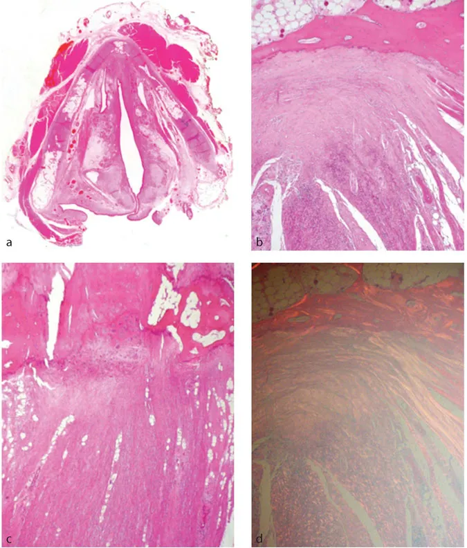
Figure 1.4a–d Broyle’s ligament. The anterior commissure tendon fibres can be seen inserting into cartilage. The arrangement of the collagen fibres is well demonstrated when viewed under polarised light (d).
Thyroarytenoid muscles
The thyroarytenoid muscles originate from the anterior commissure tendon (or Broyle’s ligament) on the internal surface of the thyroid cartilage (Figures 1.4a–d) and run posteriorly to insert on the arytenoid cartilage. The majority of the muscles’ fibres run lateral to the vocal ligament and constitute the vocalis muscle. The upper division of the muscle, which varies among individuals, runs in the aryepiglottic fold, vestibular folds and lateral to the laryngeal ventricle.
The primary function of the thyroarytenoid muscle is to shorten the vocal ligament and adjust vocal fold tension during phonation. Furthermore, muscle contraction can rotate the arytenoid cartilage inwards to close the glottis.
Posterior cricoarytenoid muscles
The posterior cricoarytenoid muscles extend upwards and laterally from the posterior surface of the cricoid lamina and insert on the muscular process of the arytenoid cartilage. The posterior cricoarytenoid muscles are the only vocal fold abductors. The more horizontal superior part of these muscles rotate the arytenoid cartilage laterally, thereby abducting the vocal folds, whereas the vertical inferior fibres pull the arytenoids over the upper margin of the cricoid, which moves them apart.
Lateral cricoarytenoid muscles
The lateral cricoarytenoid muscles originate from the upper border of the arch of the cricoid cartilage and insert on the muscular process of the arytenoid. They rotate the arytenoid medially to close the glottis.
Interarytenoid muscle
The interarytenoid muscle is the only unpaired intrinsic muscle. It runs posteriorly between the muscular processes of the arytenoids. Its main function is to adduct the vocal folds by approximating them.
Cricothyroid muscles
The cricothyroid muscles originate from the anterolateral surface of the cricoid cartilage arch. The fibres pass superior and dorsal to insert on the oblique ridge of the thyroid cartilage. While all the other intrinsic larynx muscles are innervated by the recurrent laryngeal nerve, the cricothyroid is innervated by the external branch of the superior laryngeal nerve.
Joints of the larynx
Cricothyroid joint
The inferior horn of the thyroid cartilage articulates with the cricoid cartilage at an encapsulated synovial joint. The cricothyroid joint allows both rotation and glide movement. Contraction of the cricothyroid muscles leads to forward and downward tilting of the thyroid cartilage and a simultaneous upward movement of the cricoid.
Cricoarytenoid joint
The cricoarytenoid joint (CAJ) is a synovial articulation between the base of the arytenoid cartilage and the lamina of the cricoid cartilage. The joint has a loose capsule inserting at a distance from the cartilage articulation surface margin, allowing a wider range of movement.9 The arytenoid cartilage can glide forwards and backwards over the cricoid lamina, rock medial and lateral and rotate around its axis. The capsule of the CAJ is reinforced by the posterior cricoarytenoid ligament. Desp...