From Neuroscience to Neurology
eBook - ePub

From Neuroscience to Neurology

Neuroscience, Molecular Medicine, and the Therapeutic Transformation of Neurology

Stephen Waxman

Buch teilen
  1. 552 Seiten
  2. English
  3. ePUB (handyfreundlich)
  4. Über iOS und Android verfügbar
eBook - ePub

From Neuroscience to Neurology

Neuroscience, Molecular Medicine, and the Therapeutic Transformation of Neurology

Stephen Waxman

Angaben zum Buch
Buchvorschau
Inhaltsverzeichnis
Quellenangaben

Über dieses Buch

The field of neurology is being transformed, from a therapeutically nihilistic discipline with few effective treatments, to a therapeutic specialty which offers new, effective treatments for disorders of the brain and spinal cord. This remarkable transformation has bridged neuroscience, molecular medicine, and clinical investigation, and represents a major triumph for biomedical research. This book, which contains chapters by more than 29 internationally recognized authorities who have made major contributions to neurotherapeutics, tells the stories of how new treatments for disabling disorders of the nervous system, such as stroke, multiple sclerosis, Parkinson's disease, and migraine, were developed, and explores evolving themes and technologies that offer hope for even more effective treatments and ultimately cures for currently untreatable disorders of the brain and spinal cord. The first part of this book reviews the development of new therapies in neurology, from their inception in terms of basic science to their introduction into the clinical world. It also explores evolving themes and new technologies. This book will be of interest to everyone – clinicians and basic scientists alike – interested in diseases of the brain and spinal cord, and in the quest for new treatments for these disorders.* Presents the evolution of the field of neurology into a therapeutic discipline * Discusses lessons learned from past successes and applications to ongoing work* Explores the future of this field

Häufig gestellte Fragen

Wie kann ich mein Abo kündigen?
Gehe einfach zum Kontobereich in den Einstellungen und klicke auf „Abo kündigen“ – ganz einfach. Nachdem du gekündigt hast, bleibt deine Mitgliedschaft für den verbleibenden Abozeitraum, den du bereits bezahlt hast, aktiv. Mehr Informationen hier.
(Wie) Kann ich Bücher herunterladen?
Derzeit stehen all unsere auf Mobilgeräte reagierenden ePub-Bücher zum Download über die App zur Verfügung. Die meisten unserer PDFs stehen ebenfalls zum Download bereit; wir arbeiten daran, auch die übrigen PDFs zum Download anzubieten, bei denen dies aktuell noch nicht möglich ist. Weitere Informationen hier.
Welcher Unterschied besteht bei den Preisen zwischen den Aboplänen?
Mit beiden Aboplänen erhältst du vollen Zugang zur Bibliothek und allen Funktionen von Perlego. Die einzigen Unterschiede bestehen im Preis und dem Abozeitraum: Mit dem Jahresabo sparst du auf 12 Monate gerechnet im Vergleich zum Monatsabo rund 30 %.
Was ist Perlego?
Wir sind ein Online-Abodienst für Lehrbücher, bei dem du für weniger als den Preis eines einzelnen Buches pro Monat Zugang zu einer ganzen Online-Bibliothek erhältst. Mit über 1 Million Büchern zu über 1.000 verschiedenen Themen haben wir bestimmt alles, was du brauchst! Weitere Informationen hier.
Unterstützt Perlego Text-zu-Sprache?
Achte auf das Symbol zum Vorlesen in deinem nächsten Buch, um zu sehen, ob du es dir auch anhören kannst. Bei diesem Tool wird dir Text laut vorgelesen, wobei der Text beim Vorlesen auch grafisch hervorgehoben wird. Du kannst das Vorlesen jederzeit anhalten, beschleunigen und verlangsamen. Weitere Informationen hier.
Ist From Neuroscience to Neurology als Online-PDF/ePub verfügbar?
Ja, du hast Zugang zu From Neuroscience to Neurology von Stephen Waxman im PDF- und/oder ePub-Format sowie zu anderen beliebten Büchern aus Medicine & Neurology. Aus unserem Katalog stehen dir über 1 Million Bücher zur Verfügung.

Information

Jahr
2004
ISBN
9780080506982
Part I
Clinical Rewards From Neuroscience, Molecular Medicine, and Translational Research
Chapter 1

Seeing the Brain So We Can Save It: The Evolution of Magnetic Resonance Imaging as a Clinical Tool

Peter D. Schellinger, MD, PhD
Steven Warach, MD, PhD

Publisher Summary

Magnetic resonance imaging (MRI) has emerged as a clinical tool that has an impact on neurology. This chapter illustrates this transformation through three classical neurological diseases, namely, acute ischemic stroke, multiple sclerosis, and brain tumors. The advent of new MRI techniques such as diffusion- and perfusion-weighted imaging (DWI and PWI) has added another dimension to diagnostic imaging in stroke. Furthermore, MRI has changed not only clinical management because of a higher diagnostic yield but also daily neurological and neurosurgical practice. Not all tumors are candidates for surgical resection; however, confirmation of the pathology is critical for treatment decisions or enrollment in a clinical trial. Imaging and the development of intraoperative MRI suites have played an increasing role in assisting with biopsy guidance as part of frameless navigation systems. It is exciting to observe that, with an increasing number of clinical therapeutic trials being designed; MRI may not only function as a diagnostic tool but may also have prognostic strength and thus serve as a surrogate endpoint for the development of new therapies.
“MRI researchers win Nobel Prize for medicine” – CBC News 10/2003
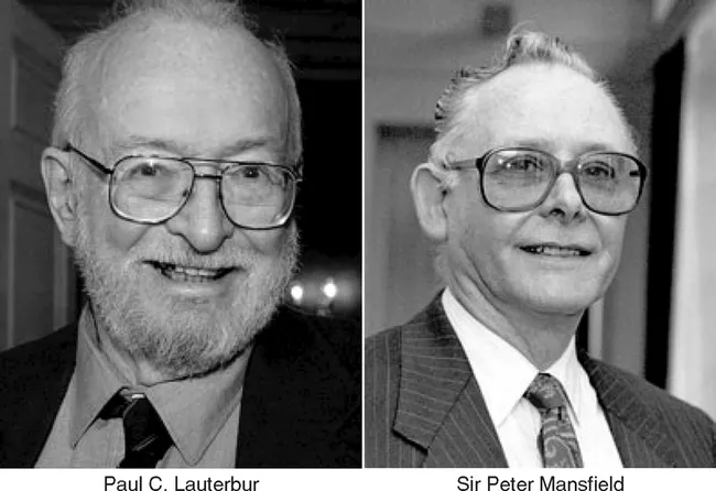
STOCKHOLM, SWEDEN -Two scientists whose discoveries led to the development of medical imaging of the body’s inner organs have won the 2003 Nobel Prize for medicine. The Swedish Academy said the discoveries of American Paul C. Lauterbur and Briton Sir Peter Mansfield represent “a breakthrough in medical diagnostics and research.” The last time the medicine prize was awarded in the field of diagnostics was for the development of computer assisted tomography in 1979. (See Figure 1.1.)
image

FIGURE 1.1 Paul C. Lauterbur and Sir Peter Mansfield.
The last, but most important, step for a new technology is its establishment in clinical practice. The evolution from tentative reports and early research activity to the definition of possible clinical benefits and acceptance into practice (Jackson, 2001) is often protracted in complex technologies such as magnetic resonance imaging (MRI). Lauterbur (1973) published his landmark article about the derivation of position-dependent information by nuclear magnetic resonance and magnetic gradients in the early 1970s. Despite the work that followed by Sir Peter Mansfield (Mansfield and Maudsley, 1976, 1977) and other groups, the first clinical MRI system was not available until 1984 followed in 1987 by the first paramagnetic MRI contrast agent. The last 20 years have realized several advances in computer technology and software development, and these changes have continuously improved the accessibility and quality of MRI. The earliest scans—T2-weighted, T1-weighted, and (proton density) PD-weighted sequence without contrast—took hours to complete. Today, multiparametric protocols can assess within 15 minutes the most complex pathophysiological processes, which has allowed a dramatic shift in the evaluation and treatment of neurological illness. As the clinical discipline of neurology has evolved from a diagnostic to a therapeutic specialty, MRI is being transformed into a clinical tool that has an impact on neurology at the bedside. This chapter illustrates this transformation through three classical neurological diseases that have gained growing interest with regard to therapeutic options only within the last 30 years: acute ischemic stroke, multiple sclerosis, and brain tumors. It is exciting to observe that, with an increasing number of clinical therapeutic trials being designed, MRI may not only function as a diagnostic tool but may also have prognostic strength and thus serve as a surrogate endpoint for the development of new therapies (Warach, 2001b).

ADVANCES IN MRI

Once a technology is accepted into clinical practice, it is inevitable that the demands of clinical specialties stimulate the evolution of the technology and the extension of its functionality to new applications (Jackson, 2001). For instance, interest in functional imaging led to developments such as higher gradients, allowing rapid and repetitive image acquisition with high spatial and temporal resolution. Echo planar imaging (EPI) was found useful for expanding the capabilities of MRI in the routine acquisition of T2-weighted images (Heiland et al., 1998; Pierpaoli et al., 1996; Warach et al., 1995) and allowing formerly time-consuming sequences such as fat- (STIR, short tau inversion recovery) and fluid-suppressed sequences (FLAIR, fluid attenuated inversion recovery) to become practical in the clinical setting (Bydder and Young, 1985; De Coene et al., 1992). FLAIR images have substantially improved the diagnostic yield in diseases such as stroke and multiple sclerosis, where chronic vascular lesions and foci of demyelination are in close proximity to cerebrospinal fluid (CSF) spaces (Hajnal et al., 1992). Other examples of technical progress stimulated by clinical needs include magnetic resonance angiography (Brant-Zawadzki and Heiserman, 1997; Fujita et al., 1994; Johnson et al., 1994) and magnetization transfer imaging (Haehnel et al., 2000; Knauth et al., 1996; Kurki et al., 1994).
Two techniques that have received much attention in the last 15 years are diffusion- and perfusion-weighted imaging (DWI and PWI) (Heiland et al., 1997; Moseley et al., 1991; Ostergaard et al., 1996; Warach et al., 1992). The phenomenon of indirectly measuring brownian molecular motion of water with DWI was first described in 1965 (Stejskal and Tanner, 1965). The measurement of water diffusion renders pathophysiological tissue information, especially in ischemic, inflammatory, and neoplastic disease, that cannot be obtained with standard MRI sequences (Le Bihan et al., 1986; Le Bihan et al., 1992). In addition to the qualitative measurements of diffusion, the apparent diffusion coefficient (ADC) can be quantified from measurements at different b-values (Tanner and Stejskal, 1968) on a per pixel basis, within a region of interest, or to generate ADC parameter maps. For structures, such as nerve fibers or tracts, that have a predominant direction in space, the ADC depends on the direction of the diffusion gradient. Whereas water diffusion is only mildly impaired in the longitudinal direction, water diffusion in a perpendicular direction to the main axis is decreased (Basser and Pierpaoli, 1996); this is referred to as anisotropy. In the brain, anisotropy is most pronounced in the white matter, especially in densely packed fiber structures such as the corpus callosum and the corona radiata. The effects of anisotropy can be used to obtain information of the regional fascicular or fiber tract anatomy, which may be helpful for neurosurgical procedures. The expansion of these principles has led to the development of diffusion tensor imaging, which has been used to characterize myelination and demyelination (Le Bihan et al., 2001; Neil et al., 1998; Ono et al., 1995; Pierpaoli et al., 1996).
PWI uses a dynamic bolus (paramagnetic contrast) tracking technique to derive concentration time curves, allowing the qualitative assessment of various hemodynamic parameters relative to areas of normal parenchyma (Rosen et al., 1989, 1990; Villringer et al., 1988). Typical parameters are mean transit time (MTT), cerebral blood volume (CBV), cerebral blood flow, and time to peak (TTP). The actual quantification of hemodynamic information has been limited by postprocessing capabilities and methods of calculating the arterial input function. Nevertheless, areas of relative hypoperfusion can be reliably demonstrated by current methods, which is of special relevance in ischemic stroke (Schlaug et al., 1999). Other applications, such as functional imaging with the BOLD (blood oxygen level dependence) technique and spectroscopic imaging, exceed the scope of this chapter. It suffices to say that substantial technical and programming refinements have supplied neuroscience with a comprehensive diagnostic armament to guide our daily clinical practice. The growing interest in neurology for physiological data, the development of new treatment paradigms, and the definition of parameters that predict disease course will cont...

Inhaltsverzeichnis