
eBook - ePub
International Review of Experimental Pathology
Kidney Disease
G. W. Richter, G. W. Richter
This is a test
Buch teilen
- 410 Seiten
- English
- ePUB (handyfreundlich)
- Über iOS und Android verfügbar
eBook - ePub
International Review of Experimental Pathology
Kidney Disease
G. W. Richter, G. W. Richter
Angaben zum Buch
Buchvorschau
Inhaltsverzeichnis
Quellenangaben
Über dieses Buch
International Review of Experimental Pathology, Volume 30, is organized around the theme of renal disease. The choice of renal disease reflects both the author's personal interest and the realization that there is a need for such a collection of reviews in this area. There are many new books on renal pathology, but almost all have a clinical rather than experimental orientation. The book opens with a chapter on the pathogenesis of experimentally induced renal papillary necrosis and upper urothelial carcinoma. Subsequent chapters deal with the use of cell cultures in the study of renal diseases; mechanisms of cyclosporine nephrotoxicity in humans and animal systems; spontaneously occurring renal diseases in laboratory animals; and the use of video microscopy to define the reactivity of the renal microvasculature and the hydraulic permeability of the glomerular capillaries. This book will be of interest to a diverse group of readers interested in renal disease. This broad spectrum of potential readership is reflected in the list of contributors which includes, in addition to pathologists, nephrologists, anatomists, veterinarians, and experimental chemists. This volume will also be of interest to transplant surgeons and to pediatricians specializing in renal disease.
Häufig gestellte Fragen
Wie kann ich mein Abo kündigen?
Gehe einfach zum Kontobereich in den Einstellungen und klicke auf „Abo kündigen“ – ganz einfach. Nachdem du gekündigt hast, bleibt deine Mitgliedschaft für den verbleibenden Abozeitraum, den du bereits bezahlt hast, aktiv. Mehr Informationen hier.
(Wie) Kann ich Bücher herunterladen?
Derzeit stehen all unsere auf Mobilgeräte reagierenden ePub-Bücher zum Download über die App zur Verfügung. Die meisten unserer PDFs stehen ebenfalls zum Download bereit; wir arbeiten daran, auch die übrigen PDFs zum Download anzubieten, bei denen dies aktuell noch nicht möglich ist. Weitere Informationen hier.
Welcher Unterschied besteht bei den Preisen zwischen den Aboplänen?
Mit beiden Aboplänen erhältst du vollen Zugang zur Bibliothek und allen Funktionen von Perlego. Die einzigen Unterschiede bestehen im Preis und dem Abozeitraum: Mit dem Jahresabo sparst du auf 12 Monate gerechnet im Vergleich zum Monatsabo rund 30 %.
Was ist Perlego?
Wir sind ein Online-Abodienst für Lehrbücher, bei dem du für weniger als den Preis eines einzelnen Buches pro Monat Zugang zu einer ganzen Online-Bibliothek erhältst. Mit über 1 Million Büchern zu über 1.000 verschiedenen Themen haben wir bestimmt alles, was du brauchst! Weitere Informationen hier.
Unterstützt Perlego Text-zu-Sprache?
Achte auf das Symbol zum Vorlesen in deinem nächsten Buch, um zu sehen, ob du es dir auch anhören kannst. Bei diesem Tool wird dir Text laut vorgelesen, wobei der Text beim Vorlesen auch grafisch hervorgehoben wird. Du kannst das Vorlesen jederzeit anhalten, beschleunigen und verlangsamen. Weitere Informationen hier.
Ist International Review of Experimental Pathology als Online-PDF/ePub verfügbar?
Ja, du hast Zugang zu International Review of Experimental Pathology von G. W. Richter, G. W. Richter im PDF- und/oder ePub-Format sowie zu anderen beliebten Büchern aus Medizin & Krankheiten & Allergien. Aus unserem Katalog stehen dir über 1 Million Bücher zur Verfügung.
Information
Thema
MedizinExperimentally Induced Renal Papillary Necrosis and Upper Urothelial Carcinoma
PETER H. BACH and NEILL J. GREGG, Nephrotoxicity Research Group, Toxicology Unit, Robens Institute of Industrial and Environmental Health and Safety, University of Surrey, Surrey, Guildford GU2 5XH, England
Publisher Summary
This chapter discusses experimentally induced renal papillary necrosis and upper urothelial carcinoma. It presents several morphological, histochemical, and functional data to support several distinct series of pathological changes following the administration of 2-bromoethanamine (BEA). The earliest histochemical changes take place in the medullary matrix that appears to undergo depolymerization. The renal medullary interstitial cells are the first cell type to undergo degenerative change that is rapidly followed by damage to the delicate elements of the medulla. The collecting ducts and endothelial changes are late and generally follow the necrosis of other anatomical regions of the medulla. At present, the lipid changes in the medulla are not well understood, but they are similar to those already reported in human analgesic abusers. The early subtle degenerative changes in the proximal tubule do not appear to be central to the development of the papillary lesion, but the subsequent exfoliation of the brush border and proximal tubular cells are important components of the protein casts that begin to form in the distal nephron. These subsequently appear to play at least some role in the development of functional changes that cause marked proximal tubular dilatation. The chapter illustrates the time course of the major pathophysiological changes associated with the development of RPN, and its secondary consequences of cortical degeneration and upper urothelial hyperplasia.
I Introduction
The etiology of renal papillary necrosis (RPN) in humans has been associated with the long-term abuse of analgesics and therapeutic doses of nonsteroidal antiinflammatory drugs (NSAID). However, the lesion has not been clearly defined in terms of the exact causative agent(s), how much (of each) was taken to cause a lesion, and over what period. The primary pathogenesis and the role of other complicating factors are also not clearly understood, nor have the secondary pathophysiological consequences of RPN been adequately interrelated, despite the fact that chronic renal failure and upper urothelial carcinoma are frequently associated with analgesic abuse (Bach and Bridges, 1985).
The understanding of the pathophysiology of a chronically developing renal lesion in humans is a major problem in those conditions where the etiology has been clearly defined, because of the strong likelihood of concurrent and complicating secondary (and unrelated disease) factors. There are important anatomical and functional differences between the kidneys of most animals and humans (Mudge, 1982; Stolte and Alt, 1980). The use of experimental models has generally shown a number of very important clinical and morphological differences; therefore, the use of these models has often limited the understanding of similar conditions in humans.
Although RPN (and upper urothelial carcinoma) are examples of renal disease developing chronically in humans, it has been possible to study a number of chemicals that induce these lesions rapidly in experimental animals. These models (Bach and Hardy, 1985; Bach and Bridges, 1985) all have the important pathophysiological hallmarks of the lesion that has been described in humans (Burry, 1968; Burry et al., 1977; Rosner, 1976; Bach and Bridges, 1985). The use of these experimental models has therefore fortuitously provided a way to study the development of papillary necrosis and the progression to a series of renal changes similar to those seen in human analgesic abusers. These models are also allowing the interrelationship between the primary lesion and its secondary consequences to be defined in terms of biochemical mechanisms. An understanding of the molecular genesis of this syndrome may be highly relevant to improved clinical management of RPN and upper urothelial carcinoma in humans.
II Renal Papillary Necrosis and Upper Urothelial Carcinoma in Humans
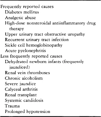
RPN was first described over 100 years ago (Turner, 1885). It is a lesion that may have a number of different causes (Table I), but most often when encountered in the clinical environment before the 1950s, was due to diabetes mellitus or sickle cell disease (Mandel, 1952). The most frequent cause of RPN since then (and in current clinical experience) is chronic, inappropriate, high-dose analgesic intake, especially the addiction to mixed analgesics over a number of years. Therapeutic closes of NSAID may also induce RPN (Nanra and Kincaid-Smith, 1972; Burry et al., 1977; Prescott, 1979, 1982; Bach and Bridges, 1985).
TABLE I
CAUSES OF RENAL PAPILLARY NECROSIS IN HUMANS
Initially, all of the mixed analgesics that were associated with the pyelonephritis seen in urology clinics contained phenacetin, and the condition was dubbed “phenacetin kidney” (Spuhler and Zollinger, 1953). Subsequently, however, it become apparent that other analgesics had the potential to cause RPN (Gilman, 1964). The early confusion over the cause of RPN, and the fact that most patients abused, or were prescribed, mixed analgesics and/or a number of different NSAID, also served to obscure case history data that might have provided vital information for the more accurate identification of which analgesics and/or NSAID had the greatest potential to cause the lesion (Cove-Smith and Knapp, 1978; Nanra and Kincaid-Smith, 1975; Nanra et al., 1980). The early failure to realize that phenacetin was not the sole cause of RPN shaped the dogma that resulted in the withdrawal of this drug from the market (Shelley, 1967, 1978). This, it was assumed, would remove the major etiological factor in the genesis of the lesion. When acetaminophen (paracetamol) replaced phenacetin in mixed analgesic preparations the incidence of RPN was expected to drop (Gault et al., 1968; Duggin, 1977; Kincaid-Smith, 1979). The occurrence of the lesion did not, however, decrease in those circumstances where the abuse of mixed analgesics continued (Prescott, 1979, 1982), although some decreases have been attributed to the withdrawal of phenacetin and extensive educational programs to discourage the abuse of mixed analgesics (Wilson and Gault, 1982). A variety of indirect evidence (Table II...