Helen Whitwell, Christopher Milroy, Daniel du Plessis, Helen Whitwell, Christopher Milroy, Daniel du Plessis
This is a test
This is a test
Buch teilen
218 Seiten
English
ePUB (handyfreundlich)
Über iOS und Android verfügbar
eBook - ePub
Forensic Neuropathology
Helen Whitwell, Christopher Milroy, Daniel du Plessis, Helen Whitwell, Christopher Milroy, Daniel du Plessis
Angaben zum Buch
Buchvorschau
Inhaltsverzeichnis
Quellenangaben
Über dieses Buch
Forensic neuropathology is an important specialty within forensic pathology. In addition to traumatic brain injury in the adult and child, forensic neuropathologists must also consider the role of natural disease within the forensic setting such as cerebrovascular disease, as well as neurotoxicology. Focusing on difficulties that arise in the medico-legal context, the chapters include techniques for the post-mortem examination of the brain and related structures. Forensic pathologists, neuropathologists, general pathologists, clinical forensic specialists as well as neuroscientists, neurologists and neurosurgeons will all find useful information. In addition, members of the legal profession have found this an important reference work.
Chapters have been extensively revised and new content includes
- Updates on pathological aspects of head injury including infant head injury with ocular pathology
- Clinical aspects of head injury and spinal injury including a new chapter on neuroradiology
Reviews of the First Edition
This outstanding book is unique. Well-illustrated with high-quality colour photographs and line drawings, it reads well. Strongly recommended for trainees in histopathology, neuropathology, paediatric pathology, and forensic pathology, as well as for consultants practising in these fields.
Häufig gestellte Fragen
Wie kann ich mein Abo kündigen?
Gehe einfach zum Kontobereich in den Einstellungen und klicke auf „Abo kündigen“ – ganz einfach. Nachdem du gekündigt hast, bleibt deine Mitgliedschaft für den verbleibenden Abozeitraum, den du bereits bezahlt hast, aktiv. Mehr Informationen hier.
(Wie) Kann ich Bücher herunterladen?
Derzeit stehen all unsere auf Mobilgeräte reagierenden ePub-Bücher zum Download über die App zur Verfügung. Die meisten unserer PDFs stehen ebenfalls zum Download bereit; wir arbeiten daran, auch die übrigen PDFs zum Download anzubieten, bei denen dies aktuell noch nicht möglich ist. Weitere Informationen hier.
Welcher Unterschied besteht bei den Preisen zwischen den Aboplänen?
Mit beiden Aboplänen erhältst du vollen Zugang zur Bibliothek und allen Funktionen von Perlego. Die einzigen Unterschiede bestehen im Preis und dem Abozeitraum: Mit dem Jahresabo sparst du auf 12 Monate gerechnet im Vergleich zum Monatsabo rund 30 %.
Was ist Perlego?
Wir sind ein Online-Abodienst für Lehrbücher, bei dem du für weniger als den Preis eines einzelnen Buches pro Monat Zugang zu einer ganzen Online-Bibliothek erhältst. Mit über 1 Million Büchern zu über 1.000 verschiedenen Themen haben wir bestimmt alles, was du brauchst! Weitere Informationen hier.
Unterstützt Perlego Text-zu-Sprache?
Achte auf das Symbol zum Vorlesen in deinem nächsten Buch, um zu sehen, ob du es dir auch anhören kannst. Bei diesem Tool wird dir Text laut vorgelesen, wobei der Text beim Vorlesen auch grafisch hervorgehoben wird. Du kannst das Vorlesen jederzeit anhalten, beschleunigen und verlangsamen. Weitere Informationen hier.
Ist Forensic Neuropathology als Online-PDF/ePub verfügbar?
Ja, du hast Zugang zu Forensic Neuropathology von Helen Whitwell, Christopher Milroy, Daniel du Plessis, Helen Whitwell, Christopher Milroy, Daniel du Plessis im PDF- und/oder ePub-Format sowie zu anderen beliebten Büchern aus Droit & Science médico-légale. Aus unserem Katalog stehen dir über 1 Million Bücher zur Verfügung.
The surface anatomy and features of the head and neck are derived from skeletal and soft tissue structures covered by skin and connective tissue that covers the underlying bony skull. In some areas, particularly the face, the skin is thin, allowing easy palpation of underlying skeletal features. It is also highly mobile as a result of the presence of a number of small but extensive subcutaneous muscles of facial expression, all supplied by a single cranial nerve (facial). The scalp is, in contrast, relatively tough and hidden from view in most individuals by hair. Within the skull, which acts as a protective shield, are the brain and brainstem, with its associated covering of tissue layers and blood vessels. The anatomy is complex and this chapter can only provide an overview of the principal features, with craniofacial anatomy included to link anatomically with the underlying structures, including the neuroanatomy, which may be of particular significance in penetrating or blunt head trauma.
The external face
The anatomical surface features of the face are never totally symmetrical. As facial expressions have evolved as a communication method, the underlying anatomy has evolved from the functional need. The skin is often marked by moles and freckles and may also present with scars consequential to cuts and other trauma.
The shape and size of the hairline vary with race and the eyebrows can also be highly variable.
The nose comprises underlying cartilaginous and bony structures. In the coronal section, the nose is triangular in shape. The external nares (nostrils) are protected by coarse hairs (vibrissae) and serve to filter air entering the nose. The anterior part of the nose is composed of flexible underlying fibrocartilage.
Inferior to the nose is the mouth, surrounded by the lips. Here, the size and shape of the mouth are very variable, both within races and between different racial groups. The lips have non-keratinised epithelium and thus appear pink as a result of the underlying blood vessels.
The eyes are set within the bony orbits of the skull but protected by the rim of bone. Their individual position relative to the nose is variable and can be close-set or wide set in normal individuals. Attached within the orbits are the muscles that control eye movement while, superficially, the paired eyelids cover and protect the eye from potential damage. The lids normally permit only a portion of white sclera to appear laterally, with the transparent conjunctiva and cornea that cover the pigmented iris seen medially. The upper lid normally overlaps the iris, but the sclera may be seen between the iris and the lower lid. The shape of the lids can vary between individuals; in particular, the elevator of the upper lid can be weak or damaged, leading to a drooping appearance.
Ethnic differences are often prominent. Mongoloid epicanthic folds and other minor folds of skin in the medial aspect of the orbit should be noted. Conditions such as exophthalmos associated with hyperthyroidism can result in prominent eyes. Facial fractures affecting the maxilla and inferior margin of the orbit can lead to a sinking of the eyeball. The shape of the lids themselves can lead to a wide range of different appearances of the eye within the orbit.
The internal facial structures
Internally, the muscles of facial expression and their nerve and vascular supplies contribute to the facial structures. In addition, the parotid gland is located within the lateral parts of the cheeks.
The facial expression muscles are supplied by the facial (cranial VII) nerve. Their function is to control and support the structures and openings in the face, such as the eyes and mouth. In humans, their functionality serves an important role in non-verbal communication as well as aiding actions such as screwing up the eyes and chewing. The mouth is surrounded by the sphincteric orbicularis oris muscle into which merge the fibres of the buccinator, the muscle of the cheek. The buccinator contracts during chewing and serves to prevent trapping of food within the space between the gums and teeth; it also acts to raise the pressure of air expelled by musicians playing wind instruments or by whistling. The orbicularis oculi surrounds the eye and serves to function in two ways. First, fibres that surround the eye serve to screw the eye up because they are attached to the bone on the medial aspect of the orbit. Second, the palpebral fibres attach to the lateral palpebral raphe and serve to close the eye when blinking. Additional fibres are attached to the lacrimal sac and serve to dilate the sac and keep the puncta in contact with the eyeball.
The facial nerve enters the face by passing through the tough fibrous capsule of the parotid gland and can be damaged during surgical procedures to that gland.
The nose
The nose, as the upper part of the respiratory tract, is located superior to the hard palate and contains the organ of smell. It is divided into right and left nasal cavities by the nasal septum, with each nasal cavity having an olfactory and a respiratory area.
The external nose varies considerably in size and shape in individuals and races because of differences in the nasal cartilage structure. The inferior aspect is composed of two openings called the nares (nostrils), each separated from the other by the nasal septum. The nasal bones, the frontal processes of the maxillae, the nasal part of the frontal bone and the bony part of the nasal septum form the skeletal components of the nose, whereas five main cartilages form the cartilaginous nose. These are two lateral cartilages, two alar cartilages and a septal cartilage that articulates with the bony septum.
The nasal cavities open through the choanae into the nasopharynx at the posterior. The nasal mucosa is bound closely to the periosteum and perichondrium of the nasal bones and cartilages, and lines the nasal cavities, except for the vestibule which is lined with skin. The olfactory area lies superior within the cavity and is the organ of smell, with its nerve fibres passing through the cribriform plate to enter the olfactory bulbs, which lie against the inferior surface of the frontal lobe of the brain.
The narrow, curved roof of the nasal cavity is divided into frontonasal, ethmoidal and sphenoidal parts, named by adjacent bones. The wide floor is formed by the horizontal plate of the palatine bone and the palatine process of the maxilla. Medially, the wall is the nasal septum, comprising the vomer, perpendicular plate of the ethmoid, septal cartilage and the nasal crests of the maxillary and palatine bones. The lateral walls of the nasal cavity are made up of three nasal conchae or scroll bones, each forming a roof over a meatus connecting the nasal cavity to a sinus or the orbit. The superior meatus is between the superior and middle conchae, into which orifices from the posterior ethmoidal sinuses open. The middle meatus, inferior to the middle conchae, communicates with the frontal sinus via the frontonasal duct and the maxillary sinus at its posterior end. The inferior meatus is inferolateral to the inferior conchae and receives the nasolacrimal duct from the lacrimal sac into its anterior portion.
The nose receives arterial blood from many branches, including the sphenopalatine artery, ethmoidal arteries and the facial artery. Kiesselbach's area, found on the anterior nasal septum, is rich in capillaries and is the site of profuse nose bleeding. The nerve supply of the nasal mucosa is by the maxillary nerve, nasal branches of the greater palatine nerve and the anterior ethmoidal nerves, and branches of the nasociliary nerve.
The paranasal sinuses are air-filled extensions of the nasal cavity within the frontal, maxillary, sphenoid and ethmoid bones and are named according to each bone. The ethmoidal sinuses consist of ethmoidal cells located within the ethmoid bone between the orbit and nose. The sphenoid air sinuses are unevenly divided like the frontal air sinuses and separated by a bony septum. They occupy the body of the sphenoid bone and are separated by thin bone from the optic chiasma, the pituitary gland, the internal carotid arteries and the cavernous sinuses.
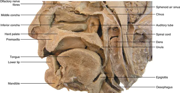
The maxillary sinuses are large pyramidal cavities within the maxillae. Their floor is formed by the alveolar part of the maxilla, with the roots of the maxillary teeth, particularly the first two molars, creating conical elevations (Figure 1.1).
Figure 1.1 The nasal cavity and associated structures.
The oral cavity
The oral cavity consists of the oral vestibule and the oral cavity. The vestibule is the space between the lips and cheeks and the teeth and gums, and communicates with the exterior through the orifice of mouth, the size of which is controlled by muscles, including the orbicularis oris.
The oral cavity lies posterior and medial to the upper and lower dental arches and is limited posteriorly by the terminal groove of the tongue and palatoglossal arches and anteriorly and laterally by the maxillary and mandibular arches containing the teeth. The roof is formed by the hard and soft palate, which also forms the floor of the nasal cavities. Posteriorly, the oral cavity communicates with the oropharynx. If the mouth is closed, the tongue fills the space of the oral cavity.
The hard palate forms the anterior component of the roof of the oral cavity, with its cavity filled by the resting tongue when it is at rest and formed by the palatine processes of the maxillae and the horizontal plates of the palatine bones. The incisive fossa and the greater and lesser palatine foramina open on the oral aspect of the hard palate.
The soft palate is the muscular posterior part, attached to the posterior border of the hard palate and extending as a posteroinferiorly curved free margin that terminates in the uvula. It is strengthened by a palatine aponeurosis formed by the expanded tendon of the tensor veli palatini and is attached to the posterior margin of the hard palate. Laterally, it is continuous with the wall of the pharynx and joined to the pharynx and tongue by the palatopharyngeal and palatoglossal arches. The masses of lymphoid tissue forming the palatine tonsil lie within the tonsillar fossa, bounded by the palatoglossal and palatopharyngeal arches and the tongue.
The orbit
The orbit is a pyramidal, bony cavity in the face. It contains and protects the eye with its associated muscles, nerves and vessels and the lacrimal apparatus. The roof is formed by the orbital part of the frontal bone, separating the orbit from the anterior cranial fossa and containing a small fossa for the lacrimal gland. The lesser wing of the sphenoid contributes to the roof at its apex. The medial wall is formed by the thin bone of the ethmoid, frontal, lacrimal and sphenoid bones. It is indented by the fossa of the lacrimal sac and nasolacrimal duct. The lateral wall comprises the frontal process of the zygomatic bone and the greater wing of the sphenoid, and is vulnerable to direct trauma. It serves to separate the orbit from the tem...