eBook - ePub
Pediatric Dermatology in Skin of Color
A Practical Guide
Manish K Shah, Preeti K Sheth
This is a test
- 320 Seiten
- English
- ePUB (handyfreundlich)
- Über iOS und Android verfügbar
eBook - ePub
Pediatric Dermatology in Skin of Color
A Practical Guide
Manish K Shah, Preeti K Sheth
Angaben zum Buch
Buchvorschau
Inhaltsverzeichnis
Quellenangaben
Über dieses Buch
This book focusses on the clinical aspects and management of pediatric skin disorders, especially seen in darker skin types. It includes unique conditions that the authors have encountered in their lifetime with their independent observations and approach to management. Original high-quality images are used to illustrate most dermatoses described in the book enabling a strong visual impression of the discussed diseases. It hopes to provide readers with a blend of evidence and experience based pediatric dermatology. This book aims to be a hands-on manual that can be referred to during a busy practice as it discusses the practical approach to dermatoses.
Key Features
-
- Focusses on darker skin types.
-
- Examines unusual presentations with detailed clinical features.
-
- Discusses the ways to differentiate between similar-appearing diseases.
-
- Explores approaches to therapy, especially in resource-poor settings.
-
- Covers topics with high quality illustrations.
Häufig gestellte Fragen
Wie kann ich mein Abo kündigen?
Gehe einfach zum Kontobereich in den Einstellungen und klicke auf „Abo kündigen“ – ganz einfach. Nachdem du gekündigt hast, bleibt deine Mitgliedschaft für den verbleibenden Abozeitraum, den du bereits bezahlt hast, aktiv. Mehr Informationen hier.
(Wie) Kann ich Bücher herunterladen?
Derzeit stehen all unsere auf Mobilgeräte reagierenden ePub-Bücher zum Download über die App zur Verfügung. Die meisten unserer PDFs stehen ebenfalls zum Download bereit; wir arbeiten daran, auch die übrigen PDFs zum Download anzubieten, bei denen dies aktuell noch nicht möglich ist. Weitere Informationen hier.
Welcher Unterschied besteht bei den Preisen zwischen den Aboplänen?
Mit beiden Aboplänen erhältst du vollen Zugang zur Bibliothek und allen Funktionen von Perlego. Die einzigen Unterschiede bestehen im Preis und dem Abozeitraum: Mit dem Jahresabo sparst du auf 12 Monate gerechnet im Vergleich zum Monatsabo rund 30 %.
Was ist Perlego?
Wir sind ein Online-Abodienst für Lehrbücher, bei dem du für weniger als den Preis eines einzelnen Buches pro Monat Zugang zu einer ganzen Online-Bibliothek erhältst. Mit über 1 Million Büchern zu über 1.000 verschiedenen Themen haben wir bestimmt alles, was du brauchst! Weitere Informationen hier.
Unterstützt Perlego Text-zu-Sprache?
Achte auf das Symbol zum Vorlesen in deinem nächsten Buch, um zu sehen, ob du es dir auch anhören kannst. Bei diesem Tool wird dir Text laut vorgelesen, wobei der Text beim Vorlesen auch grafisch hervorgehoben wird. Du kannst das Vorlesen jederzeit anhalten, beschleunigen und verlangsamen. Weitere Informationen hier.
Ist Pediatric Dermatology in Skin of Color als Online-PDF/ePub verfügbar?
Ja, du hast Zugang zu Pediatric Dermatology in Skin of Color von Manish K Shah, Preeti K Sheth im PDF- und/oder ePub-Format sowie zu anderen beliebten Büchern aus Medicine & Medical Theory, Practice & Reference. Aus unserem Katalog stehen dir über 1 Million Bücher zur Verfügung.
Information
1
PRINCIPLES OF DIAGNOSIS
Lesionology
Lesions can be classified as primary (appear de novo) and secondary (develop due to a change in an already existing lesion).
Primary lesions
- Macule: A circumscribed alteration in the color or texture of the skin that is not elevated or depressed below the surface of the skin.
It may be
- Hypopigmented, e.g., pityriasis alba, pityriasis versicolor
- Hyperpigmented, e.g., lentigines, freckles
- Depigmented, e.g., vitiligo (Figure 1.1a)
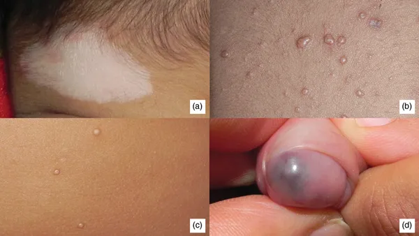 FIGURE 1.1(a) Depigmented macule of vitiligo. (b) Flesh-colored papule of molluscum contagiosum.(c) Clear fluid-filled vesicles of varicella. (d) Blue vascular nodule on the penis.
 - Erythematous, e.g., exanthems, drug rash
- Purpuric, e.g., thrombocytopenic purpura
- Papule: A circumscribed palpable elevation of the skin less than 0.5 cm in diameter.
It may be
- White, e.g., milium
- Flesh-colored, e.g., molluscum contagiosum (Figure 1.1b)
- Red, e.g., eczema
- Yellowish, e.g., xanthoma
- Yellowish brown, e.g., lupus vulgaris
- Black, e.g., melanoma
- Nodule: A solid mass in the skin that is either seen as an elevation or can be palpated. It is more than 0.5 cm but less than 2 cm in size (Figure 1.1d).
- E.g.: lupus vulgaris, erythema nodosum
- They may be soft, e.g., neurofibroma.
- They may be hard, e.g., calcinosis cutis.
- Tumors: Soft or firm masses more than 2 cm in size, which can be inflammatory or noninflammatory, malignant or nonmalignant.
- Vesicle: Visible accumulation of fluid beneath the skin, less than 0.5 cm in size. The fluid may be
- Clear, e.g., chicken pox, herpes simplex (Figure 1.1c)
- Hemorrhagic, e.g., herpes zoster
- Cloudy, e.g., impetigo
- Pustule: Visible accumulation of free pus beneath the skin, measuring less than 0.5 cm in diameter. This may be
- Follicular, e.g., folliculitis (Figure 1.2a)
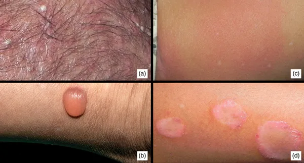 FIGURE 1.2(a) Pustules on the scalp. (b) Single bulla. (c) Urticarial wheal. (d) Plaque of psoriasis.
 - Nonfollicular, e.g., impetigo, candidiasis
- Bulla: Visible accumulation of fluid beneath the skin, more than 0.5 cm in size. The fluid may be clear, hemorrhagic, or purulent.
Bullae may be tense, e.g., bullous pemphigoid (Figure 1.2b), or they may be flaccid, e.g., pemphigus.
- Wheal: Evanescent, edematous, erythematous or pale, flat elevation of various sizes, e.g., urticaria, urticarial vasculitis (Figure 1.2c).
- Patch: Alteration in skin color or texture measuring more than 0.5 cm, that is not elevated or depressed. One can argue that a patch is a large macule and need not be classified separately. Examples of patches include:
- Hypopigmented, e.g., leprosy
- Hyperpigmented, e.g., fixed drug eruption
- Plaque: Circumscribed elevated area of the skin more than 1 cm in size such that the horizontal dimensions are more than the vertical dimension of the lesion.
Plaques can be formed by coalescence of papules, e.g., psoriasis, or nodules, e.g., granuloma annulare. These are elevated plaques (Figure 1.2d).
Plaques of morphea are depressed.
Secondary lesions
- Scale: Abnormal accumulation or disordered shedding of the stratum corneum, which appears as visible flakes on the skin.
Types of scales are seen:
- Collarette scales: fine, peripherally attached and centrally detached scale at the edges of inflammatory lesions, i.e., pityriasis rosea (Figure 1.3a).
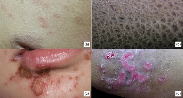 FIGURE 1.3(a) Collarette scaling in pityriasis rosea. (b) Thick fish-like scales in X-linkedichthyosis. (c) Honey-colored crusts in impetigo. (d) Excoriations in a case of atopicdermatitis.
 - Branny scales, e.g., pityriasis versicolor, pityriasis alba.
- Ichthyotic scales: large brown polygonal scales (like on the body of a fish), e.g., ichthyosis vulgaris (Figure 1.3b).
- Silvery, micaceous (mica-like that looks sparkling), thick scales, e.g., psoriasis.
- Crust: Dried up exudates consisting of pus, serum, or dead inflammatory cells or just dried serous fluid. Sometimes dried blood.
Types:
- Dry, golden-yellow, soft, friable, and superficial crust: impetigo contagiosa (Figure 1.3c)
- Yellow, greasy crust: favus
- Thick, hard, and tough crust: third-degree burns
- Lamellated, elevated, brown crust: syphilis
- Erosion: Loss of epidermis, mostly after rupture of vesicles. There is no dermal damage. Healing occurs without or with minimal scarring.
- Excoriation: Abrasion by mechanical means, which usually involves only the epidermis. This is usually linear and is a sign of scratching. Excoriations are seen in scabies, dermatitis herpetiformis, and eczemas (Figure 1.3d).
- Ulcer: Breach in the continuity of skin or mucous membrane resulting from loss of dermis as well as epidermis, with slow molecular death. Ulcers always heal with scarring. Examples are traumatic ulcers and neuropathic(trophic) ulcers (Figure 1.4a).
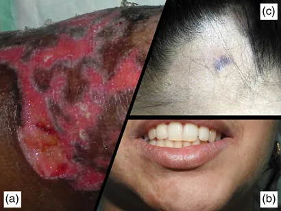 FIGURE 1.4(a) Ulcer of pyoderma gangrenosum. (b) Atrophy of right mandibular region in morphea.(c) Lichenification in a patch of alopecia areata.
 - Atrophy: Secondary change in the skin characterized by loss of normal ...
Inhaltsverzeichnis
Zitierstile für Pediatric Dermatology in Skin of Color
APA 6 Citation
Shah, M., & Sheth, P. (2021). Pediatric Dermatology in Skin of Color (1st ed.). CRC Press. Retrieved from https://www.perlego.com/book/2527386/pediatric-dermatology-in-skin-of-color-a-practical-guide-pdf (Original work published 2021)
Chicago Citation
Shah, Manish, and Preeti Sheth. (2021) 2021. Pediatric Dermatology in Skin of Color. 1st ed. CRC Press. https://www.perlego.com/book/2527386/pediatric-dermatology-in-skin-of-color-a-practical-guide-pdf.
Harvard Citation
Shah, M. and Sheth, P. (2021) Pediatric Dermatology in Skin of Color. 1st edn. CRC Press. Available at: https://www.perlego.com/book/2527386/pediatric-dermatology-in-skin-of-color-a-practical-guide-pdf (Accessed: 15 October 2022).
MLA 7 Citation
Shah, Manish, and Preeti Sheth. Pediatric Dermatology in Skin of Color. 1st ed. CRC Press, 2021. Web. 15 Oct. 2022.