
eBook - ePub
Atlas of Cardiac Surgical Techniques E-Book
Frank Sellke, Marc Ruel
This is a test
Buch teilen
- 496 Seiten
- English
- ePUB (handyfreundlich)
- Über iOS und Android verfügbar
eBook - ePub
Atlas of Cardiac Surgical Techniques E-Book
Frank Sellke, Marc Ruel
Angaben zum Buch
Buchvorschau
Inhaltsverzeichnis
Quellenangaben
Über dieses Buch
Get expert, step-by-step guidance on a wide variety of both open and interventional cardiac surgical techniques. Atlas of Cardiac Surgical Techniques, 2nd Edition, helps you expand your surgical repertoire and hone your skills with a vividly illustrated, easy-to-navigate text and pearls and pitfalls throughout. This revised atlas covers the surgical procedures you need to master, including minimally invasive techniques, robotic surgery, aortic dissection, and much more.
- Seven brand-new chapters cover Hybrid Coronary Revascularization, Aortic Valve Repair Techniques, Transcatheter Aortic Valve Replacement, Robotic Mitral Valve Surgery, Surgery for Hypertrophic Cardiomyopathy, Approaches and Techniques to Extra-Corporeal Membrane Oxygenation, and Pulmonary Endarterectomy.
- Multiple new contributing authors offer a fresh perspective in their areas of expertise.
- A consistent chapter format guides you quickly from surgical anatomy and preoperative considerations through operative steps and postoperative care.
- More than 400 full-color images, line drawings, and intraoperative photographs clearly depict the step-by-step progression of procedures.
Häufig gestellte Fragen
Wie kann ich mein Abo kündigen?
Gehe einfach zum Kontobereich in den Einstellungen und klicke auf „Abo kündigen“ – ganz einfach. Nachdem du gekündigt hast, bleibt deine Mitgliedschaft für den verbleibenden Abozeitraum, den du bereits bezahlt hast, aktiv. Mehr Informationen hier.
(Wie) Kann ich Bücher herunterladen?
Derzeit stehen all unsere auf Mobilgeräte reagierenden ePub-Bücher zum Download über die App zur Verfügung. Die meisten unserer PDFs stehen ebenfalls zum Download bereit; wir arbeiten daran, auch die übrigen PDFs zum Download anzubieten, bei denen dies aktuell noch nicht möglich ist. Weitere Informationen hier.
Welcher Unterschied besteht bei den Preisen zwischen den Aboplänen?
Mit beiden Aboplänen erhältst du vollen Zugang zur Bibliothek und allen Funktionen von Perlego. Die einzigen Unterschiede bestehen im Preis und dem Abozeitraum: Mit dem Jahresabo sparst du auf 12 Monate gerechnet im Vergleich zum Monatsabo rund 30 %.
Was ist Perlego?
Wir sind ein Online-Abodienst für Lehrbücher, bei dem du für weniger als den Preis eines einzelnen Buches pro Monat Zugang zu einer ganzen Online-Bibliothek erhältst. Mit über 1 Million Büchern zu über 1.000 verschiedenen Themen haben wir bestimmt alles, was du brauchst! Weitere Informationen hier.
Unterstützt Perlego Text-zu-Sprache?
Achte auf das Symbol zum Vorlesen in deinem nächsten Buch, um zu sehen, ob du es dir auch anhören kannst. Bei diesem Tool wird dir Text laut vorgelesen, wobei der Text beim Vorlesen auch grafisch hervorgehoben wird. Du kannst das Vorlesen jederzeit anhalten, beschleunigen und verlangsamen. Weitere Informationen hier.
Ist Atlas of Cardiac Surgical Techniques E-Book als Online-PDF/ePub verfügbar?
Ja, du hast Zugang zu Atlas of Cardiac Surgical Techniques E-Book von Frank Sellke, Marc Ruel im PDF- und/oder ePub-Format sowie zu anderen beliebten Büchern aus Medizin & Chirurgie & chirurgische Medizin. Aus unserem Katalog stehen dir über 1 Million Bücher zur Verfügung.
Information
Section III
Operations for Valvular Heart Disease
Chapter 9
Aortic Valve Replacement
Afshin Ehsan, Frank W. Sellke
Introductory Considerations
Step 1 Surgical Anatomy
- ◆ The aortic valve is the last valve in the heart through which the blood is pumped before it goes to the body. The purpose of the aortic valve is to prevent backflow of blood from the aorta into the left ventricle.
- ◆ The normal aortic valve is tricuspid, with left coronary, right coronary, and noncoronary leaflets. Each leaflet is supported by a fibrous skeleton with a shallow U-shaped configuration. The portion of this skeleton that supports the left coronary leaflet is continuous with the anterior leaflet of the mitral valve, forming the aortic-mitral curtain (annulus fibrosa).
- ◆ Each leaflet is attached just beneath their corresponding sinus of Valsalva. The sinuses of Valsalva are slight dilations of the aorta above the valve that act to create the vortex of blood required for valve closure. The sinuses end at the sinotubular junction, which is the narrowest portion of the ascending aorta.
- ◆ The left main coronary artery arises from the left sinus of Valsalva. Its ostium lies directly posterior, below the level of the sinotubular junction. The left main coronary artery runs to the left, beneath the pulmonary artery. The right coronary ostium is an anterior structure located above the right coronary cusp. Its location tends to be more variable than that of the left main coronary artery.
- ◆ The ventricular septum is located beneath the right coronary cusp and contains the atrioventricular conduction system, which passes below the noncoronary cusp near the right-noncoronary commissure (Fig. 9.1).
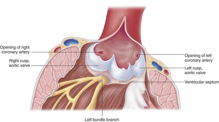 Figure 9.1
Step 2 Preoperative Considerations
Indications for Aortic Valve Replacement for Aortic Stenosis
- ◆ In the vast majority of adults, aortic valve replacement (AVR) is the only effective treatment for severe aortic stenosis (AS). Although there is some lack of agreement about the optimal timing of surgery, particularly in asymptomatic patients, it is possible to develop rational guidelines for most patients.
- ◆ In the absence of serious comorbid conditions, AVR is indicated in virtually all symptomatic patients with severe AS. There are many ways in which AVR benefits these patients. These depend partly on the patient's left ventricular (LV) function. The outcome is similar in patients with normal LV function and in those with moderate ventricular dysfunction. The depressed ejection fraction in many of these patients is caused by excessive afterload, and LV function improves after AVR. If LV dysfunction is not caused by afterload mismatch, improvement in LV function and resolution of symptoms may not be complete after valve replacement,1 but survival is still improved in this setting.2
- ◆ Symptomatic patients with angina, dyspnea, or syncope exhibit symptomatic improvement and an increase in survival after AVR.1-6
- ◆ In patients who have severe AS, even those with a low transvalvular pressure gradient, AVR results in hemodynamic improvement and better overall patient functional status.
- ◆ In summary, symptomatic patients with severe AS should undergo AVR. These patients will have improved LV function, reduced or resolved symptoms, and increased survival.
- ◆ Many clinicians are reluctant to proceed with AVR in an asymptomatic patient, whereas others are concerned about conservative treatment of a patient with severe AS. Insertion of a prosthetic aortic valve is associated with low perioperative morbidity and mortality. Despite this, some difference of opinion persists among clinicians regarding the indications for corrective surgery in asymptomatic patients. Irreversible myocardial depression or fibrosis may develop during a prolonged asymptomatic stage, and this may preclude an optimal outcome.5,7 Still others attempt to identify patients who may be at especially high risk of sudden death without surgery, although evidence supporting this approach is limited. Patients in this subgroup include those who have an abnormal response to exercise (e.g., hypotension), those with LV systolic dysfunction, those with marked or excessive LV hypertrophy, and those with evidence of very severe AS.
- ◆ We recommend that asymptomatic patients with an aortic valve area of less than 0.8 cm2 undergo valve replacement. Similarly, any evidence of impaired LV function (e.g., decreased ejection fraction, LV dilation, or significantly elevated LV diastolic pressure at rest or with exercise) is an indication for AVR. In the absence of symptoms, a peak aortic gradient of 70 mm Hg may be an indication for surgery, but this is controversial.
- ◆ Patients with moderate or more AS (mean gradient of 20 mm Hg or higher), with or without symptoms, who are undergoing coronary artery bypass grafting should undergo AVR at the time of the revascularization procedure.
- ◆ Similarly, patients with moderate or more severe AS undergoing surgery on other valves (e.g., mitral valve repair) or the aortic root should also undergo AVR as part of the surgical procedure.
Indications for Aortic Valve Replacement in Aortic Regurgitation
- ◆ AVR is recommended for patients with severe regurgitation in the presence of symptoms or any evidence of pathologic LV remodeling (e.g., impairment of LV function, LV dilation, significant elevation of LV end-diastolic pressure).
- ◆ Symptomatic patients with advanced LV dysfunction (ejection fraction < 0.25 or end-systolic dimension > 60 mm) present difficult management issues. Some patients manifest meaningful recovery of LV function after operation, but many will have developed irreversible myocardial changes. The mortality rate associated with valve replacement approaches 10% in these patients, and the postoperative mortality rate over the subsequent few years is high.
- ◆ AVR should be considered more strongly for patients with New York Heart Association (NYHA) functional class II and III symptoms, especially if symptoms and evidence of LV dysfunction are of recent onset, and intensive short-term therapy with vasodilators, diuretics, or intravenous positive inotropic agents results in substantial improvement in hemodynamics or systolic function. However...