Radiology, Lasers, Nanoparticles and Prosthetics
eBook - ePub

Radiology, Lasers, Nanoparticles and Prosthetics

Hartmut Zabel

  1. 343 Seiten
  2. English
  3. ePUB (handyfreundlich)
  4. Über iOS und Android verfügbar
eBook - ePub

Radiology, Lasers, Nanoparticles and Prosthetics

Hartmut Zabel

Angaben zum Buch
Buchvorschau
Inhaltsverzeichnis
Quellenangaben

Über dieses Buch

Order the Set Medical Physics and save almost 25€.

Medical Physics covers the applied branch of physics concerned with the application of concepts and methods of physics to diagnostics and therapeutics of human diseases. This second volume in a series of two complements the imaging modalities presented in the first volume by those methods, which use ionizing radiation. The first chapters in part A on Radiography provide a solid background on radiation sources, interaction of radiation with matter, and dosimetry for the safe handling of radiation before introducing x-ray radiography, scintigraphy, SPECT and PET.

The second part B on Radiotherapy starts from basic information on the life cycle of cells, radiation response of healthy and tumorous cells. In subsequent chapters the main methods of radiation treatment are presented, in particular x-ray radiotherapy, proton and neutron radiation therapy, and brachytherapy. The last part C, Diagnostics and Therapeutics beyond Radiology, covers laser applications, multifunctional nanoparticles and prosthetics.

The present volume

  • introduces the physical background on ionizing radiation, the biological effectiveness of radiation, as well as radiation based methods for diagnostics and therapeutics.
  • covers the second part of the entire field of medical physics, including imaging methods with the use of ionizing radiation; radiation therapy with photons, protons, and neutrons; laser methods, nanomedicine and prosthetics.
  • provides an introduction for Bachelor students to the main concepts of Medical Physics during their fi rst semesters guiding them to further specialized and advanced literature.
  • contains many questions & answers related to the content of each chapter.
  • is also available as a set together with Volume 1.

Contents

Part A: Radiography
X-ray generation
Nuclei and isotopes
Interaction of radiation with matter
Radiation detection and protection
X-ray radiography
Scintigraphy
Positron emission tomography

Part B: Radiotherapy
Cell cycle and cancer
X-ray radiotherapy
Charged particle radiotherapy
Neutron radiotherapy
Brachytherapy

Part C: Diagnostics and therapeutics beyond radiology
Laser applications in medicine
Nanoparticles for nanomedical applications
Prosthetics

Häufig gestellte Fragen

Wie kann ich mein Abo kündigen?
Gehe einfach zum Kontobereich in den Einstellungen und klicke auf „Abo kündigen“ – ganz einfach. Nachdem du gekündigt hast, bleibt deine Mitgliedschaft für den verbleibenden Abozeitraum, den du bereits bezahlt hast, aktiv. Mehr Informationen hier.
(Wie) Kann ich Bücher herunterladen?
Derzeit stehen all unsere auf Mobilgeräte reagierenden ePub-Bücher zum Download über die App zur Verfügung. Die meisten unserer PDFs stehen ebenfalls zum Download bereit; wir arbeiten daran, auch die übrigen PDFs zum Download anzubieten, bei denen dies aktuell noch nicht möglich ist. Weitere Informationen hier.
Welcher Unterschied besteht bei den Preisen zwischen den Aboplänen?
Mit beiden Aboplänen erhältst du vollen Zugang zur Bibliothek und allen Funktionen von Perlego. Die einzigen Unterschiede bestehen im Preis und dem Abozeitraum: Mit dem Jahresabo sparst du auf 12 Monate gerechnet im Vergleich zum Monatsabo rund 30 %.
Was ist Perlego?
Wir sind ein Online-Abodienst für Lehrbücher, bei dem du für weniger als den Preis eines einzelnen Buches pro Monat Zugang zu einer ganzen Online-Bibliothek erhältst. Mit über 1 Million Büchern zu über 1.000 verschiedenen Themen haben wir bestimmt alles, was du brauchst! Weitere Informationen hier.
Unterstützt Perlego Text-zu-Sprache?
Achte auf das Symbol zum Vorlesen in deinem nächsten Buch, um zu sehen, ob du es dir auch anhören kannst. Bei diesem Tool wird dir Text laut vorgelesen, wobei der Text beim Vorlesen auch grafisch hervorgehoben wird. Du kannst das Vorlesen jederzeit anhalten, beschleunigen und verlangsamen. Weitere Informationen hier.
Ist Radiology, Lasers, Nanoparticles and Prosthetics als Online-PDF/ePub verfügbar?
Ja, du hast Zugang zu Radiology, Lasers, Nanoparticles and Prosthetics von Hartmut Zabel im PDF- und/oder ePub-Format sowie zu anderen beliebten Büchern aus Biowissenschaften & Biophysik. Aus unserem Katalog stehen dir über 1 Million Bücher zur Verfügung.

Information

Jahr
2017
ISBN
9783110553178

Part A:Radiography

1X-ray generation

1.1Introduction

In 1895 Wilhelm Conrad Röntgen discovered a new type of radiation, which he called x-rays. He was uncertain what they were, but he noticed that they were able to penetrate opaque matter. To demonstrate this he took x-ray images of his wife’s hand, the first x-ray images ever, which made him and the new method instantly famous worldwide. Since then x-rays have been known to the general public mainly for their medical use. Röntgen was the first awardee of the Physics Nobel Prize in 1901. However, medical x-ray imaging is only one of many other uses of x-rays, the others include x-ray scattering, x-ray spectroscopy, and x-ray microscopy in all fields of science and technology. X-rays from low energies to high energies are so omnipresent that a world without x-rays is hard to imagine. Without x-rays we probably would not know about the helical structure of DNA, the complex folding of proteins such as myoglobin and hemoglobin, the rich structure and functionality of ribonucleic acid (RNA), and many others.
X-rays are electromagnetic (EM) waves with energies ranging from 50 eV up to several MeV, corresponding to wavelengths λ from 25nm (50 eV) down to 0.0012 nm (1 MeV). The conversion factor derived from the equation for the energy of photons E = hf = hc/λ is:
λ(nm)= 1240eVnm E(eV)
where h = 6.623 × 10⁻³⁴ Js is the Planck constant, c = 299792458m/s ≈ 3 × 10⁸ m/s−1 is the vacuum velocity of EM waves, and f is the frequency.
At the lower energy end x-rays overlap with far ultraviolet radiation. At the upper energy scale they overlap with γ-radiation. It is not primarily the energy or the respective wavelength that characterizes x-rays; it is the method by which x-rays are produced. Three kinds of x-ray production can be distinguished:
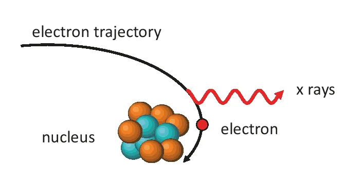
  1. bremsstrahlung, radiation produced by deacceleration of high energy electrons;
  2. characteristic radiation, occurring after excitation of core shell electrons of atoms;
  3. synchrotron radiation emitted by radial acceleration of electrons in high energy storage rings.
One can simplify this characterization and reduce it to two main effects: acceleration and excitation. Bremsstrahlung and synchrotron radiation are due to acceleration of electrons: deacceleration (stopping) of high energy electrons in a target in one case and radial acceleration of electrons on a circular orbit in the other case. Characteristic radiation is due to excitation of atomic core shell electrons. γ-radiation, although overlapping in energy with x-rays, is reserved as decay product of radioactive isotopes.
For medical x-ray diagnostics (radiography) and for x-ray cancer treatment (radiotherapy) only bremsstrahlung is used. Their specifications are, however, very different. Hence the same x-ray equipment cannot be employed for both applications. X-ray diagnostics requires bremsstrahlung up to about 150 keV, whereas x-ray radiotherapy entails x-ray energies up to 25 MeV. In this chapter we will introduce basic concepts of x-ray production for both applications, radiography and radiotherapy. X-ray radiography is then presented in Chapter 5 and x-ray radiotherapy is discussed in Chapter 9.

1.2X-ray production

1.2.1Bremsstrahlung
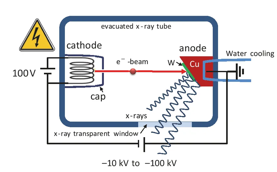
Standard x-ray tubes, which are frequently used in research laboratories, use a high voltage difference between cathode and anode for accelerating free electrons over a short distance in vacuum. These x-ray glass tubes are permanently evacuated and sealed off like traditional light bulbs. The principle features of such a tube are shown in Fig. 1.1. The cathode is connected to a high negative voltage supply between −10 kV and −100 kV and the anode is grounded. A tungsten filament in the cathode is heated to very high temperatures just below melting temperature of the wire in order to generate free electrons via thermionic emission. The current going through the filament of the cathode Icathode controls the filament temperature and consequently also the rate of electrons emitted into the vacuum tube. Electrons entering at the high negative potential into the vacuum are immediately accelerated towards the anode. A cap with a small aperture surrounding the filament, called a Wehnelt cylinder, acts as an electrostatic lens that keeps the electrons from straying. Obviously there is a close connection between the current heating the filament Icathode and the anode current Ianode hitting the target.
Fig. 1.1: Schematic of an x-ray tube.
Fig. 1.2: Generation of bremsstrahlung in the Coulomb potential of target nuclei.
The anode (target) consists of a water cooled Cu block coated with some metal film. Tungsten is usually chosen for medical applications. In the target material the free elect...

Inhaltsverzeichnis