eBook - ePub
Essential Clinical Oral Biology
Stephen Creanor, Stephen Creanor
This is a test
Buch teilen
- English
- ePUB (handyfreundlich)
- Über iOS und Android verfügbar
eBook - ePub
Essential Clinical Oral Biology
Stephen Creanor, Stephen Creanor
Angaben zum Buch
Buchvorschau
Inhaltsverzeichnis
Quellenangaben
Über dieses Buch
Essential Clinical Oral Biology is an accessible guide to oral biology, introducing the scientific knowledge necessary to succeed in clinical practice.
- Student-friendly layout with clinical photographs throughout
- Each chapter has clearly defined key topics and learning objectives
- Covers the essentials: what you need to know and why
- Companion website featuring interactive MCQs, teaching presentations and downloadable images
Häufig gestellte Fragen
Wie kann ich mein Abo kündigen?
Gehe einfach zum Kontobereich in den Einstellungen und klicke auf „Abo kündigen“ – ganz einfach. Nachdem du gekündigt hast, bleibt deine Mitgliedschaft für den verbleibenden Abozeitraum, den du bereits bezahlt hast, aktiv. Mehr Informationen hier.
(Wie) Kann ich Bücher herunterladen?
Derzeit stehen all unsere auf Mobilgeräte reagierenden ePub-Bücher zum Download über die App zur Verfügung. Die meisten unserer PDFs stehen ebenfalls zum Download bereit; wir arbeiten daran, auch die übrigen PDFs zum Download anzubieten, bei denen dies aktuell noch nicht möglich ist. Weitere Informationen hier.
Welcher Unterschied besteht bei den Preisen zwischen den Aboplänen?
Mit beiden Aboplänen erhältst du vollen Zugang zur Bibliothek und allen Funktionen von Perlego. Die einzigen Unterschiede bestehen im Preis und dem Abozeitraum: Mit dem Jahresabo sparst du auf 12 Monate gerechnet im Vergleich zum Monatsabo rund 30 %.
Was ist Perlego?
Wir sind ein Online-Abodienst für Lehrbücher, bei dem du für weniger als den Preis eines einzelnen Buches pro Monat Zugang zu einer ganzen Online-Bibliothek erhältst. Mit über 1 Million Büchern zu über 1.000 verschiedenen Themen haben wir bestimmt alles, was du brauchst! Weitere Informationen hier.
Unterstützt Perlego Text-zu-Sprache?
Achte auf das Symbol zum Vorlesen in deinem nächsten Buch, um zu sehen, ob du es dir auch anhören kannst. Bei diesem Tool wird dir Text laut vorgelesen, wobei der Text beim Vorlesen auch grafisch hervorgehoben wird. Du kannst das Vorlesen jederzeit anhalten, beschleunigen und verlangsamen. Weitere Informationen hier.
Ist Essential Clinical Oral Biology als Online-PDF/ePub verfügbar?
Ja, du hast Zugang zu Essential Clinical Oral Biology von Stephen Creanor, Stephen Creanor im PDF- und/oder ePub-Format sowie zu anderen beliebten Büchern aus Medizin & Mundhygiene & Zahnchirurgie. Aus unserem Katalog stehen dir über 1 Million Bücher zur Verfügung.
Information
Chapter 1
An Introduction to the Human Dentition
Stephen Creanor
Key Topics
- Overview
- Descriptive terms applied to the human dentition
- Tooth nomenclature and the FDI tooth index
Learning Objectives
- To be familiar with the terms describing the various aspects of the dentition
- To be aware of the indices used in the charting of teeth and in particular the FDI tooth index
Overview
This chapter will introduce you to a series of terms which are applied to the various surfaces of the human dentition. Such terms are used constantly within the clinical scenario to describe the exact location of dental disease and the extent of dental restorations and so on. You must become familiar with these terms – you will be expected to know them in clinic. You should be familiar with the major characteristics of each tooth type, to enable you to identify the following:
- Is the tooth permanent (secondary) or primary (deciduous)?
- Has the tooth come from the upper or lower arch?
- Is the tooth a central or lateral (incisor), or a first or second (premolar) or a first, second or third (molar)?
- Is the tooth from the right or left side of the mouth?
- What is the tooth type (incisor, canine, etc.)?
All teeth have a crown and normally have one, two or three roots. The shape of the crown and the number of roots any tooth might have are both usually governed by the site within the oral cavity from where the tooth has come. The crown of a tooth is usually the only part of a tooth that is visible from a clinical examination of the mouth (Figure 1.1). The tip of the root is called the apex and usually has one or more holes (foramen/pl. foramina) through which blood vessels and nerves pass into and out of the dental pulp.
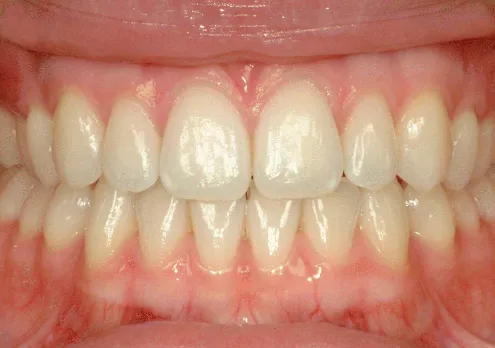
Figure 1.1 Clinical photograph of a normal healthy dentition, gingivae and oral mucosa.
(Source: Dr Rachael McKeown. Reproduced with permission of Dr McKeown)
There are two arches within the oral cavity – an upper or maxillary arch and a lower or mandibular arch (Figures 1.1 to 1.3). The upper arch may be named the maxillary arch, since the roots of the maxillary or upper teeth can be found attached to the upper jaw bone – the maxilla. Likewise, the lower arch may be named the mandibular arch, since the roots of the mandibular or lower teeth can be found attached to the lower jaw bone – the mandible. An anterior (front) view and a lateral (side) view of a human skull can be seen in Figures 1.2 and 1.3.
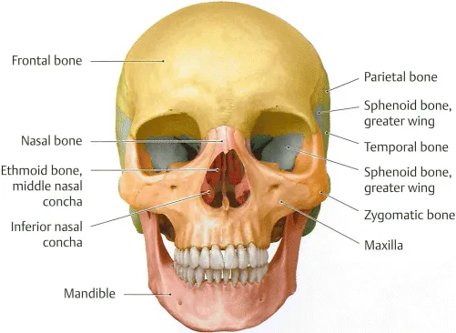
Figure 1.2 Illustrates an anterior (front) view of the human skull. From Head and Neck Anatomy for Dental Medicine. 2010. Ed. EW Baker. Thieme Medical Publishers, Inc.
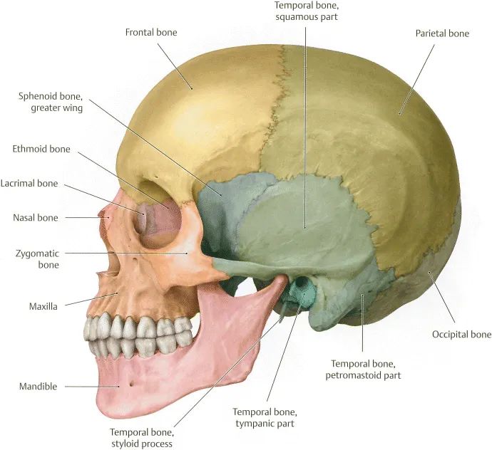
Figure 1.3 Illustrates a lateral (side) view of the human skull. From Head and Neck Anatomy for Dental Medicine. 2010. Ed. EW Baker. Thieme Medical Publishers, Inc.
The permanent dentition consists of 32 teeth:
- 8 incisors, 4 canines, 8 premolars and 12 molars The primary dentition consists of 20 teeth:
- 8 incisors, 4 canines and 8 molars
Descriptive Terms
Remember when viewing a patient, you would refer to the upper right, for example, as the patient's upper right and not your right! The midline of the two arches is a common reference point – this is the line dividing right and left central incisors – see Figures 1.1 and 1.2.
The dentition may be split into either four quadrants or six sextants. Both terms are commonly used within clinical dentistry. When the dentition is split into quadrants, each “quadrant” will normally contain a maximum of eight permanent teeth or five primary teeth – a quarter of the dentition. In the case of the permanent dentition, this is made up of two incisors and one canine (which make up the anterior teeth) and two premolars and three molars (which make up the posterior teeth). In the case of the primary dentition, this is made up of two incisors and one canine (the anterior teeth) and two molars (the posterior teeth). The quadrants are referred to as upper right (UR) and upper left (UL), and lower right (LR) and lower left (LL).
The system used in this book will be similar to the Palmer notation (see below). The quadrant will be given first, as above, where UR will be the patient's upper right quadrant, and so on. Permanent teeth are numbered 1–8, with 1 being the permanent central incisor and 8 being the permanent third molar. Primary (or deciduous) teeth are referred to as A–E, with A being the primary central incisor and E being the primary second molar.
Within some clinical disciplines, the dentition is split into six sextants. Each “sextant” may contain approximately one sixth of the teeth – the mandible and the maxilla each contain...