A timely, accessible survey of the multidisciplinary field of bioanalytical chemistry
Provides an all in one approach for both beginners and experts, from a broad range of backgrounds, covering introductions, theory, advanced concepts and diverse applications for each method
Each chapter progresses from basic concepts to applications involving real samples
Includes three new chapters on Biomimetic Materials, Lab-on-Chip, and Analytical Methods
Contains end-of-chapter problems and an appendix with selected answers
Häufig gestellte Fragen
Wie kann ich mein Abo kündigen?
Gehe einfach zum Kontobereich in den Einstellungen und klicke auf „Abo kündigen“ – ganz einfach. Nachdem du gekündigt hast, bleibt deine Mitgliedschaft für den verbleibenden Abozeitraum, den du bereits bezahlt hast, aktiv. Mehr Informationen hier.
(Wie) Kann ich Bücher herunterladen?
Derzeit stehen all unsere auf Mobilgeräte reagierenden ePub-Bücher zum Download über die App zur Verfügung. Die meisten unserer PDFs stehen ebenfalls zum Download bereit; wir arbeiten daran, auch die übrigen PDFs zum Download anzubieten, bei denen dies aktuell noch nicht möglich ist. Weitere Informationen hier.
Welcher Unterschied besteht bei den Preisen zwischen den Aboplänen?
Mit beiden Aboplänen erhältst du vollen Zugang zur Bibliothek und allen Funktionen von Perlego. Die einzigen Unterschiede bestehen im Preis und dem Abozeitraum: Mit dem Jahresabo sparst du auf 12 Monate gerechnet im Vergleich zum Monatsabo rund 30 %.
Was ist Perlego?
Wir sind ein Online-Abodienst für Lehrbücher, bei dem du für weniger als den Preis eines einzelnen Buches pro Monat Zugang zu einer ganzen Online-Bibliothek erhältst. Mit über 1 Million Büchern zu über 1.000 verschiedenen Themen haben wir bestimmt alles, was du brauchst! Weitere Informationen hier.
Unterstützt Perlego Text-zu-Sprache?
Achte auf das Symbol zum Vorlesen in deinem nächsten Buch, um zu sehen, ob du es dir auch anhören kannst. Bei diesem Tool wird dir Text laut vorgelesen, wobei der Text beim Vorlesen auch grafisch hervorgehoben wird. Du kannst das Vorlesen jederzeit anhalten, beschleunigen und verlangsamen. Weitere Informationen hier.
Ist Bioanalytical Chemistry als Online-PDF/ePub verfügbar?
Ja, du hast Zugang zu Bioanalytical Chemistry von Susan R. Mikkelsen, Eduardo Cortón im PDF- und/oder ePub-Format sowie zu anderen beliebten Büchern aus Biological Sciences & Biochemistry. Aus unserem Katalog stehen dir über 1 Million Bücher zur Verfügung.
This chapter introduces the basic principles underlying many common methods of signal transduction. This term is used to describe the conversion of one type of energy to another. Generally, analytical specialists use the term transducer to describe the conversion of a concentration (or mass) into a useful electronic signal, which is ultimately almost always a voltage. This voltage is related to the concentration (or mass) of the analyte, or species of interest, in the original sample. The species that can be measured by one or more of these methods is not always the analyte itself; for example, if the analyte is an enzyme or other catalytic species, the depletion of reactants or accumulation of products is assessed based on their own unique properties.
Transduction can be accomplished in many different ways, and the choice of the best method depends on which of many possible physical properties are exhibited by the measured species. In this chapter, we consider the three main types of transduction that are widely used in instrumental methods in bioanalytical chemistry. The conversion of light into current is performed by photodiodes or photomultipliers, and this current is then electronically converted into a voltage that is proportional to the intensity of the light. Electrochemical and surface plasmon resonance transducers convert chemical energy into a measured voltage or into a current that is subsequently converted to a voltage. Scintillation counters, used in many radiochemical methods, first convert beta-particle radioactivity to light, and the light is detected using photodiodes or photomultipliers. Thermal transducers, used for calorimetry, convert heat into current (and then voltage).
Considerations for the choice of a transduction method include the uniqueness of the various measurable properties of the measured species, since it is often present in a complicated sample matrix. The matrix is the surrounding environment, and includes all other components present in the sample. Matrix components can interfere with measurements in direct or indirect ways: a matrix component may exhibit a similar physical property to the analyte, and interfere with analyte measurement; also, a matrix component may interact with the analyte, changing the nature of its physical property and/or the magnitude of its resulting signal.
This chapter is intended as an introduction and brief review of common transduction methods used in bioanalytical chemistry. More detailed descriptions of applications and instrumental variations will be found within specific chapters of this book, where more specialized adaptations are described for specific assay methods.
The reader is referred to two excellent analytical chemistry textbooks for greater depth of coverage of most of the basic descriptions given in this chapter, as well as two excellent review articles for more information on thermal measurement methods, listed at the end of the chapter.
1.2 Optical Measurements
The majority of quantitative optical methods make use of light that is either absorbed or emitted in the ultraviolet and visible regions of the electromagnetic spectrum. These regions formally correspond to wavelengths of 1.0 × 10⁻⁸ to 7.8 × 10⁻⁷ m, and are more commonly expressed in nm units (10 to 780 nm). The far UV region, also called the vacuum UV region, is generally not analytically useful, but the near UV and the visible regions are widely used.
The colours that surround us result mainly from wavelength-selective visible light absorption by molecules present in the items that we see. However, differences between species, and between individuals within a species, cause the wavelength range of visible light, and the colours within this range, to be perceived differently. Common examples are bumblebees, that have blue-shifted visible ranges, and hummingbirds, that have red-shifted ranges. For this reason, standard wavelength ranges have been defined for the different colours of the visible spectrum. For example, blue light is defined as the 440–470 nm range, and if blue light is absorbed, its complementary colour, orange, is observed. Similarly, if green light (500–520 nm) is absorbed, purple is the observed colour. Many compounds absorb light at multiple wavelengths, and it is the combination of complementary colours that we observe.
The relationship between wavelength, frequency and energy of light is shown below:
(1.1)
where E is the energy of the light, h is Planck's constant (6.626 × 10⁻³⁴ J·s), υ is the frequency of the light (s−1), λ is the wavelength of the light (m), and c is the speed of light (2.998 × 10⁸ m/s in a vacuum, and this number is divided by the refractive index n for any other medium). This relationship connects the two key concepts that light is both a particle (a photon with energy E) and a wave, with frequency υ and wavelength λ.
In the visible and near UV regions of the spectrum, molecules absorb and emit light as their electronic configurations change. For example, electrons convert between paired and unpaired states, or between bonding and non-/antibonding orbitals. These conversions are accompanied by energy gains or losses as the molecule absorbs or emits a photon. Depending on molecular structure, as well as many other factors including solvent, pH and temperature, fixed electronic energy levels exist, and only photons of particular energies (wavelengths) can be absorbed or emitted. Associated with each electronic energy level are vibrational and rotational energy levels, which are separated by much smaller energy differences. Isolated vibrational or rotational transitions can be made to occur using infrared or microwave radiation, which have much lower energy. But the electronic transitions that occur in the UV-visible region are accompanied by vibrational and rotational transitions, and this means that a range of wavelengths can be absorbed by molecules, shown in Eq. 1.2:
(1.2)
where, for a given electronic transition, the total energy ΔET of the photons absorbed is the sum of the energy required for the electronic transition itself, ΔEElec, which is fixed, plus the energy changes associated with multiple possible vibrational and rotational transitions, ΔEVib and ΔERot. This means that, for any given electronic transition, molecules absorb or emit a fairly wide range of wavelengths, centered on a wavelength of maximal absorption or emission. For molecules absorbing or emitting light in the near UV and visible regions, the range of wavelengths can be as large as 100 nm for a given electronic transition, because of these accompanying vibrational and rotational transitions.
1.2.1 UV-Visible Absorbance
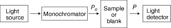
A simple spectrophotometer, an instrument for measuring absorbance, consists of a light source, a monochromator (or filter), a sample compartment and a light detector, all of which are enclosed to prevent interference from ambient light. These components are shown as a block diagram in Figure 1.1. Typically, the light source is a tungsten filament lamp (for the visible region) and/or a deuterium lamp (for the UV region); both of these sources emit continuous radiation over a wide range of wavelengths. Wavelength selection can be accomplished using filters, for repetitive fixed-wavelength measurements, or a monochromator containing a diffraction grating or prism, that allows adjustment of wavelength as well as wavelength scanning. The quality of the filter or monochromator determines the width of the wavelength range in the light beam that exits the device and is directed into the sample. Analyte solutions are contained in a cuvette (or cell) made of a material that is transparent to the wavelength(s) of interest, such as quartz, glass or polystyrene. Light detection may be accomplished using a photomultiplier tube, a photodiode, or a photodiode array (in which the spatial distribution of light of different wavelengths allows nearly instantaneous acquisition of a complete spectrum).
Figure 1.1 Block diagram of a simple UV-Vis absorption spectrophotometer.
Many variations of this simple design have been introduced for specialized applications. For example, dedicated instruments may employ an inexpensive light-emitting diode as the light source, a c...