The Respiratory System at a Glance
Jeremy P. T. Ward, Jane Ward, Richard M. Leach
- English
- ePUB (handyfreundlich)
- Über iOS und Android verfügbar
The Respiratory System at a Glance
Jeremy P. T. Ward, Jane Ward, Richard M. Leach
Über dieses Buch
The Respiratory System at a Glance has been thoroughly updated in line with current practice guidelines and new techniques to provide a highly illustrated and comprehensive guide to normal lung structure and function, as well as associated pathophysiology. Each topic has been fully revised and is accompanied by clear diagrams to encapsulate essential knowledge. Reflecting changes to the content, teaching and assessment methods used in medical education, this new edition now includes more information on acid base and its clinical ramifications, further detail on defence mechanisms and immunology, and also features online access to clinical cases and flashcards. The Respiratory System at a Glance:
•Integrates basic and clinical science – ideal for integrated and systems-based courses
•Includes both the pathophysiology and clinical aspects of the respiratory system
•Is fully revised and updated to reflect current practice guidelines and new therapies
•Provides online clinical cases, brand new flashcards, and MCQs
• Includes a companion website at www.ataglanceseries.com/respiratory featuring interactive multiple choice questions and digital flashcards
Häufig gestellte Fragen
Information
Part 1
Structure and function
Chapters
- 1 Structure of the respiratory system: lungs, airways and dead space
- 2 The thoracic cage and respiratory muscles
- 3 Pressures and volumes during normal breathing
- 4 Gas laws
- 5 Diffusion
- 6 Lung mechanics: elastic forces
- 7 Lung mechanics: airway resistance
- 8 Carriage of oxygen
- 9 Carriage of carbon dioxide
- 10 Acid–base balance
- 11 Acid–base disorders
- 12 Control of breathing I: chemical mechanisms
- 13 Control of breathing II: neural mechanisms
- 14 Pulmonary circulation and anatomical right-to-left shunts
- 15 Ventilation–perfusion mismatching
- 16 Exercise, altitude and diving
- 17 Development of the respiratory system and birth
- 18 Complications of development and congenital disease
- 19 Lung defence mechanisms
- 20 Immunology of the lung
1
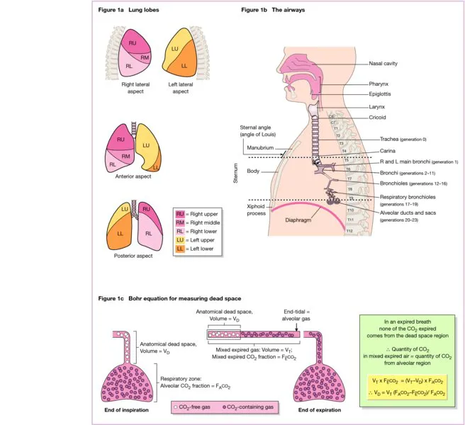
Structure of the respiratory system: lungs, airways and dead space
Lungs