Instant Anatomy presents anatomy and anatomical relationships in a simple, unique, schematic manner to aid the speedy understanding and retrieval of anatomical facts. It shows structuressuch as nerves and blood vessels in their entirety, unlike the partial, regional presentations given in most textbooks. Covering the major aspects of anatomy, each section presents the relevant structures in double page spreads, with clear, full-colour diagrams on the left and concise text for each structure on the right. This new fifth edition includes more surface anatomy such asnew myotome maps, bones of the hands and feet, principles of movement at shoulder and hip and images to clarify the understanding of the inguinal region and the lesser sac of the stomach. Ideal for use alongside a core anatomy textbook, Instant Anatomy is the perfect quick reference guide for medical students, surgeons, radiologists and those in many other specialties.The companion website at www.instantanatomy.net with its podcasts andwide ranging multiple choice questionsprovide invaluable exam preparation.
Häufig gestellte Fragen
Wie kann ich mein Abo kündigen?
Gehe einfach zum Kontobereich in den Einstellungen und klicke auf „Abo kündigen“ – ganz einfach. Nachdem du gekündigt hast, bleibt deine Mitgliedschaft für den verbleibenden Abozeitraum, den du bereits bezahlt hast, aktiv. Mehr Informationen hier.
(Wie) Kann ich Bücher herunterladen?
Derzeit stehen all unsere auf Mobilgeräte reagierenden ePub-Bücher zum Download über die App zur Verfügung. Die meisten unserer PDFs stehen ebenfalls zum Download bereit; wir arbeiten daran, auch die übrigen PDFs zum Download anzubieten, bei denen dies aktuell noch nicht möglich ist. Weitere Informationen hier.
Welcher Unterschied besteht bei den Preisen zwischen den Aboplänen?
Mit beiden Aboplänen erhältst du vollen Zugang zur Bibliothek und allen Funktionen von Perlego. Die einzigen Unterschiede bestehen im Preis und dem Abozeitraum: Mit dem Jahresabo sparst du auf 12 Monate gerechnet im Vergleich zum Monatsabo rund 30 %.
Was ist Perlego?
Wir sind ein Online-Abodienst für Lehrbücher, bei dem du für weniger als den Preis eines einzelnen Buches pro Monat Zugang zu einer ganzen Online-Bibliothek erhältst. Mit über 1 Million Büchern zu über 1.000 verschiedenen Themen haben wir bestimmt alles, was du brauchst! Weitere Informationen hier.
Unterstützt Perlego Text-zu-Sprache?
Achte auf das Symbol zum Vorlesen in deinem nächsten Buch, um zu sehen, ob du es dir auch anhören kannst. Bei diesem Tool wird dir Text laut vorgelesen, wobei der Text beim Vorlesen auch grafisch hervorgehoben wird. Du kannst das Vorlesen jederzeit anhalten, beschleunigen und verlangsamen. Weitere Informationen hier.
Ist Instant Anatomy als Online-PDF/ePub verfügbar?
Ja, du hast Zugang zu Instant Anatomy von Robert H. Whitaker, Neil R. Borley im PDF- und/oder ePub-Format sowie zu anderen beliebten Büchern aus Medicina & Anatomía. Aus unserem Katalog stehen dir über 1 Million Bücher zur Verfügung.
Internal carotid artery, vertebrobasilar system & circle of Willis
Ophthalmic artery
External carotid artery
Maxillary artery
Middle meningeal artery
Subclavian artery
Axillary artery
Brachial artery
Radial artery
Ulnar artery
Thoracic (descending) aorta
Abdominal aorta
External iliac artery
Coeliac trunk
Superior mesenteric artery
Inferior mesenteric artery
Internal iliac artery
Femoral artery
Popliteal artery
Anterior tibial artery
Posterior tibial artery
Fibular (peroneal) artery
Arterial anastomoses around scapula
Arterial anastomoses around hip
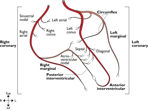
Coronary arteries
Coronary Arteries
From: Ascending aorta
To: Myocardium
Right coronary artery. Originates from the anterior aortic sinus. It passes anteriorly between the pulmonary trunk and the right auricle to reach the atrioventricular sulcus in which it runs down the anterior surface of the right cardiac border and then onto the inferior surface of the heart. It terminates at the junction of the atrioventricular sulcus and the posterior interventricular groove by anastomosing with the circumflex branch of the left coronary artery and giving off the posterior interventricular (posterior descending) artery. It supplies the right atrium and part of the left atrium, the sinuatrial node in 60% of cases, the right ventricle, the posterior part of the interventricular septum and the atrioventricular node in 80% of cases.
Left coronary artery. Arises from the left posterior aortic sinus. It passes laterally, posterior to the pulmonary trunk and anterior to the left auricle to reach the atrioventricular groove where it divides into an anterior interventricular (formally left anterior descending) artery and circumflex branches. The circumflex artery runs in the atrioventricular sulcus around the left border of the heart to anastomose with the right coronary artery. The anterior interventricular artery descends on the anterior surface of the heart in the anterior interventricular groove and around the apex of the heart into the posterior interventricular groove where it anastomoses with the posterior interventricular branch of the right coronary artery. The left coronary artery supplies the left atrium, left ventricle, anterior interventricular septum, sinuatrial node in 40% of cases and the atrioventricular node in 20%.
Dominance. In approximately 10% of hearts the posterior interventricular artery arises from the circumflex artery (left coronary) and then most of the left ventricle and interventricular septum are supplied by the left coronary artery. The heart is said to have left cardiac dominance.
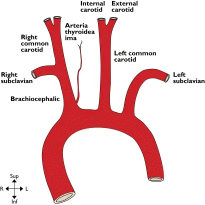
Ascending & arch of aorta
Ascending & Arch of Aorta
From: Left ventricle
To: Descending aorta
Ascending aorta. Arises at the vestibule of the left ventricle at the level of the third left costal cartilage and passes upwards and slightly to the right to a point behind the sternum at the level of the manubriosternal joint (second costal cartilage) where it becomes the arch of the aorta. It is enclosed in fibrous and serous pericardium. Anterior to it are the right auricle, the infundibulum of the right ventricle and pulmonary trunk. Posterior, lie the left atrium, the right pulmonary artery and right main bronchus. To the left lie the pulmonary trunk and the left auricle. To the right are the superior vena cava and the right atrium.
Arch of aorta. The arch begins posterior to the manubriosternal joint at the level of the second costal cartilage and passes posterior and to the left, over the left main bronchus to end at the left side of the body of T4 vertebra. Its highest level is the mid-point of the manubrium sterni and at this level its three main branches emerge. Anterior and to the left of the arch are (from anterior to posterior) the left phrenic nerve, vagal and sympathetic contributions to the cardiac plexus, and the left vagus. Also, the left superior intercostal vein runs forwards on the arch anterior to the vagus and posterior to the phrenic nerve. Lateral to all these structures are the pleura and left lung. Posterior and to the right of the arch are the trachea, deep cardiac plexus, left recurrent laryngeal nerve, oesophagus, thoracic duct and the body of T4. Inferior to the arch are the pulmonary bifurcation, the left main bronchus, the ligamentum arteriosum and the left recurrent laryngeal nerve. From its superior surface emerge the brachiocephalic artery, the left common carotid and left subclavian arteries. Within the adventitia of the ascending and arch of the aorta lie baro- and chemoreceptors.
Brachiocephalic artery. Arises from the convexity of the aortic arch behind the manubrium sterni and passes upwards and posteriorly to the right. It divides into the right subclavian and right common carotid arteries posterior to the right sternoclavicular joint. Anterior to it are the left brachiocephalic vein with the right inferior thyroid vein entering it, and the thymic remnants. The artery initially lies anterior to the trachea and then passes to lie on its right lateral side. On the right of the artery are the right brachiocephalic vein, upper part of the superior vena cava, the pleura and the cardiac branches of the vagus. The main vagal trunk is more posterolateral. At the origin of the brachiocephalic artery the left common carotid artery lies posteriorly on its left.
Ascending & arch of aorta
Common carotid arteries. The right common carotid artery arises from the brachiocephalic artery as it divides posterior to the right sternoclavicular joint, whilst the left common carotid arises from the convexity of the aortic arch. Both end as the arteries bifurcate at the level of the upper border of the thyroid cartilage (C4).
Left common carotid artery (thorax). Lying anterior to the thoracic part of this artery are the left brachiocephalic vein and the thymic remnant. Posterior to it in its lower part are the left subclavian artery and the trachea whilst further superiorly there is the left recurrent laryngeal nerve, the thoracic duct and the left side of the oesophagus. On its right at its origin is the brachioceph...