Chest X-Rays for Medical Students
eBook - ePub

Chest X-Rays for Medical Students

Christopher Clarke, Anthony Dux

Partager le livre
  1. English
  2. ePUB (adapté aux mobiles)
  3. Disponible sur iOS et Android
eBook - ePub

Chest X-Rays for Medical Students

Christopher Clarke, Anthony Dux

DĂ©tails du livre
Aperçu du livre
Table des matiĂšres
Citations

À propos de ce livre

Chest X-rays for Medical Students offers a fresh analytical approach to identifying chest abnormalities, helping medical students, junior doctors, and nurses understand the underlying physics and basic anatomical and pathological details of X-ray images of the chest. The authors provide a memorable framework for analysing and presenting chest radiographs, with each radiograph appearing twice in a side-by-side comparison, one as seen in a clinical setting and the second highlighting the pathology.

This new second edition includes significant revisions, improved annotations of X-rays, expanded pathologies, and numerous additional high-quality images. A comprehensive one-stop guide to learning chest radiograph interpretation, this book:

  • Aligns with the latest Royal College of Radiologists' Undergraduate Radiology Curriculum
  • Offers guidance on how to formulate normal findings
  • Features self-assessment tests, presentation exercises, and varied examples
  • Includes sections on radiograph quality X-ray hazards and precautions

Chest X-rays for Medical Students is an ideal study guide and clinical reference for any medical student, junior doctor, nurse or radiographer.

Foire aux questions

Comment puis-je résilier mon abonnement ?
Il vous suffit de vous rendre dans la section compte dans paramĂštres et de cliquer sur « RĂ©silier l’abonnement ». C’est aussi simple que cela ! Une fois que vous aurez rĂ©siliĂ© votre abonnement, il restera actif pour le reste de la pĂ©riode pour laquelle vous avez payĂ©. DĂ©couvrez-en plus ici.
Puis-je / comment puis-je télécharger des livres ?
Pour le moment, tous nos livres en format ePub adaptĂ©s aux mobiles peuvent ĂȘtre tĂ©lĂ©chargĂ©s via l’application. La plupart de nos PDF sont Ă©galement disponibles en tĂ©lĂ©chargement et les autres seront tĂ©lĂ©chargeables trĂšs prochainement. DĂ©couvrez-en plus ici.
Quelle est la différence entre les formules tarifaires ?
Les deux abonnements vous donnent un accĂšs complet Ă  la bibliothĂšque et Ă  toutes les fonctionnalitĂ©s de Perlego. Les seules diffĂ©rences sont les tarifs ainsi que la pĂ©riode d’abonnement : avec l’abonnement annuel, vous Ă©conomiserez environ 30 % par rapport Ă  12 mois d’abonnement mensuel.
Qu’est-ce que Perlego ?
Nous sommes un service d’abonnement Ă  des ouvrages universitaires en ligne, oĂč vous pouvez accĂ©der Ă  toute une bibliothĂšque pour un prix infĂ©rieur Ă  celui d’un seul livre par mois. Avec plus d’un million de livres sur plus de 1 000 sujets, nous avons ce qu’il vous faut ! DĂ©couvrez-en plus ici.
Prenez-vous en charge la synthÚse vocale ?
Recherchez le symbole Écouter sur votre prochain livre pour voir si vous pouvez l’écouter. L’outil Écouter lit le texte Ă  haute voix pour vous, en surlignant le passage qui est en cours de lecture. Vous pouvez le mettre sur pause, l’accĂ©lĂ©rer ou le ralentir. DĂ©couvrez-en plus ici.
Est-ce que Chest X-Rays for Medical Students est un PDF/ePUB en ligne ?
Oui, vous pouvez accĂ©der Ă  Chest X-Rays for Medical Students par Christopher Clarke, Anthony Dux en format PDF et/ou ePUB ainsi qu’à d’autres livres populaires dans Medicine et Radiology, Radiotherapy & Nuclear Medicine. Nous disposons de plus d’un million d’ouvrages Ă  dĂ©couvrir dans notre catalogue.

Informations

Éditeur
Wiley-Blackwell
Année
2020
ISBN
9781119504122

II
The ABCDE of chest X‐rays

It is important to use a systematic approach when looking at a chest radiograph. The following ABCDE approach is easy to remember, so when it comes to your exams and you have a moment of panic after being asked to talk about a chest X‐ray, you can stick to these basics even if you don’t have a clue what’s going on!
A is for Airway:
  • Look at the trachea and right and left main bronchi.
B is for Breathing:
  • Look to see if the lungs are uniformly expanded and compare the lung fields.
  • Look around the edges of each lung.
  • Look at the costophrenic angles and the four silhouettes.
C is for Circulation:
  • Look at the cardiac size.
  • Look at the great vessels (pulmonary vessels and aorta).
  • Look at the mediastinum and hila.
D is for Disability:
  • Look for a fracture, especially of the ribs or shoulder girdle.
E is for Everything else (review areas):
  • Look for gas under the diaphragm.
  • Look for surgical emphysema.
  • Look for both breast shadows.
  • Look for foreign bodies and medical interventions.

6
A – Airway

How to review the airway
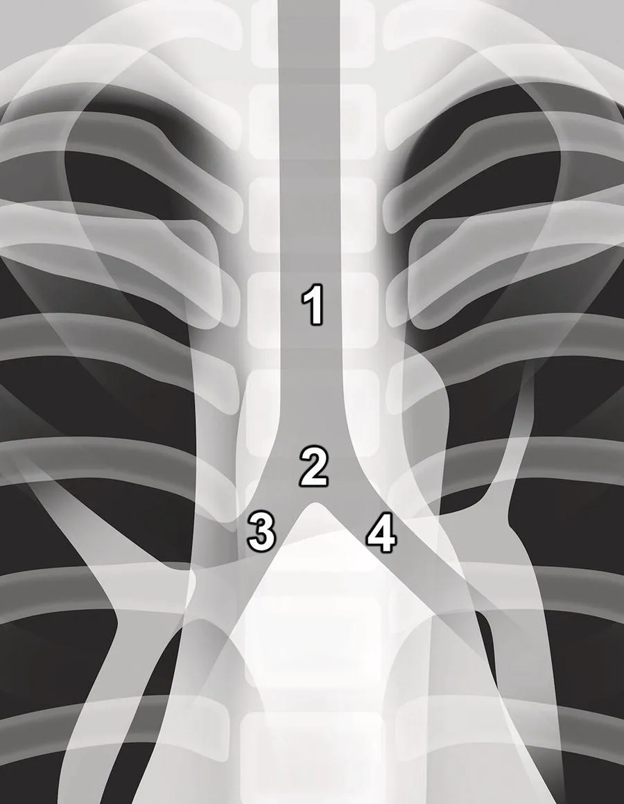
Start at the top and follow the trachea (1) inferiorly. It should lie in the midline. It divides at the carina (2) to give off the right main bronchus (3) and left main bronchus (4) (Figure 6.1). On a normal radiograph you will often not be able to visualise the more distal airway branches.
Photo depicts the airways of the lung.
Figure 6.1 The airways of the lung.

What to look for

  • Tracheal deviation
p. 24
  • Carinal angle
p. 25

Tracheal deviation

The trachea is considered to be deviated if a portion, anywhere along its length, is completely to the left or right of the midline (the midline being the centre of the vertebral column as indicated by the spinous processes).
Note: Be sure to check that the radiograph is not rotated, as a rotated radiograph can give the impression of tracheal deviation when the trachea is actually central.
If you suspect that the trachea is deviated, look for a possible cause.
  • Deviated towards diseased side (conditions that pull the trachea):
    • lung collapse;
    • pneumonectomy (removal of a lung) or lobectomy (removal of just one lobe);
    • unilateral fibrosis;
    • agenesis of lung (also called lung aplasia – complete absence of a whole lung and its bronchus).
  • Deviated away from diseased side (conditions that push the trachea):
    • tension pneumothorax;
    • massive pleural effusion;
    • mediastinal masses;
    • para‐tracheal masses.
The mos...

Table des matiĂšres