Point of Care Ultrasound Made Easy
eBook - ePub

Point of Care Ultrasound Made Easy

John McCafferty, James M Forsyth, John McCafferty, James M Forsyth

Partager le livre
  1. 154 pages
  2. English
  3. ePUB (adapté aux mobiles)
  4. Disponible sur iOS et Android
eBook - ePub

Point of Care Ultrasound Made Easy

John McCafferty, James M Forsyth, John McCafferty, James M Forsyth

DĂ©tails du livre
Aperçu du livre
Table des matiĂšres
Citations

À propos de ce livre

Point of Care Ultrasound Made Easy (POCUSME) is an exciting and innovative book that aims to teach all healthcare professionals how to do simple and clinically relevant ultrasound scanning at the point of care. This book will help you solve clinical problems at the bedside across a range of specialty areas, including: trauma, emergency medicine, respiratory medicine, cardiology, general surgery, otolaryngology and vascular surgery. Straightforward and practical, and designed for clinicians who are generally unfamiliar with ultrasound scanning, it will make a positive difference to your clinical practice, and help improve the delivery of optimised patient care. So read the book, grab an ultrasound machine, and please embrace the Point of Care Ultrasound movement!

Foire aux questions

Comment puis-je résilier mon abonnement ?
Il vous suffit de vous rendre dans la section compte dans paramĂštres et de cliquer sur « RĂ©silier l’abonnement ». C’est aussi simple que cela ! Une fois que vous aurez rĂ©siliĂ© votre abonnement, il restera actif pour le reste de la pĂ©riode pour laquelle vous avez payĂ©. DĂ©couvrez-en plus ici.
Puis-je / comment puis-je télécharger des livres ?
Pour le moment, tous nos livres en format ePub adaptĂ©s aux mobiles peuvent ĂȘtre tĂ©lĂ©chargĂ©s via l’application. La plupart de nos PDF sont Ă©galement disponibles en tĂ©lĂ©chargement et les autres seront tĂ©lĂ©chargeables trĂšs prochainement. DĂ©couvrez-en plus ici.
Quelle est la différence entre les formules tarifaires ?
Les deux abonnements vous donnent un accĂšs complet Ă  la bibliothĂšque et Ă  toutes les fonctionnalitĂ©s de Perlego. Les seules diffĂ©rences sont les tarifs ainsi que la pĂ©riode d’abonnement : avec l’abonnement annuel, vous Ă©conomiserez environ 30 % par rapport Ă  12 mois d’abonnement mensuel.
Qu’est-ce que Perlego ?
Nous sommes un service d’abonnement Ă  des ouvrages universitaires en ligne, oĂč vous pouvez accĂ©der Ă  toute une bibliothĂšque pour un prix infĂ©rieur Ă  celui d’un seul livre par mois. Avec plus d’un million de livres sur plus de 1 000 sujets, nous avons ce qu’il vous faut ! DĂ©couvrez-en plus ici.
Prenez-vous en charge la synthÚse vocale ?
Recherchez le symbole Écouter sur votre prochain livre pour voir si vous pouvez l’écouter. L’outil Écouter lit le texte Ă  haute voix pour vous, en surlignant le passage qui est en cours de lecture. Vous pouvez le mettre sur pause, l’accĂ©lĂ©rer ou le ralentir. DĂ©couvrez-en plus ici.
Est-ce que Point of Care Ultrasound Made Easy est un PDF/ePUB en ligne ?
Oui, vous pouvez accĂ©der Ă  Point of Care Ultrasound Made Easy par John McCafferty, James M Forsyth, John McCafferty, James M Forsyth en format PDF et/ou ePUB ainsi qu’à d’autres livres populaires dans MĂ©decine et MĂ©decine clinique. Nous disposons de plus d’un million d’ouvrages Ă  dĂ©couvrir dans notre catalogue.

Informations

Éditeur
CRC Press
Année
2020
ISBN
9781000039511
Édition
1
Part I
Background to Point-of-Care Ultrasound Made Easy (POCUSME)
Chapter 1
How Does Ultrasound Work?
John McCafferty
Learning Objectives
What Is Ultrasound?
Properties of Ultrasound in Tissues
Ultrasound Imaging
Image Acquisition
Scanning Modes
Optimising Imaging
Other Functions
Ultrasound Devices Used in Point of Care
Ultrasound Safety
Learning Objectives
â—ŸUnderstand the basic physics behind medical ultrasound imaging.
â—ŸUnderstand the hardware components of a medical ultrasound scanner.
â—ŸUnderstand the different imaging modes: B-mode, M-mode and Doppler.
â—ŸKnow how to acquire an ultrasound image, appreciating the imaging conventions.
â—ŸKnow how to optimise your ultrasound image.
â—ŸAppreciate your choices of point-of-care ultrasound (POCUS) device.
â—ŸHave an awareness of safety factors in POCUS.
What Is Ultrasound?
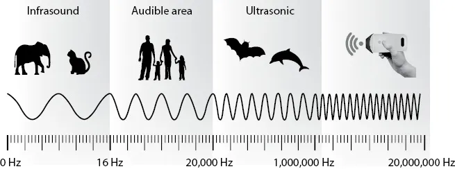
Ultrasound waves are mechanical energy that are transmitted through a medium from repetitive periodic oscillations of a transducer. The number of such cycles per second is termed the frequency of the ultrasound signal. Ultrasound refers to sound propagated at frequencies higher than the audible spectrum for humans (>20 KHz) (see Figure 1.1). Typically, medical ultrasound imaging operates in the range 3.5–20 MHz.
129146.webp
Figure 1.1 The frequency spectrum of sound waves with reference to the medical ultrasound range.
Properties of Ultrasound in Tissues
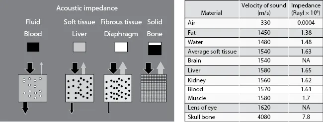
A B-mode ultrasound image is constructed from echoes generated by reflection of ultrasound from boundaries and scattering from small inhomogeneities within the tissue structures. Reflection occurs at the boundaries between two mediums of different acoustic impedance such as those between organs. Here, acoustic impedance is defined as the product of density of the tissue and the speed of sound within the tissue. The amplitude of the ultrasound wave that is reflected back into the tissue/organ or transmitted across a tissue/organ boundary is dependent on the change in acoustic impedance. If the change is small, the majority of the ultrasound will be transmitted and little reflected; if the change is large, the majority will be reflected back and little transmitted. For example, the medium of fluid or water has low impedance and will conduct the ultrasound wave, whereas bone is a poor conductor and will reflect the ultrasound wave back towards its source (see Figure 1.2).
129181.webp
Figure 1.2 Impedance values of typical tissues seen in the body. Air has very low impedance, meaning the ultrasound wave is strongly reflected at an air/tissue interface. This is why ultrasound gel is used when scanning a patient to avoid air interfaces between the transducer and the patient’s skin.
Small inhomogeneities within tissues/organs result in scattering of the ultrasound beam. This scattering is dependent on the size of the scatterer relative to the frequency/wavelength of the ultrasound beam. However, the scattering from these small structures (including red blood cells) is much smaller than typical reflections from boundaries so that within a B-mode image, boundaries are associated with brighter echoes compared to the less bright echoes generated by scattering within soft tissue.
Ultrasound Imaging
Ultrasound imaging uses the differences in the acoustic properties of tissues to build a picture of the structures within the body. The ultrasound signal is generated by a transducer that contains an array of tiny piezoelectric crystals which when excited by an electrical signal vibrate at a set frequency and transmit an ultrasound signal. The transducer is also a receiver, detecting the re...

Table des matiĂšres