Medical Microbiology Illustrated
eBook - ePub

Medical Microbiology Illustrated

S. H. Gillespie

  1. 296 pages
  2. English
  3. ePUB (adapté aux mobiles)
  4. Disponible sur iOS et Android
eBook - ePub

Medical Microbiology Illustrated

S. H. Gillespie

DĂ©tails du livre
Aperçu du livre
Table des matiĂšres
Citations

À propos de ce livre

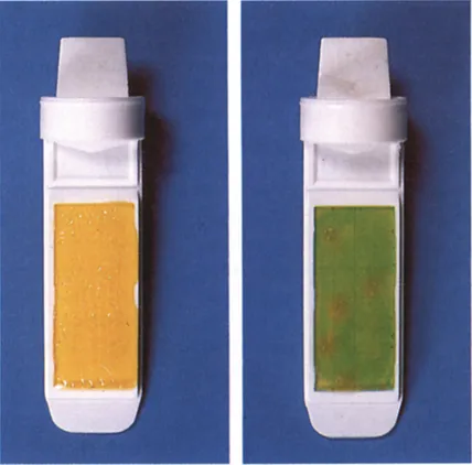
Medical Microbiology Illustrated presents a detailed description of epidemiology, and the biology of micro-organisms. It discusses the pathogenicity and virulence of microbial agents. It addresses the intrinsic susceptibility or immunity to antimicrobial agents. Some of the topics covered in the book are the types of gram-positive cocci; diverse group of aerobic gram-positive bacilli; classification and clinical importance of erysipelothrix rhusiopathiae; pathogenesis of mycobacterial infection; classification of parasitic infections which manifest with fever; collection of blood for culture and control of substances hazardous to health. The classification and clinical importance of neisseriaceae is fully covered. The definition and pathogenicity of haemophilus are discussed in detail. The text describes in depth the classification and clinical importance of spiral bacteria. The isolation and identification of fungi are completely presented. A chapter is devoted to the laboratory and serological diagnosis of systemic fungal infections. The book can provide useful information to microbiologists, physicians, laboratory scientists, students, and researchers.

Foire aux questions

Comment puis-je résilier mon abonnement ?
Il vous suffit de vous rendre dans la section compte dans paramĂštres et de cliquer sur « RĂ©silier l’abonnement ». C’est aussi simple que cela ! Une fois que vous aurez rĂ©siliĂ© votre abonnement, il restera actif pour le reste de la pĂ©riode pour laquelle vous avez payĂ©. DĂ©couvrez-en plus ici.
Puis-je / comment puis-je télécharger des livres ?
Pour le moment, tous nos livres en format ePub adaptĂ©s aux mobiles peuvent ĂȘtre tĂ©lĂ©chargĂ©s via l’application. La plupart de nos PDF sont Ă©galement disponibles en tĂ©lĂ©chargement et les autres seront tĂ©lĂ©chargeables trĂšs prochainement. DĂ©couvrez-en plus ici.
Quelle est la différence entre les formules tarifaires ?
Les deux abonnements vous donnent un accĂšs complet Ă  la bibliothĂšque et Ă  toutes les fonctionnalitĂ©s de Perlego. Les seules diffĂ©rences sont les tarifs ainsi que la pĂ©riode d’abonnement : avec l’abonnement annuel, vous Ă©conomiserez environ 30 % par rapport Ă  12 mois d’abonnement mensuel.
Qu’est-ce que Perlego ?
Nous sommes un service d’abonnement Ă  des ouvrages universitaires en ligne, oĂč vous pouvez accĂ©der Ă  toute une bibliothĂšque pour un prix infĂ©rieur Ă  celui d’un seul livre par mois. Avec plus d’un million de livres sur plus de 1 000 sujets, nous avons ce qu’il vous faut ! DĂ©couvrez-en plus ici.
Prenez-vous en charge la synthÚse vocale ?
Recherchez le symbole Écouter sur votre prochain livre pour voir si vous pouvez l’écouter. L’outil Écouter lit le texte Ă  haute voix pour vous, en surlignant le passage qui est en cours de lecture. Vous pouvez le mettre sur pause, l’accĂ©lĂ©rer ou le ralentir. DĂ©couvrez-en plus ici.
Est-ce que Medical Microbiology Illustrated est un PDF/ePUB en ligne ?
Oui, vous pouvez accĂ©der Ă  Medical Microbiology Illustrated par S. H. Gillespie en format PDF et/ou ePUB ainsi qu’à d’autres livres populaires dans Ciencias biolĂłgicas et MicrobiologĂ­a. Nous disposons de plus d’un million d’ouvrages Ă  dĂ©couvrir dans notre catalogue.

Informations

Année
2014
ISBN
9781483193632
1

Introduction to clinical microbiology

Publisher Summary

This chapter discusses the process of microbiological diagnosis from the initial clinical encounter to the completion of the diagnostic and therapeutic process. The process of making a microbiological diagnosis has many component steps. The process begins when the clinician meets the patient, takes a history, and performs a clinical examination. The clinician forms a hypothesis about the cause of the patient’s complaint: the differential diagnosis. With this hypothesis, a diagnostic is constructed that will include laboratory, radiological, and other investigations. Relevant specimens are obtained and transported to the laboratory. It is essential that this process is closely controlled as most microbiological specimens are highly perishable. When they are received in the laboratory, the specimens should be carefully documented and urgent specimens identified for rapid processing. They should then be investigated according to the standard laboratory procedures. A report is drawn up that may indicate the presence or absence of human pathogens and, where relevant, their susceptibility to anti-microbial agents. These results are communicated to the clinician together with further advice from the clinical microbiologist, who may request further specimens, modifications to the treatment regimen, or clinical and laboratory follow-up.

Introduction

A detailed understanding of epidemiology, and the biology of micro-organisms is required if patients with infectious diseases are to be adequately treated. Knowledge of the normal resident microbial flora facilitates the evaluation of the significance of individual organisms isolated from clinical specimens. An understanding of the pathogenicity and virulence will assist in predicting the likely behaviour of potential pathogens. In the same way, knowledge of intrinsic susceptibility or resistance to antimicrobial agents will be valuable in planning effective antimicrobial chemotherapy. This introductory chapter discusses the process of microbiological diagnosis from the initial clinical encounter to the completion of the diagnostic and therapeutic process.

The process of microbiological diagnosis
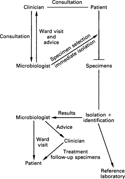
The process of making a microbiological diagnosis has many component steps. All of these must be understood and optimized if the laboratory is to collaborate fully with the clinician in achieving the best outcome for the patient.
The process begins when the clinician meets the patient, takes a history and performs a clinical examination. The clinician forms a hypothesis about the cause of the patient’s complaint: the differential diagnosis. With this hypothesis a diagnostic plan must be constructed which will include laboratory, radiological and other investigations. At this point the clinical microbiologist may be contacted and discussions may lead to the development of the differential diagnosis. The clinical microbiologist can also advise on the most appropriate investigations, how the specimens should be sent and may also give advice on empirical therapy.
Relevant specimens are obtained and transported to the laboratory. It is essential that this process is closely controlled as most microbiological specimens are highly perishable. When they are received in the laboratory the specimens should be carefully documented and urgent specimens identified for rapid processing. They should then be investigated according to the standard laboratory procedures. A report is drawn up which may indicate the presence or absence of human pathogens and, where relevant, their susceptibility to antimicrobial agents. These results are communicated to the clinician, together with further advice from the clinical microbiologist, who may request further specimens, modifications to the treatment regimen or clinical and laboratory follow up. This is summarized in Figure 1.1.
image

Figure 1.1 Role and function of a clinical microbiology service

Specimens

The quality of microbiological specimens is crucial to the value of the results obtained. For example the possibility of isolating an organism is much reduced if antibiotics are given before a specimen is obtained.

Specimen types

Specimens may be divided into two groups: those from normally sterile sites, and those containing normal bacterial flora. It is important to classify them in this way as it affects the way in which they are processed, culture results are evaluated and reports issued.
Specimens from sites which are normally sterile are inoculated into enrichment media. This protects fastidious pathogens that may be present in small numbers, provides all essential nutrients, and allows rapid multiplication so that enough organisms will be available for identification and susceptibility testing. By contrast, in specimens from a site with a normal flora, selective agents must be used to suppress the growth of commensal organisms which would obscure any pathogens. Thus for specimens from sterile sites amplification is the aim whereas for sites with a normal flora selection is the aim.
The isolation of an organism from a site which is normally sterile is likely to be clinically important. Organisms may be present as a result of contamination during the clinical procedure required to collect it or during laboratory processing. Before an organism is dismissed as a contaminant, careful evaluation must be made of the clinical details usually by a visit to the patient.
In many instances, the interpretation of bacterial isolates is made more complex when important pathogenic organisms form part of the normal flora in asymptomatic subjects. Streptococcus pneumoniae may be found in the nasopharynx of up to 30% of healthy adults. Sputum specimens are contaminated with pharyngeal secretions which may therefore contain this organism. To overcome this problem quantitative techniques have been evolved which assume that potential pathogens are present in smaller numbers when they are acting as commensals than when they are acting as pathogens (see p. 181).

Specimen choice

Advice must be given to clinical colleagues on the most appropriate specimens for the investigation of each infective condition: for example in gonorrhoea in females an endocervical swab should be sent rather than a high vaginal swab as the former has a much higher diagnostic yield.
In the investigation of acute lower respiratory tract infection where the patient is unable to produce a satisfactory sputum specimen, more invasive techniques such as broncho-alveolar lavage may be necessary. When an abscess is drained surgically, specimens of pus should be sent in a sterile container or a sterile syringe rather than on a bacteriological swab. Pathological material may dry rapidly causing fastidious organisms to die. Swabs maximize the exposure of bacteria to drying and atmospheric oxygen resulting in a reduction in diagnostic yield; some organisms are inhibited by materials in the swab itself.

Containers

Labelled, sterile containers should be available for collection of microbiological specimens.
These should be of different sizes, depending on the specimen (i.e. a screw-capped container for cerebrospinal fluid (CSF), a wide-mouth container for mid-stream specimens of urine (MSU)). Stool pots may have a scoop incorporated in the lid to make specimen collection more aesthetic. Bottles should have a label which prompt the clinician to record essential information (i.e. name, hospital number, sex, date of birth) so that they can be reliably identified. Sodium borate can be added to urine containers to inhibit bacterial multiplication. A ‘dip slide container’ may be provided in which there is a paddle coated on both sides with an agar medium (usually MacConkey and cysteine lactose electrolyte deficient (CLED) agars). The paddle is dipped in the urine which is then discarded and the paddle replaced in the container. Organisms survive well on these media at ambient temperature while in transit to the laboratory where they are subsequently incubated at 37°C. Colony counts will be available on the following day and identification and susceptibility tests can be performed by subculture (Fig. 1.2).
image

Figure 1.2 (a and b) The dipslide has two types of media on alternate sides of the paddle, MacConkey and CLED in this case. By comparing the number of colonies with a diagram the approximate number of organisms can be estimated
Some perishable specimens are collected directly into a transport or culture medium and these require careful stock control to ensure that these media are kept in an optimum condition. Some workers consider that blood cultures collected into blood culture medium which is held at 37°C produces a higher diagnostic yield as this is thought to prevent ‘cold shock’ which retards growth. If this strategy is adopted, the microbiologist must provide blood culture bottles ...

Table des matiĂšres