Cartilage Biochemistry and Morphology
The hyaline cartilage could be regarded as a composite gel with relatively low percentage chondrocytes (5%) embedded in a rich extracellular matrix consisting in negatively charged hydrophilic proteoglycans constrained by a three-dimensional collagen network.
The negatively charged proteoglycans have the ability to form large aggregates, which can bind water molecules within the positively charged collagen fibrils, thus generating a high osmotic pressure within the gel.
The collagen fibers are responsible for the structure of cartilage and consist mainly of collagen type II. They are highly cross-linked via collagen type IX fibers.2
Chondrocytes are the producers of the surrounding ground substance: matrix.
The cells have different appearances depending on where in the cartilage they are situated. The cells in the top layer appear flattened, whereas the cells in the deeper layer are more rounded and aligned along vertically orientated type II collagen.3
Collagen is the most important scaffolding material in the body, existing in several types. The major type in hyaline cartilage is named type II. It is built by three identical polypeptide alpha-chains. These chains are coiled to form a triple-helix and are produced by the chondrocyte in the form of procollagen. Outside the cell, this procollagen is transformed to tropocollagen, and these molecules aggregate to form the much larger molecule: collagen.
In the hyaline cartilage there also exist minor collagens like types IX, XI, V, and VI. Type IX contributes with covalent cross-linking of the type II fibrils, whereas type XI is thought to control the diameter of type II fibrils. The collagen gives the cartilage its strength and tensile stiffness.
Proteoglycans are large protein-polysaccharide molecules making up 5% to 10% of the wet weight of the cartilage.4 They are composed by chains of the glycosaminoglycans keratan sulfate and chondroitin sulfate covalently bound to a central protein core molecule. Large aggregates are formed with several proteoglycan monomers via a link protein connecting the central protein cores to a chain of hyaluronic acid.
All the components of the proteoglycan aggregates are synthesized by the chondrocytes.
The proteoglycans are unevenly distributed throughout the cartilage layers with the highest concentration in the middle part and the lowest concentration in the superficial layers.5 The proteoglycans give the cartilage its elasticity and resilience.
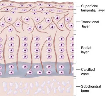
There is a difference in cartilage composition between the cartilage surface and the subchondral bone plate. These structural differences give rise to four separate layers or zones (see Fig. 1-1).
In the top zone, the superficial zone, there is first a cell-free fibril-layer, called the lamina splendens.6 Beneath this thin layer, chondrocytes are dispersed in an elongated manner parallel to the surface, reflecting as well the tangential orientation of the collagen fibers. This is the tangential layer.
In the second zone, often called the transitional layer, the cells are larger, rounded, and randomly distributed between the oblique-oriented collagen fiber. In the third zone, the chondrocytes are even larger and arranged in typical columns because of the radial collagen fiber courses, the radial zone.
The fourth layer, finally, which is mineralized, is called the calcified zone. There exists a visible border between the third and fourth zone, the tidemark with a special affinity for basic dyes (e.g., toluidine blue).
The calcified zone provides an important transition to the less resilient subchondral bone. For a long time this was regarded more or less as an inactive zone, until Hunziker (1992)7 noted that also the chondrocytes here could take up and incorporate (35S) sulfate into the pericellular and territorial matrix. Hunziker speculated that, following trauma, the metabolic activity here becomes temporarily impaired.7
Regarding experimental animals, it is important to know that it is only in adult animals that the division into zone I to zone III is possible.8 In the immature animal, the cells are more randomly distributed with a gradient in cell size from the top to the calcified zone, with the cells in the deeper parts being largest. Thus, the articular cartilage organization during prepubertal growth imitates the structure of the growth plate, and during that time the biomechanical properties of the cartilage change with an increase in stiffness and in shearing and compressive resistance.7,9
Metabolic Events in the Cartilage
Under normal conditions, the components of the matrix have a slow turnover. The collagen has the slowest turnover rate compared to the much faster turnover of the proteoglycans.
The majority of the proteoglycans have a life span of about 600 days, but a small proportion of the proteoglycans in adult cartilage act as a fast fraction with a half-life of about 8 days. The proteoglycans are thus also more vulnerable to enzymatic degradation.10,11
The chondrocytes secrete different enzymes called metalloproteinases (collagenases, gelatinases, and stromelysin), which all control the degree of degradation. The degradation of proteoglycans is followed by an increased synthesis...