The electrophoresis techniques are used in medicine, biochemistry, analytical chemistry, and biology to separate soluble and insoluble proteins, nucleic acids, chromosomes, viruses, as well as lysosomes, mitochondria, ribosomes and other cell organelles, red cells, tissue cells, and parasites. This book provides a view over the old electrophoresis techniques, as well as the recent developments in electrophoresis.
Electrophoresis Fundamentals is based on the recent book Electrophoresis: Theory and Practice published in 2020 by De Gruyter. The previous book combines theory and technical applications with troubleshooting and problem solving. While Electrophoresis is intended for specialists, Electrophoresis Fundamentals is a book for laboratory technicians, students, biochemists, general practitioners, and more.
Foire aux questions
Comment puis-je résilier mon abonnement ?
Il vous suffit de vous rendre dans la section compte dans paramètres et de cliquer sur « Résilier l’abonnement ». C’est aussi simple que cela ! Une fois que vous aurez résilié votre abonnement, il restera actif pour le reste de la période pour laquelle vous avez payé. Découvrez-en plus ici.
Puis-je / comment puis-je télécharger des livres ?
Pour le moment, tous nos livres en format ePub adaptés aux mobiles peuvent être téléchargés via l’application. La plupart de nos PDF sont également disponibles en téléchargement et les autres seront téléchargeables très prochainement. Découvrez-en plus ici.
Quelle est la différence entre les formules tarifaires ?
Les deux abonnements vous donnent un accès complet à la bibliothèque et à toutes les fonctionnalités de Perlego. Les seules différences sont les tarifs ainsi que la période d’abonnement : avec l’abonnement annuel, vous économiserez environ 30 % par rapport à 12 mois d’abonnement mensuel.
Qu’est-ce que Perlego ?
Nous sommes un service d’abonnement à des ouvrages universitaires en ligne, où vous pouvez accéder à toute une bibliothèque pour un prix inférieur à celui d’un seul livre par mois. Avec plus d’un million de livres sur plus de 1 000 sujets, nous avons ce qu’il vous faut ! Découvrez-en plus ici.
Prenez-vous en charge la synthèse vocale ?
Recherchez le symbole Écouter sur votre prochain livre pour voir si vous pouvez l’écouter. L’outil Écouter lit le texte à haute voix pour vous, en surlignant le passage qui est en cours de lecture. Vous pouvez le mettre sur pause, l’accélérer ou le ralentir. Découvrez-en plus ici.
Est-ce que Electrophoresis Fundamentals est un PDF/ePUB en ligne ?
Oui, vous pouvez accéder à Electrophoresis Fundamentals par Budin Michov en format PDF et/ou ePUB ainsi qu’à d’autres livres populaires dans Biowissenschaften et Biotechnologie. Nous disposons de plus d’un million d’ouvrages à découvrir dans notre catalogue.
The term electrophoresis means moving of charged dissolved particles in an electric field, which causes their resolution depending on their velocities and interaction with the separation medium [1, 2, 3]. In its current form, the electrophoresis is connected with the studies of Tiselius [4] in the 1930s.
The electrophoresis of positively charged particles (cations) is called cataphoresis; the electrophoresis of negatively charged particles (anions) is called anaphoresis.
The electrophoresis is used for resolving of proteins and nucleic acids, chromosomes, viruses, cell membranes (plasma, lysosomal, nuclear, and other), cell organelles (mitochondria, ribosomes), cells (red cells, tissue cells, and parasites), etc., it takes place in biochemistry, proteomic and genomic studies, forensics, molecular biology, and microbiology. By electrophoresis, more than the half of all separations and almost all separations of blood proteins and DNA are carried out.
Overview on electrophoresis
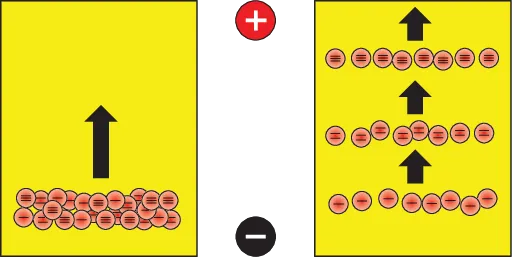
Proteins and nucleic acids form polyions. In an electric field, the positively charged polyions move toward the negative pole, while the negatively charged polyions move toward the positive pole (Figure 1.1).
Left – start of electrophoresis; Right – end of electrophoresis.
Figure 1.1: Electrophoresis of unipolar particles carrying different electric charges.
Commonly, polyions that are to be resolved are applied onto a separation medium, which in its turn is placed into an electrophoresis cell that is connected to a power supply. When electric current is turned on, the larger and less charged polyions move slower through the medium, while the smaller and more charged polyions move faster.
Mostly, electrophoresis is carried out in agarose or polyacrylamide gels, soaked with buffers. Other separation media are: starch gel, cellulose acetate, and paper. Today they have lost their actuality. Electrophoresis can also be carried out in buffers only. The buffers are electrolyte solutions, which maintain constant pH values, e.g. TRIS-borate buffer, TRIS-histidinate buffer, Goods buffers, and more.
Since proteins and nucleic acids are mostly colorless, their movement through the gel cannot be followed during electrophoresis. Therefore, tracking dyes are usually included in the sample buffer. At electrophoresis of negatively charged proteins, Bromophenol blue, Xylene cyanol, which runs slower than Bromophenol blue, or Orange G are used; at electrophoresis of positively charged proteins Bromocresol green or Methylene blue are used.
Proteins can be separated also in pH gradients, where they stop at their isoelectric (pI) points. This electrophoresis, called isoelectric focusing, can be carried out in two types of pH gradients: produced by carrier ampholytes, or by immobilines.
The electrophoresis takes place in horizontal or vertical separation cells. The electrodes can be placed on the gel or in separate electrode tanks.
Beside electrophoresis cells and power supplies, thermostats for reserving the resolving medium temperature, and densitometers or scanners for analyzing the pherograms (the resolved polyions in the medium) are used. Semi-automated electrophoresis devices are also offered on the market.
The electrophoretic velocity of a polyion is proportional to its effective mobility and the electric field strength . In its turn, the effective mobility depends on the total electric charge of the polyion and is inversely proportional to the viscosity of the separation medium. The total electric charge is determined by the buffer pH value; and the viscosity of the resolving medium depends mainly on the medium structure and temperature.
The electric field strength is equal to the ratio between the electric voltage and distance between the two electrodes. Since the distance remains constant during the electrophoresis, the polyion velocity depends only on the electric voltage.
Electrophoresis should be carried out at voltage and electric current, when the heating could be drawn out from the electrophoresis cell.
After electrophoresis, blotting of proteins and nucleic acids can be made. Using this technique, the resolved bands can be immobilized onto blot membranes and treated afterward. The blotting methods are carried out in four steps: electrophoretic separation of proteins or nucleic acid fragments; their transfer and immobilization onto blot membranes; binding of analytical probes to the blotted substances; and visualization of the blotted bands.
The blotting of DNA bands is called Southern blotting, the blotting of RNA bands is called Northern blotting, and the blotting of protein bands is called Western blotting. The blotting membrane consists usually of nitrocellulose, nylon or polyvinylidene difluoride (PVDF).
Separation media
The electrophoresis can be carried out in a solution, but the diffusion there is too strong. In order to limit the diffusion, solid media are used. The earliest solid medium was cellulose contained in the filter paper. The paper electrophoresis was invented by Kunkel and Tiselius [5] in 1951. Cellulose acetate membranes were the next step in the electrophoresis progress. Today most common are agarose and polyacrylamide gel electrophoresis.
Electrophoresis resolution and sharpness
The resolution of electrophoresis is referred to the ability of electrophoresis to separate two sample components from each other. It depends on the Gaussian profiles of the bands and is calculated by dividing the distance between the centers of adjacent bands by their average bandwidths.
The sharpness of electrophoresis is referred to as the reciprocal value of the bandwidth; narrow the bands, the higher the sharpness.
Detection of resolved bands
The majority of polyion bands are not visible to the naked eye, with a few exceptions. Direct optical detection of resolved bands can be applied, for example, to hemoglobins (which are red colored). Therefore, a couple of methods are created for detection, localization, and quantitation of separated bands.
The detection of resolved polyions is carried out directly or indirectly. The direct detection is performed in the resolving medium by nonspecific or specific staining, enzyme-substrate reactions, immune precipitation, autoradiography, and fluorography. The indirect detection is performed by immune printing or blotting.
The proteins can be stained in gels. Common dye is Coomassie brilliant blue, which can detect 0.3 μg of protein in a spot. DNA is detected usually by fluorescent intercalating of ethidium bromide. Both proteins and DNA can react with silver ions to form black bands. The silver methods are 100 times more sensitive than the other staining methods and can detect 2 ng of protein in a spot.
The process of staining is followed by destaining of the gel background to remove unbound dye. In some cases, placing the gel on an ultraviolet (UV) lightbox or under a UV lamp can reveal UV absorbing bands, or UV fluorescence bands.
If radioactive atoms have been incorporated in the polyions prior to electrophoresis, their position in the gel or membrane can be determined by autoradiography. For this purpose, the gel or membrane is placed on a photographic film or overlaid with it and let to expose in the dark and cold to show the positions of the radioactive bands.
The most modern methods of both detection and characterization of resolved proteins involve mass spectrometry.
The electrophoresis results can be recorded with a computer operated camera, and the intensity of a band or spot of interest can be compared against markers in the same gel, using specialized software.
After electrophoresis, the gels with bands can be saved in dry forms.
References
[1]Lyklema J. Fund Interface Colloid Sci, 1995, 2, 3, 208. →
[2]Hunter RJ. Foundations of Colloid Science, 2nd edn, Oxford University Press, Oxford, 2001. →
[3]Russel WB, Saville DA, Schowalter WR. Colloidal Dispersions. Cambridge University Press, Cambridge, 1989. →
[4]Tiselius A. Trans Faraday Soc, 1937, 33, 524–531. →
[5]Kunkel HG, Tiselius A. Electrophoresis of proteins on filter paper, J Gen Physiol, 1951, 35, 89–118. →