
eBook - ePub
Evolution of Neurosensory Cells and Systems
Gene regulation and cellular networks and processes
Bernd Fritzsch, Karen Elliott, Bernd Fritzsch, Karen L. Elliott
This is a test
Partager le livre
- 344 pages
- English
- ePUB (adapté aux mobiles)
- Disponible sur iOS et Android
eBook - ePub
Evolution of Neurosensory Cells and Systems
Gene regulation and cellular networks and processes
Bernd Fritzsch, Karen Elliott, Bernd Fritzsch, Karen L. Elliott
Détails du livre
Aperçu du livre
Table des matières
Citations
À propos de ce livre
This book is an overview of primary sensory maps of vertebrates, characterized by continuous and discrete properties. The eight primary sensory maps of vertebrates have unique features and use distinct molecular cues, cell cycle exit, and activity combinations during development, regeneration, and plasticity. As an introduction and overview, the book provides a short overview for all eight sensory senses and presents through evolution and gene regulatory networks, the molecular cues needed for sensory processing. Independent contributions are included for olfactory, vision, trigeminal, taste, vestibular, auditory, lateral line, and electroreception.
Foire aux questions
Comment puis-je résilier mon abonnement ?
Il vous suffit de vous rendre dans la section compte dans paramètres et de cliquer sur « Résilier l’abonnement ». C’est aussi simple que cela ! Une fois que vous aurez résilié votre abonnement, il restera actif pour le reste de la période pour laquelle vous avez payé. Découvrez-en plus ici.
Puis-je / comment puis-je télécharger des livres ?
Pour le moment, tous nos livres en format ePub adaptés aux mobiles peuvent être téléchargés via l’application. La plupart de nos PDF sont également disponibles en téléchargement et les autres seront téléchargeables très prochainement. Découvrez-en plus ici.
Quelle est la différence entre les formules tarifaires ?
Les deux abonnements vous donnent un accès complet à la bibliothèque et à toutes les fonctionnalités de Perlego. Les seules différences sont les tarifs ainsi que la période d’abonnement : avec l’abonnement annuel, vous économiserez environ 30 % par rapport à 12 mois d’abonnement mensuel.
Qu’est-ce que Perlego ?
Nous sommes un service d’abonnement à des ouvrages universitaires en ligne, où vous pouvez accéder à toute une bibliothèque pour un prix inférieur à celui d’un seul livre par mois. Avec plus d’un million de livres sur plus de 1 000 sujets, nous avons ce qu’il vous faut ! Découvrez-en plus ici.
Prenez-vous en charge la synthèse vocale ?
Recherchez le symbole Écouter sur votre prochain livre pour voir si vous pouvez l’écouter. L’outil Écouter lit le texte à haute voix pour vous, en surlignant le passage qui est en cours de lecture. Vous pouvez le mettre sur pause, l’accélérer ou le ralentir. Découvrez-en plus ici.
Est-ce que Evolution of Neurosensory Cells and Systems est un PDF/ePUB en ligne ?
Oui, vous pouvez accéder à Evolution of Neurosensory Cells and Systems par Bernd Fritzsch, Karen Elliott, Bernd Fritzsch, Karen L. Elliott en format PDF et/ou ePUB ainsi qu’à d’autres livres populaires dans Scienze biologiche et Biologia. Nous disposons de plus d’un million d’ouvrages à découvrir dans notre catalogue.
Informations
1The SensesPerspectives from Brain, Sensory Ganglia, and Sensory Cell Development in Vertebrates
Bernd Fritzsch, Karen L. Elliott
DOI: 10.1201/9781003092810-1
CONTENTS
- 1.1 Introduction of Primary Neurosensory Organization
- 1.2 Neural Induction
- 1.2.1 Formation of the Neural Plate and Neural Tube
- 1.2.2 Patterning of the Neural Tube
- 1.3 Placode and Neural Crest Development into Sensory Cells and Neurons
- 1.3.1 Olfactory Receptors are Found on Olfactory Sensory Neurons Which Project Directly to the Forebrain
- 1.3.2 The Retina and Lens have Different Embryonic Origins
- 1.3.3 Cranial Ganglion Neurons Develop from Both Placodes and Neural Crest
- 1.3.4 Otic Placode Development uses Common and Unique Gene Regulatory Networks
- 1.3.5 Hair Cells of the Inner Ear, Lateral Line, and Electroreception have a Shared Developmental Program
- 1.4 Summary and Conclusion
- Acknowledgements
- References
1.1 INTRODUCTION OF PRIMARY NEUROSENSORY ORGANIZATION
Responding to an environment is a demand placed on all organisms and necessitates the ability to sense external stimuli. For organisms that have complex interactions with their environment, such as vertebrates, this ability has been evolutionary optimized to ensure precise methods of discriminating sensory information. Such optimization has been driven by coordinated development of a centralized processing center that is connected to peripheral sensory organs by way of peripheral neurons. Many studies have investigated the origins of the nervous system (Layden, 2019). Current evidence suggests a common ancestor for cnidarians and bilaterian nervous systems (Galliot et al., 2009). In chordates, the central nervous system (CNS) develops from the dorsal ectoderm. However, the urbilaterian ancestor likely had a similar organization to protostomes with a ventrally positioned central nervous system (Gerhart, 2000). The evolution of deuterostomes saw an inversion of that body plan. Multiple ideas have been proposed to explain these differing dorso-ventral developmental inversion schemes and originated primarily from comparative data on neuron, heart, and mouth location (Arendt and Nübler-Jung, 1997; Fritzsch et al., 2017; Gee, 2007). Among protostomes, the central nervous system is located ventrally, whereas the heart is found dorsally in the body (Gerhart, 2000). In contrast, among deuterostomes, the central nervous system is located dorsally and the heart is ventral (Gerhart, 2000). The ancestral deuterostome mouth is hypothesized to have originally formed dorsally following the inversion of the body plan, suggesting the mouth had to migrate ventrally (Lacalli, 2008). Closer examination showed that this inversion of body plan may have happened in different steps, as shown in the lateral positioning of the mouth in developing lancelets, which migrates ventrally during development (Kaji et al., 2016; Lacalli, 2008). The tunicates, which are the sister group to vertebrates, form their mouth directly from the neuropore, the opening at the rostral end of the neural tube (Veeman and Reeves, 2015), confirming the independent connection of the neuropores with the opening of the gut (Veeman et al., 2010) to forming a new ventral opening of vertebrates (Figure 1.1). Movement of the deuterostome mouth ventrally allows for the entire gut to remain ventrally, whereas in protostomes, the gut passes from the ventral mouth through an opening in the brain to then run dorsally in the organism (Gerhart, 2000). Elimination of the gut passing through the brain in deuterostomes would have allowed for the evolution of larger brains in vertebrates and thus acquisition of more complex processing centers. Gaining these new and more complex sensory processing occurred following duplication and diversification of gene regulatory networks (Fritzsch and Elliott, 2017).
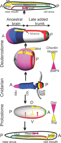
FIGURE 1.1 Understanding evolution explains how Cnidarians, with polarized Wnt expression (green) and the ‘heart’ (Nkx, lilac) is transformed independently to generate a ring-to-rod transformation by downregulation of BMP2/4 in Deuterostomes. This contrasts with a ventral midline (red, Sog) that results a dorsal signaling (green, Dpp) in Protostomes. Cnidarians start with Wnt expression at the posterior part, followed by Bra, Gsc, Otx, and Nkx (most anterior). Deuterostome development follows the Spemann organizer that will induce a symmetry break by Wnt and suppression of BMP2/4 to generate a strip of ectoderm that produces the brain and spinal cord, followed by convergent extension. Earliest expression of heart genes (Nkx) is anterior to the head in hagfish and migrates ventrally during vertebrate development; a master reorganization consistent with heart homology between Deuterostomes and Cnidarians. Note the unique opening of the neuropore (NP) in chordates. Nearly identical genes are present among Protostomes, but they have a different expression of Dpp (the BMP homolog): In contrast to being downregulated in Deuterostomes under dorsal expressing genes (Chordin, Noggin, lilac), they are upregulated from dorsal Dpp (lilac, Protostomes) and specify the dorsal expression of the heart. The A/P is different, flipping the old mouth in Protostomes into the old anus in Deuterostomes. (Modified from Layden, 2019; Meinhardt, 2015b.)
Development from a single cell to a complex adult individual foremost requires establishing the two body axes: antero-posterior and dorso-ventral (Meinhardt, 2015b). Several genes are involved in this two-step process (Figure 1.1): First, factors define the antero-posterior organization. Wnt signaling has been shown to be a key component in establishing the antero-posterior axis across various organisms, including Cnidarians (Hobmayer et al., 2000; Meinhardt, 2015b). Also, Hox genes become expressed in a time-dependent, segmental pattern along the antero-posterior axis to further specify regions along the length of this axis (Wacker et al., 2004). Second, additional factors establish the dorso-ventral organization. Bilaterally-symmetrical protostomes and deuterostomes express Chordin and Noggin dorsally, which inhibit the expression of BMPs (Meinhardt, 2015a). In addition, BMPs antagonize the expression of Chordin (Lele et al., 2001; Xue et al., 2014). Thus, Chordin and Noggin define dorsal and BMPs define ventral. In protostomes, Chordin and BMP are referred to as short gastrulation (Sog) and decapentaplegic (Dpp), respectively (Meinhardt, 2015a). Thus in Drosophila and other protostomes, ventral identity is defined by Sog and dorsal by Dpp (Heingård and Janssen, 2020). Like Chordin, Sog is an antagonist of Dpp expression (Akiyama-Od...