
eBook - ePub
Surface Electromyography
Physiology, Engineering, and Applications
Roberto Merletti, Dario Farina, Roberto Merletti, Dario Farina
This is a test
Partager le livre
- English
- ePUB (adapté aux mobiles)
- Disponible sur iOS et Android
eBook - ePub
Surface Electromyography
Physiology, Engineering, and Applications
Roberto Merletti, Dario Farina, Roberto Merletti, Dario Farina
Détails du livre
Aperçu du livre
Table des matières
Citations
À propos de ce livre
Reflects on developments in noninvasive electromyography, and includes advances and applications in signal detection, processing and interpretation
- Addresses EMG imaging technology together with the issue of decomposition of surface EMG
- Includes advanced single and multi-channel techniques for information extraction from surface EMG signals
- Presents the analysis and information extraction of surface EMG at various scales, from motor units to the concept of muscle synergies.
Foire aux questions
Comment puis-je résilier mon abonnement ?
Il vous suffit de vous rendre dans la section compte dans paramètres et de cliquer sur « Résilier l’abonnement ». C’est aussi simple que cela ! Une fois que vous aurez résilié votre abonnement, il restera actif pour le reste de la période pour laquelle vous avez payé. Découvrez-en plus ici.
Puis-je / comment puis-je télécharger des livres ?
Pour le moment, tous nos livres en format ePub adaptés aux mobiles peuvent être téléchargés via l’application. La plupart de nos PDF sont également disponibles en téléchargement et les autres seront téléchargeables très prochainement. Découvrez-en plus ici.
Quelle est la différence entre les formules tarifaires ?
Les deux abonnements vous donnent un accès complet à la bibliothèque et à toutes les fonctionnalités de Perlego. Les seules différences sont les tarifs ainsi que la période d’abonnement : avec l’abonnement annuel, vous économiserez environ 30 % par rapport à 12 mois d’abonnement mensuel.
Qu’est-ce que Perlego ?
Nous sommes un service d’abonnement à des ouvrages universitaires en ligne, où vous pouvez accéder à toute une bibliothèque pour un prix inférieur à celui d’un seul livre par mois. Avec plus d’un million de livres sur plus de 1 000 sujets, nous avons ce qu’il vous faut ! Découvrez-en plus ici.
Prenez-vous en charge la synthèse vocale ?
Recherchez le symbole Écouter sur votre prochain livre pour voir si vous pouvez l’écouter. L’outil Écouter lit le texte à haute voix pour vous, en surlignant le passage qui est en cours de lecture. Vous pouvez le mettre sur pause, l’accélérer ou le ralentir. Découvrez-en plus ici.
Est-ce que Surface Electromyography est un PDF/ePUB en ligne ?
Oui, vous pouvez accéder à Surface Electromyography par Roberto Merletti, Dario Farina, Roberto Merletti, Dario Farina en format PDF et/ou ePUB ainsi qu’à d’autres livres populaires dans Scienze biologiche et Biotecnologia. Nous disposons de plus d’un million d’ouvrages à découvrir dans notre catalogue.
Informations
1
Physiology of Muscle Activation and Force Generation
R. M. Enoka1 and J. Duchateau2
1Department of Integrative Physiology, University of Colorado, Boulder, Colorado
2Laboratory of Applied Biology and Neurophysiology, ULB Neuroscience Institute (UNI), Université Libre de Bruxelles (ULB), Brussels, Belgium
1.1 Introduction
To extract information about the control of movement by the nervous system from electromyographic (EMG) signals, it is necessary to understand the processes underlying both the generation of the activation signal and the torques exerted by the involved muscles. As a foundation for the subsequent chapters in this book, the goal of this chapter is to describe the physiology of muscle activation and force generation. We discuss the anatomy of the final common pathway from the nervous system to muscle, the electrical properties of motor neurons and muscle fibers, the contractile properties of muscle fibers and motor units, the concept of motor unit types, and the control of muscle force by modulating the recruitment and rate coding of motor unit activity.
1.2 Anatomy of a Motor Unit
The basic functional unit of the neuromuscular system is the motor unit. It comprises a motor neuron, including its dendrites and axon, and the muscle fibers innervated by the axon [28]. The motor neuron is located in the ventral horn of the spinal cord or brain stem where it receives sensory and descending inputs from other parts of the nervous system. The axon of each motor neuron exits the spinal cord through the ventral root, or through a cranial nerve in the brain stem, and projects in a peripheral nerve to its target muscle and the muscle fibers it innervates. Because the generation of an action potential by a motor neuron typically results in the generation of action potentials in all of the muscle fibers belonging to the motor unit, EMG recordings of muscle fiber action potentials provide information about the activation of motor neurons in the spinal cord or brain stem.
1.2.1 Motor Nucleus
The population of motor neurons that innervate a single muscle is known as a motor nucleus or motor neuron pool [51]. The number of motor neurons in a motor nucleus ranges from a few tens to several hundred [40,58] (Table 1.1). The motor neuron pool for each muscle typically extends longitudinally for a few segments of the spinal cord (Fig. 1.1), and at each segmental level the pools for proximal muscles tend to be more ventral and lateral than those for distal muscles and the pools for anterior muscles are more lateral than those for posterior muscles [59]. Nonetheless, the extensive dendritic projections of motor neurons intermingle across motor neuron pools.
Table 1.1 Motor Neuron Locations and Numbers for Selected Forelimb Muscles
| Muscle | Spinal Location | Number |
| Biceps brachii | C5–C7 | 1051 |
| Triceps brachii | C6–T1 | 1271 |
| Flexor carpi radialis | C7–C8 | 235 |
| Extensor carpi radialis | C5–C7 | 890 |
| Flexor carpi ulnaris | C7–T1 | 314 |
| Extensor carpi ulnaris | C7–T1 | 216 |
| Extensor pollicis longus | C8–T1 | 14 |
| Abductor pollicis longus | C8–T1 | 126 |
| Flexor digitorum superficialis | C8–T1 | 306 |
| Extensor digitorum communis | C8–T1 | 273 |
| Flexor digitorum profundus | C8–T1 | 475 |
| Extensor digiti secundi proprius | C8–T1 | 87 |
| Abductor pollicis brevis and flexor pollicis brevis | C8–T1 | 115 |
| Adductor pollicis | C8–T1 | 370 |
| First dorsal interosseus | C8–T1 | 172 |
| Lateral lumbricalis | C8–T1 | 57 |
| Data are from Jenny and Inukai [58] and are listed as pairs of antagonistic muscles. | ||
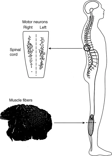
Figure 1.1 Muscle force is controlled by a population of motor units (motor unit pool) located in the spinal cord with each motor unit innervating a number of muscle fibers (muscle unit). The muscle fibers belonging to a single muscle unit are indicated by the white dots in the cross-sectional view of the muscle. A typical motor unit pool spans several spinal segments, and muscle units are usually limited to discrete parts of the muscle. Modified from Enoka [30] with permission.
1.2.2 Muscle Fibers
The muscle fibers innervated by a single axon are known as the muscle unit (Fig. 1.1), the size of which varies across each motor unit pool. The motor units first recruited during a voluntary contraction innervate fewer muscle fibers and hence have smaller muscle units than those that are recruited later in the contraction. Most motor units in a muscle have small muscle units and only a few have large muscle units [76,102,107] (Fig. 1.2A). Based on the association between muscle unit size and maximal motor unit force, Enoka and Fuglevand [32] estimated the innervation numbers (muscle unit size) for the 120 motor units in a human hand muscle (first dorsal interosseus) ranged from 21 to 1770 (Fig. 1.2B). Similar relations likely exist for most muscles [51]. Due to the exponential distribution of innervation number across a motor unit pool, it is necessary to distinguish between the number of motor unit action potentials discharged from the spinal cord and the number of muscle fiber action potentials recorded in the muscle with EMG electrodes. This distinction is indicated with the term “neural drive” to denote the motor unit action potentials and “muscle activation” to indicate the muscle fiber action potentials [26,31,36].
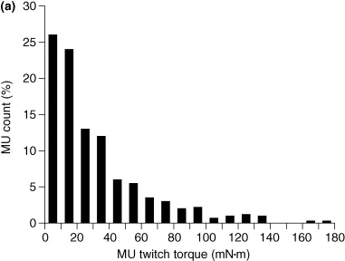
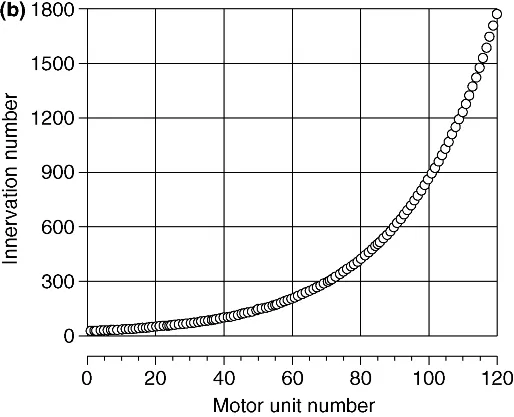
Figure 1.2 Variation in muscle unit size across the motor unit pool. (a) Distribution of motor unit (MU) twitch torques for 528 motor units in the tibialis anterior muscle of 10 subjects [107]. (b) Estimated distribution of innervation numbers across the 120 motor units comprising the first dorsal interosseus muscle [32].
The fibers in each muscle unit are located in a subvolume of the muscle and intermingle with the fibers of other muscle units (Fig. 1.1). The spatial distribution of the fibers belonging to a muscle unit is referred to as the motor unit territory. Counts of muscle unit fibers indicate that motor unit territories can occupy from 10% to 70% of the cross-sectional area of a muscle and that the density of muscle unit fibers ranges from 3 to 20 per 100 muscle fibers [51]. Moreover, the fibers of a single muscle unit often do not extend from one end of the muscle to the other, but instead terminate within a muscle fascicle [48,108]. As a consequence of muscle unit anatomy, the forces generated by individual muscle fibers must be transmitted through various layers of connective tissues before reaching the skeleton and contributing to the movement. Such interactions attenuate the unique contribution of individual fibers to the net muscle force during a movement and thereby reduce the influence of differences in contractile properties among muscle fibers.
1.3 Motor Neuron
The motor unit is classically considered to be the final common pathway in that sensory and descending inputs converge onto a single neuron that discharges an activation signal to the muscle fibers it innervates [28]. The motor neuron has extensive dendritic branches that receive up to 50,000 synaptic contacts with each contact capable of eliciting inward or outward currents across the membrane...