Biological Sciences
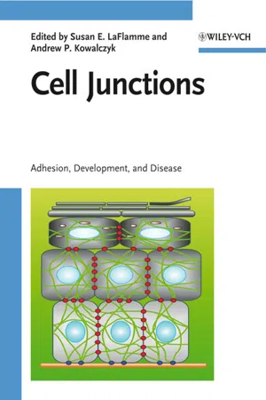
Cell Junctions
Cell junctions are specialized structures that connect neighboring cells in tissues. They play a crucial role in maintaining tissue integrity, communication between cells, and controlling the passage of molecules. There are several types of cell junctions, including tight junctions, gap junctions, and adherens junctions, each with specific functions in cell adhesion, signaling, and barrier formation.
Written by Perlego with AI-assistance
Related key terms
1 of 5
11 Key excerpts on "Cell Junctions"
 eBook - PDF
Cell Junctions
Adhesion, Development and Disease
- Susan E. La Flamme, Andrew P. Kowalczyk, Susan E. La Flamme, Andrew P. Kowalczyk(Authors)
- 2008(Publication Date)
- Wiley-Blackwell(Publisher)
Cells interact with one another and with the extracellu- lar matrix (ECM) through a variety of mechanisms. Often, large macromolecular complexes assemble at sites of contact in order to perform specific functions, including adhesion, communication, and to form barriers between tissue compart- ments. These complex molecular assemblies – or “Cell Junctions” – are now thought of as cellular organelles that not only mediate physical interactions, but also couple cell contact to signaling pathways that influence cell shape, cell-cycle progression, and gene expression. The manifestation of these various functions becomes appar- ent in model organisms in which genes encoding cell junction proteins have been ablated, and in the growing list of human disorders that are now known to result from gene mutations in junction components. Indeed, the phenotypes and clinical presentations of these genetic and acquired disorders are impressive in both scope and variety. The chapters of this book cover a wide range of cell–cell and cell–matrix interac- tions. The first section of the book focuses on cell–matrix interactions, including focal adhesions and hemidesmosomes. Embedded within these chapters are dis- cussions of integrin activation and cytoskeletal interactions, as well as signaling mediated by integrins and their cytoplasmic binding partners. The importance of the physical three-dimensional structure of the ECM in regulating cell behavior is underscored. The second section of the book concentrates on cell–cell interactions, including anchoring junctions such as adherens junctions and desmosomes, which utilize cadherins as the major adhesive molecules. Tight junctions and gap junctions are also highlighted, with unique insights provided into the roles of these proteins in complex epithelia and disease pathogenesis. eBook - ePub
- John P. Bilezikian, Lawrence G. Raisz, T. John Martin(Authors)
- 2008(Publication Date)
- Academic Press(Publisher)
Chapter 21. Intercellular Junctions and Cell–Cell Communication in the Skeletal System
Introduction
The organization of cells in tissues and organs is controlled by molecular programs that afford cells the ability to recognize other cells and the extracellular matrix and to communicate with their neighbors. Adhesive interactions are essential not only in embryonic development, but also in a variety of other biologic processes, including the differentiation and maintenance of tissue architecture and cell polarity, the immune response and the inflammatory process, cell division and death, tumor progression and metastases (Goodenough et al., 1996; Simon and Goodenough, 1998 ; Vleminckx and Kemler, 1999 ;Zbar et al., 2004). Cell–cell and cell–matrix adhesion are mediated by four major groups of molecules: cadherins, immunoglobulin-like molecules, integrins, and selectins. Cadherins are an integral part of adherens junctions, which along with tight junctions and desmosomes, constitute the so-called anchoring junctions, which join cells by anchorage through their cytoskeletons (Halbleib and Nelson, 2006 ). A different type of intercellular junction is the gap junction, which does not provide cell anchorage but allows direct communication via specialized intercellular channels (Goodenough et al., 1996). In addition to cell–cell adhesion and gap junc-tional communication, cell-to-cell propagation of locally generated signals, such as mechanically induced “calcium waves” can occur via short-range intercellular signaling systems that require either gap junctions or extracellular release of nucleotides and activation of purinergic receptors (Jørgensen et al., 1997).In the adult skeleton, bone remodeling occurs via repeated sequences of bone resorptive and formative cycles, which require continuous recruitment and differentiation of bone marrow precursors. The cooperative nature of bone remodeling requires efficient means of intercellular recognition and communication that allow cells to sort and migrate, synchronize their activity, equalize hormonal responses, and diffuse locally generated signals. Likewise, cell–cell interactions are critical for aggregation and condensation of immature chondro-osteoprogenitor cells and mesenchymal precursors during skeletal development (bone modeling). This chapter reviews current knowledge about the role of direct cell–cell interactions in the development and remodeling of the skeletal tissue, focusing on cell–cell adhesion via cadherins, cell–cell communication via gap junctions, and short-range calcium signals, or calcium waves, via extracellular nucleotides and purinergic receptors. eBook - PDF
- Alasdair Steven, Wolfgang Baumeister, Louise N. Johnson, Richard N. Perham(Authors)
- 2016(Publication Date)
- Garland Science(Publisher)
Eukaryotic cells communicate with each other at specialized contact regions The regions where cells meet and their plasma membranes become closely apposed are known as junctions . The term synapse is also used, mainly for junctions between neural cells. The molecular compositions and structures of junctions differ from those of the rest of the plasma membrane. Typically, they have an outer layer of transmembrane receptor proteins whose ectodomains (extracellular portions) bind to the cell on the other side of the junction, while their endodomains (intracellular portions) connect to cytoplasmic proteins and, in many cases, to the cytoskeleton. In addition to their adhesive role in coordinating multicellular bodies, junctions also inform a cell about its neighbors. Intercellular communication can take place in several ways. The binding of extracellular ligands by a receptor may induce conformational changes in its cytoplasmic domain that elicit a signaling response. Small water-soluble molecules may be exchanged between coupled cells through the aqueous channels of a gap junction (Section 11.5). Cytoplasmic vesicles may discharge their contents into the extracellular milieu, to be detected by receptors on the other side of the synapse (Section 16.3). Mechanical forces may also transmit signals between cells via the junctions. The cells that form the outer surface of an organism must be integrated cohesively to prevent the loss of material to the outside and the ingress of foreign materials, including microbial pathogens. Accordingly, peripheral cells are well endowed with junctions, of which there are four main kinds: tight junctions , adherens junctions , desmosomes , and gap junctions . There are also two kinds of junctions— focal adhesions and hemidesmosomes —that connect cells to the extracellular matrix (ECM), a network of secreted proteins and polysaccharides. eBook - ePub
Ultrastructural Pathology of the Cell and Matrix
A Text and Atlas of Physiological and Pathological Alterations in the Fine Structure of Cellular and Extracellular Components
- Feroze N. Ghadially(Author)
- 2013(Publication Date)
- Butterworth-Heinemann(Publisher)
The relatively constant interval (15–20nm wide) and the parallel orientation of the apposed lateral surfaces of such cells suggest the presence of a cohesive force operating over these surfaces. Often such apposed surfaces are amplified by the formation of a few or many complex interdigitating folds which probably play an additional role in cell-to-cell adhesion. Besides evidence of such a generalized mechanism, one also finds focal areas of specialization of the cell surface which are thought to represent zones of firmer attachment.Such specialized areas of cell surface serving to bind that surface to another cell surface or non-cellular structure are referred to as junctions. Many morphological and functional varieties of junctions have been described. While the sole function of some seems to be the provision of sites of firm attachment, others also act as watertight seals or as areas of low electrical resistance across which ions can flow. The structure of such junctions and their variations in normal and pathological states form the topic of this chapter.Structure and function of Cell Junctions
Although our knowledge about Cell Junctions stems largely from ultrastructural studies, it is worth recalling that the presence of some junctions can be appreciated with the light microscope.Perhaps the best-known example of this is the so-called ‘intercellular bridge’ found between the prickle cells of the epidermis. Such bridges present as fine processes or fibrils traversing a clear space between adjacent cells, and it is this which gives the prickly appearance to prickle cells. The belief that such bridges represent tonofibrils crossing over from the cytoplasm of one cell into the next one, or that the cytoplasm of neighbouring cells is continuous at such sites is not borne out by electron microscopy. It would appear that this phenomenon is largely a shrinkage artefact, the bridges being no more than plaques or zones of firm attachment (in the past referred to as ‘granules of Ranvier’ or ‘nodes of Bizzozero’, and now as desmosomes) which become drawn out as the cells shrink away from each other during fixation (see Plate 468 ).Plate 468 Fig. 1 . This Diagram Schematizes The Differences Between Desmosomes And Desmosome-Like Structures Which Are Fully Explained In the Text.Fig. 2 eBook - PDF
- Gerald Karp, Janet Iwasa, Wallace Marshall(Authors)
- 2021(Publication Date)
- Wiley(Publisher)
In addition to these adhesive junc- tions, epithelial cells often contain other types of Cell Junctions, called tight junctions and gap junctions, that will be discussed later in the chapter (see Sections 7.5 and 7.6). Adherens junctions are found in a variety of sites within the body. They are particularly common in epithelia, such as the lining of the intestine, where they occur as “belts” (or zonu- lae adherens) that encircle each cell near its apical surface, binding that cell to its surrounding neighbors (Figure 7.25). In an adherens junction, cells are held together by calcium- dependent linkages formed between the extracellular domains (b) AJ TJ D From Eveline E. Schneeberger and Robert D. Lynch, ECM Desmosome Intermediate filaments Hemidesmosome Tight junction Actin filaments Adherens junction Gap junction (a) Source: (b) From Eveline E. Schneeberger and Robert D. Lynch, Am. J. Physiol. 262:l648, 1992. © The American Physiological Society (APS). All rights reserved. FIGURE 7.25 An intercellular junctional complex. (a) Schematic diagram showing the junctional complex on the lateral surfaces of a simple columnar epithelial cell. The complex consists of a tight junction (zonula occludens), an adherens junction (zonula adherens), and a desmosome (macula adherens). Other desmosomes and gap junctions are located deeper along the lateral surfaces of the cells. Adherens junctions and tight junctions encircle the cell, whereas desmosomes and gap junctions are restricted to a particular site between adjacent cells. Hemidesmosomes are shown at the basal cell surface. (b) Electron micrograph of a junctional complex between two rat airway epithelial cells (TJ, tight junction; AJ, adherens junction; D, desmosome). 7.4 Interactions of Cells with Other Cells 293 of cadherin molecules that bridge the 30–nm gap between neighboring cells (Figure 7.26). eBook - PDF
- D. A. Cadenhead, J. F. Danielli, M. D. Rosenberg, D. A. Cadenhead, J. F. Danielli, M. D. Rosenberg(Authors)
- 2013(Publication Date)
- Academic Press(Publisher)
This is not to say that the genetic constitution of the cell or its state of genetic activity is unimportant. Cultured cell lines exist which lack the capability of forming certain junctions, and of course specialized cells of a given organism may have different complements of junctions. How-ever, if a cell which is forming desmosomes rapidly shows only a local inhibition of this activity when confronted locally with a foreign cell surface, then the fault must lie with the environment and not the syn-thetic activity of the cell. The fact that localized differences at the mem-brane can be critical makes it somewhat easier to imagine how the abundantly varied junctions which have been described might develop. 202 JANE OVERTON NOTE ADDED IN PROOF Since this article went to press a thorough review of mammalian Cell Junctions has appeared [McNutt, N. S., and Weinstein, R. S. (1973). Progr. Biophys. Mol. Biol. 26, 45.] REFERENCES Adams, E. C, and Hertig, A. T. (1964). J. Cell Biol. 21, 397. Allenspach, A. L., and Roth, L. E. (1967). J. Cell Biol. 33, 179. Anderson, E., and Beams, H. W. (1960). J. Ultrastruct. Res. 3, 432. Aoki, A. (1968). Exp. Cell Res. 52, 660. Armstrong, P. B. (1970). J. Cell Biol. 47, 197. Ashhurst, D. (1970). J. Cell Biol. 46, 421. Auber, J. (1963). J. Microsc. (Paris) 2, 325. Balinsky, B. I. (1959). Exp. Cell Res. 16, 429. Balinsky, B. I., and Walther, H. (1962). Ada Embryol. Morphol. Exp. 4, 261. Barros, C, and Franklin, L. E. (1968). J. Cell Biol. 37, C13. Beaulaton, J. (1968). J. Ultrastruct. Res. 23, 516. Bellairs, R. (1963). J. Embryol. Exp. Morphol. 11, 201. Benedetti, E. L., and Emmelot, P. (1965). J. Cell Biol. 26, 299. Benedetti, E. L., and Emmelot, P. (1967). J. Cell Sci. 2, 499. Benedetti, E. L., and Emmelot, P. (1968). J. Cell Biol. 38, 15. Bennett, H. S. (1969). In Handbook of Molecular Cytology (A. Lima-de-Faria, ed.), pp. 1261-1293. North-Holland Pubi. Amsterdam. Bennett, M. eBook - PDF
The Blood-Brain Barrier and Its Microenvironment
Basic Physiology to Neurological Disease
- Elga de Vries, Alexandre Prat(Authors)
- 2005(Publication Date)
- CRC Press(Publisher)
3 Tight Junctions of the Blood–Brain Barrier Gijs Kooij, Jack van Horssen, and Elga de Vries Department of Molecular Cell Biology and Immunology, VU University Medical Center, Amsterdam, The Netherlands 1. TIGHT JUNCTIONS 1.1. Introduction Multicellular organisms are primarily required to establish a distinct internal environment to maintain homeostasis. As a result, all internal and external surfaces of organs, such as skin, stomach, and intestines, are covered with various kinds of epithelia. In order to work efficiently as a barrier, inter-cellular spaces of these epithelial sheets are strictly sealed by junctional complexes. Likewise, microvascular-associated brain endothelial cells are linked by intercellular junctions. The main structures responsible for the barrier properties are tight junctions (1,2). These intercellular structures are located at the most apical section of the plasma membrane of adjacent cells (Fig. 1A), whereas adherens junctions are found in the basal part of the cell membrane. Tight junctions also act as an intramembrane fence that pre-vents the intermixing of apical and basolateral lipids in the exocytoplasmic leaflet of the plasma membrane. Points of cell–cell contact are sites where integral proteins of two adjacent membranes meet within the cellular space (Fig. 1B). The adjoining membranes make contact at intermittent points, rather than being fused over a large surface area. The integrity/permeability of tight junctions can be assessed by transendothelial electric resistance 47 (TEER) measurements. In this chapter we describe the molecular composi-tion of tight junctions and their role in several intercellular signaling pathways and leukocyte migration. 1.2. Structure The main transmembrane molecules mediating intercellular contacts are occludin and the endothelial claudin family. These proteins bind to the cytoskeleton via the zonula occludens-1, -2, and -3 complexs (ZO-1, ZO-2, and ZO-3) (Fig. 2). eBook - PDF
Karp's Cell and Molecular Biology
Concepts and Experiments
- Gerald Karp, Janet Iwasa, Wallace Marshall(Authors)
- 2016(Publication Date)
- Wiley(Publisher)
While the subject will be explored in detail in Chapter 15, it can be noted that all four types of cell‐adhesion molecules illustrated in Figure 7.21 have the potential to carry out this function. For example, integrins and cadherins can transmit sig- nals from the extracellular environment to the cytoplasm by means of linkages with the cytoskeleton and with cytosolic regulatory mol- ecules, such as protein kinases and G proteins. Protein kinases acti- vate (or inhibit) their target proteins through phosphorylation, whereas G proteins activate (or inhibit) their protein targets through physical interaction (see Figure 15.21 b). The engagement of an inte- grin with its ligand can induce a variety of responses within a cell, including changes in cytoplasmic pH or Ca 2+ concentration, protein phosphorylation, and gene expression. These changes, in turn, can alter a cell’s growth potential, migratory activity, state of differentia- tion, or survival. 7.11 Tight Junctions: Sealing the Extracellular Space A simple epithelium, like the lining of the intestine or lungs, is com- posed of a layer of cells that adhere tightly to one another to form a cellular sheet. Biologists have known for decades that when certain types of epithelia, such as frog skin or the wall of the urinary bladder, are mounted between two compartments containing different solute concentrations, very little diffusion of ions or solutes is observed across the wall of the epithelium from one compartment to the other. Given the impermeability of plasma membranes, it is not surprising that solutes cannot diffuse freely through the cells of an epithelial layer. But why aren’t they able to pass between cells by way of the paracellular pathway (as in FIGURE 7.27a )? The reason became apparent during the 1960s with the discovery of specialized contacts, called tight junctions (or zonulae occludens), between neighboring epithelial cells. eBook - PDF
- S.K. Malhotra(Author)
- 1996(Publication Date)
- Elsevier Science(Publisher)
THE MOLECULAR COMPOSITION OF THE TIGHT JU NCTION Andrew G. Howarth and Bruce R. Stevenson I. Introduction .................................... 25 II. The Tight Junction ......... . ....................... 26 A. Morphology .................................. 26 B. Paracellular Permeability ........................... 26 C. The Fence Function ........................ ~ ..... 27 D. Molecular Components of the Tight Junction ................ 27 Acknowledgments ................................. 37 References .................................... 37 I. INTRODUCTION The junctional complex is a zone of cell-cell interaction at the apical end of the lateral plasma membrane of epithelial and endothelial cells. This complex is comprised of four distinct intercellular junctions: the tight junction (zonula oc- cludens), adherens junction, desmosome, and gap junction. While each of these junctions is unique in structure and function, similarities between them are evident. For example, all contain transmembrane proteins that mediate cell-cell contact. In Advances in Structural Biology, Volume 4, pages 25-39. Copyright 9 1996 by JAI Press Inc. All rights of reproduction in any form reserved. ISBN: 1-55938-967-2. 25 26 ANDREW G. HOWARTH and BRUCER. STEVENSON the tight junction, adherens junction, and desmosome these integral membrane proteins interact on the cytoplasmic side of the plasma membrane with a complex of peripheral membrane proteins which are likely to serve a variety of functions, including linking the transmembrane components to the cytoskeleton. Given this intercellular junction structural paradigm, this paper will examine our current understanding of the tight junction. Emphasis will be placed on the tight junction protein ZO-1, which remains the most extensively studied of the tight junction proteins. eBook - ePub
- Alasdair Steven, Wolfgang Baumeister, Louise N. Johnson, Richard N. Perham(Authors)
- 2016(Publication Date)
- Garland Science(Publisher)
Figure 11.14A ) provide the linkage. These connections are discussed further below.Figure 11.14 Molecular organization of adherens junctions and desmosomes. In both junctions, apposing cadherins interact in trans through their EC1 domains. For their packing in a native desmosome, see Figure 11.16B . Desmosomal cadherins have longer cytoplasmic tails than do classical cadherins. They bind plakoglobin (pg), plakophilin (pkp), and desmoplakin connecting to IFs.Through their interactions with the cytoskeleton, junctions confer shape and mechanical strength on cells and tissues. Adherens junctions have a vital role during tissue morphogenesis, when coordinated movements of adherent cells generate distinct organs. Desmosomes in concert with IFs enable tissues to resist physical stress. Similar junctions are also found in endothelia, cardiac muscle, and neuronal synapses. It is likely that they have specializations needed for the functioning of these particular tissues, but their core components are similar to those in the better characterized epithelial junctions. Hemidesmosomes (see Figure 11.2 ) are junctions that appear to consist of one half of a desmosome interfaced with the extracellular matrix, except that their receptor molecules are integrins, as in focal adhesions (Section 11.6), rather than cadherins.Mutations in junctional proteins—particularly in desmosomal proteins—give rise to a variety of diseases. Some skin-blistering diseases, such as epidermolysis bullosa simplex (EBS), arise from mutations in proteins that link desmosomes to keratin IFs, as well as mutations in the keratins themselves (noted in Section 11.2). Other blistering diseases such as pemphigus vulgaris are caused by autoantibodies against desmosomal cadherins. The symptoms of these conditions reflect impairment of the tissue integrity through defects in cell–cell adhesion. eBook - PDF
- Gerald Karp, Janet Iwasa, Wallace Marshall(Authors)
- 2018(Publication Date)
- Wiley(Publisher)
4 Similar observa- tions were soon made on a variety of different types of epi- thelial and mesenchymal cells, including those of various mammals, indicating that these communicating junctions are widespread. Electron microscopic studies had shown that animal cells are bounded by a continuous plasma membrane. This new data suggested that the structure of this membrane was sometimes modified at sites where cells made contact with other cells. Otherwise, it would be impossible for substances to move directly from the cytoplasm of one cell into another. The discovery of Cell Junctions containing channels between closely applied cells was made in 1967 by Jean Paul Revel and M. J. Karnovsky. 5 Electron micrographs of these junctions showed a distinct cleft between adjoining cells, which led the researchers to name them “gap junctions” to distinguish them from tight junctions, where adjoining cells make direct contact. Studies were carried out to learn more about the size of the channels connecting the cytoplasms of adjacent cells. Loewenstein’s laboratory tested fluorescent probes that were linked to peptides of varying size. They found that molecules up to about 1200 daltons were able to diffuse between insect larval salivary gland cells. 6 Based on estimates of the dimen- sions of these molecules, they concluded that the effective diameter of the channel was approximately 10 to 15 Å (1.0 to 1 5 . nm), a value that matched closely with that estimated from high‐resolution micrographs of gap junctions taken with the electron microscope. 7 One of the first questions considered by Kanno and Loewenstein following their discovery of permeable inter- cellular contacts was whether these same types of contacts were present in cancer cells. The growth rates of normal cells were known to be influenced by stimuli from their environ- ment.
Index pages curate the most relevant extracts from our library of academic textbooks. They’ve been created using an in-house natural language model (NLM), each adding context and meaning to key research topics.