Biological Sciences
Dinoflagellates
Dinoflagellates are single-celled organisms that belong to the group of algae known as phytoplankton. They are characterized by two flagella, which they use for movement. Dinoflagellates are important in marine ecosystems as they are a major component of the plankton community and play a crucial role in the marine food web. Some species of dinoflagellates are bioluminescent, creating the phenomenon known as "sea sparkle."
Written by Perlego with AI-assistance
Related key terms
1 of 5
9 Key excerpts on "Dinoflagellates"
 eBook - PDF
Seafood and Freshwater Toxins
Pharmacology, Physiology, and Detection, Third Edition
- Luis M. Botana(Author)
- 2014(Publication Date)
- CRC Press(Publisher)
16 Apart from marine toxins production, Dinoflagellates can affect the full spectrum of living systems from the biochemical to the ecosystem level. 17 For example, at the ecosystem level, high concentrations of cells may interfere with light penetration and influence subsurface communities such as submerged aquatic vegetation. At the biochemical level, secondary metabolites produced by microalgae may interfere with particular cellular processes in the organism, but may not adversely affect the organ-ism as a whole. 18 Dinoflagellate morphology can be as miscellaneous and complex as of any unicellular eukaryote. A simple definition for Dinoflagellates states that they are “eukaryotic, primarily single-celled organ-isms in which the motile cell possesses two dissimilar flagella: a ribbon-like flagellum with multiple waves which beats to the cell’s left, and a more conventional flagellum with one or a few waves which beats posteriorly.” 19 Taxonomic approach for this group has traditionally been based on cytological characters. 20 The existence of large numbers of undescribed dinoflagellate species has been shown by use of molec-ular analyses in environments like marine picoplankton 21,22 or as symbionts (“zooxanthellae”) in many types of protists and invertebrates like corals. 23 20.2 Taxonomy Molecular phylogenetic analyses place Dinoflagellates in the Alveolata kingdom, 24 together with the cili-ates and apicomplexans. 25 The Alveolata constitute a diverse group of single-celled eukaryotes present in both marine and terrestrial ecosystems, the principal shared morphological feature of which is the presence of flattened vesicles (cortical alveoli) packed into a continuous layer supporting the cell mem-brane. 26 These structures have been associated by immunolocalization to a family of proteins, named alveolins, common to all alveolates. 27,28 Alveolates exhibit extremely diverse trophic strategies, including predation, photo-autotrophy, and intracellular parasitism. eBook - ePub
Oceans and Human Health
Risks and Remedies from the Seas
- Patrick J. Walsh, Sharon Smith, Lora Fleming, Helena Solo-Gabriele, William H. Gerwick(Authors)
- 2011(Publication Date)
- Academic Press(Publisher)
CHAPTER 13 Toxic DinoflagellatesKAREN A. STEIDINGER, JAN H. LANDSBERG, LEANNE J. FLEWELLING, BARBARA A. KIRKPATRICKINTRODUCTION
Dinoflagellates are single-celled microorganisms (5 μm to typically less than 1 mm) that live in aquatic environments from the Arctic to the Antarctic. Most of them are free living, but some are symbiotic (e.g., in coral species) and even parasitic on fish or in other Dinoflagellates. Half of the described species are primary producers with photosynthetic pigments. This means they contribute to the world’s atmospheric oxygen and produce organic carbon that enters the base of the oceans’ food webs. Primary production is a critical process that allows the seas to yield crops high in protein for human consumption. Phytoplankton, including Dinoflagellates, also produce (during photosynthesis) over 70% of the oxygen we breathe. The other half of Dinoflagellates species are “heterotrophic.” It is not known how many of the photosynthetic (or “auxotrophic”) species are also mixotrophic or heterotrophic, but this mode of nutrition can be advantageous (Stoecker et al ., 2006).At some point in their life cycle, Dinoflagellates are motile and can “swim” about 1 meter per hour, and therefore they can move or disperse within the water column. They have a characteristic nucleus (with condensed chromosomes) and other features (e.g., pigments and sterols) that separate them from other phytoplankton and microalgae. They are classified as protists in the Division Dinoflagellata (Fensome et al ., 1993), and typically the International Code of Botanical Nomenclature is used for this group of about 1500 to 2000 species (Gomez, 2005 ; Taylor, 1990 eBook - PDF
Marine Natural Products V1
Chemical And Biological Perspectives
- Poul Schever(Author)
- 2012(Publication Date)
- Academic Press(Publisher)
Prorocentrum micans Exuviaella marina a Based on Loeblich and Loeblich (1970). after cell division, forming united cell lines of four or more. Formerly, Dinoflagellates were classified as a phylum of protozoan, mostly because of their mobility, but in many respects they are more properly classified as algae. However, it should be noted that in some cases they are still placed in the animal category (e.g., Halstead, 1965). Classification of Dinoflagellates is accompanied by difficult problems due to the fact that they exist in different forms—motiles and cysts. A great number of dinoflagellate fossils are found only in cyst form, which makes it difficult to compare them with living motile organisms. Dinoflagellates comprise the division Pyrrhophyta (Table 1) and as such are separate from other algal phyla—Cyanophyta (blue-green), Chlorophyta (green), Phaeophyta (brown), and Rhodophyta (red). The Pyrrhophyta are further divided into 15 orders and 50 families. The total number of living dinoflagellate species is said to be over 400, most of which live in the marine environment (Sarjeant, 1974). Unlike blue-green algae or bacteria, which lack nuclei (prokaryotic), Dinoflagellates possess nuclei, but their chromosomes are very primitive and their concentration of nuclear proteins (histones) is remarkably low (Rizzo and Nooden, 1972). For these reasons, it was proposed that Dinoflagellates are evolutionary links between the prokaryotes such as the blue-green algae and eukaryotes such as the green or brown algae, and should, therefore, be called mesokaryotic (Dodge, 1965). In any event they have an interesting position on the evolutionary map. 4 Yuzuru Shimizu B. Blooms (Red Tide) The commonly used term red tide is not preferred by most experts in the field. The term arose from the fact that a massive number of organisms often appeared as red streaks spread on the surface of the water. eBook - ePub
Studies in Natural Products Chemistry
Bioactive Natural Products
- Atta-ur Rahman(Author)
- 2020(Publication Date)
- Elsevier(Publisher)
Chapter 4Marine Dinoflagellates as a source of new bioactive structures
Arnaud Cousseaua , b ; Raffaele Sianoc ; Ian Probertd ; Stéphane Bachb ; Mohamed Mehiria , ⁎a University Nice Côte d’Azur, CNRS, Nice Institute of Chemistry, UMR 7272, Marine Natural Products Team, Nice, Franceb Sorbonne Université, CNRS, UMR 8227, Integrative Biology of Marine Models (LBI2M), Physiology and Cell Fate Team, Station Biologique, Roscoff, Francec Ifremer, Center of Brest, French Institute of Research for the Exploitation of the Sea, Dynamics of Coastal Ecosystems—DYNECO/Pelagos, Plouzané, Franced UPMC, CNRS, FR2424, Roscoff Culture Collection—RCC, Station Biologique, Roscoff, France⁎ Corresponding author: email address: [email protected]Abstract
Dinoflagellates are an extremely diverse group of unicellular eukaryotes that represent an important part of marine plankton communities. Among other secondary metabolites, these microorganisms are known to produce toxins such as the saxitoxins, karlotoxins, amphidinolides, okadaic acid or brevetoxins, which are extremely toxic for marine fauna and ultimately humans who consume contaminated seafood. The extreme biodiversity of Dinoflagellates undoubtedly mirrors an inexhaustible reservoir of compounds, yet the chemodiversity of Dinoflagellates is still poorly studied, due in part to constraints linked to the difficulty of isolating and cultivating these organisms. Nevertheless, an increasing number of new dinoflagellate secondary metabolites have been described in recent years. Here, we review these new advances in the isolation and characterization of bioactive metabolites from several Dinoflagellates (Gymnodiniales, Amphidinium, Karlodinium, Karenia), Peridiniales (Vulcanodinium), Gonyaulacales (Alexandrium, Gambierdiscus, Ostreopsis), Prorocentrales (Prorocentrum) and Dinophysiales (Dinophysis). eBook - ePub
Freshwater Algae of North America
Ecology and Classification
- John D. Wehr, Robert G. Sheath, J. Patrick Kociolek(Authors)
- 2015(Publication Date)
- Academic Press(Publisher)
Chapter 17Dinoflagellates
Susan Carty1 ; Matthew W. Parrow21 Department of Biological and Environmental Sciences, Heidelberg University, Tiffin, Ohio, USA.2 Department of Biological Sciences, University of North Carolina at Charlotte, Charlotte, North Carolina, USAAbstract
Dinoflagellates are a group of unicellular protists that can be identified using the light microscope, and are (usually) recognized by their golden-brown plastids, assimilative cell with indented waist, distinctive swimming pattern, and relatively large nucleus that contains visible chromosomes. Shared characteristics of the group include: two dissimilar flagella in motile cells; an unusual genome consisting of liquid, crystal-like chromosomes; and a haplontic life cycle that often contains a non-motile, dormant cyst stage. Freshwater Dinoflagellates are diverse and may be phototrophic, predatory, or both (mixotrophic). They can be found in most aquatic habitats throughout the seasons; although flagship taxa, such as Ceratium , Peridinium , and Gymnodinium, are typically found as phytoplankton in calm, lentic waters during warmer months. This chapter updates taxonomy (extensive splitting of the genera Peridinium , Peridiniopsis , and Woloszynskia ), identification of genera via microscopy, and adds molecular information on taxa and group systematics where available.Keywords Dinoflagellate Dinophyceae Ceratium Gymnodinium PeridiniumChapter ContentsI.Introduction773II.Diversity and Morphology776A. Anatomy 776B. Life Cycle 779C. Classification 781III.Ecology and Distribution783A. Dinoflagellate Blooms 783B. Trophic States 786C. Specificity of Habitat 787D. Geographic Distribution 787IV.Collection and Preparation for Identification790A. Collection 790B. Fixation 790C. Preparation for Identification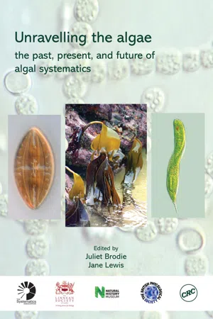 eBook - PDF
Unravelling the algae
the past, present, and future of algal systematics
- Juliet Brodie, Jane Lewis, Juliet Brodie, Jane Lewis(Authors)
- 2007(Publication Date)
- CRC Press(Publisher)
226 Acknowledgments .......................................................................................................................... 227 References ...................................................................................................................................... 227 ABSTRACT Cytologically, Dinoflagellates constitute a very aberrant protist group. Molecular data indicate that the most primitive extant species are heterotrophic and belong to the Oxyrrhinales (free-living) and Syndiniales (parasitic). The remaining species usually form a single clade in the phylogenetic trees based on ribosomal DNA sequences, but their relative relationships are poorly resolved. Classifi-cation at generic, family, and order levels is presently undergoing major changes, and characters traditionally used to characterize genera (thecate or non-thecate, position of the cingulum, distance between the two ends of the cingulum, plate formula, etc.) are being replaced with features supported by both molecular and ultrastructural data (eyespot structure, path and construction of the “apical furrow” system, details of the flagellar apparatus, etc.). Dinoflagellate chloroplasts have arisen by at least eight independent symbioses, and some of these have resulted in stable symbioses, while others are kleptoplastidic. INTRODUCTION D EFINITION OF THE GROUP The Dinoflagellates is a group of unicellular or colony-forming protists comprising approximately 2000 extant and a similar number of extinct species. The great majority (perhaps 80%) are free-living, marine, planktonic, or benthic flagellates, while 20% are from similar habitats in freshwater. 216 Unravelling the algae: the past, present and future A small number are parasitic (e.g. in copepods) or symbiotic (e.g. in corals). Estimation of species numbers is difficult as the taxonomy needs revision.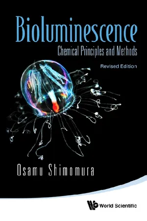 eBook - PDF
Bioluminescence: Chemical Principles And Methods (Revised Edition)
Chemical Principles and Methods
- Osamu Shimomura(Author)
- 2012(Publication Date)
- World Scientific(Publisher)
CHAPTER 8 Dinoflagellates and Other Protozoa The diverse group Protozoa consists of microscopic single-celled organisms. Of the various protozoan types, only Radiolaria and Dinoflagellata are known to contain luminous organisms (Harvey, 1952; Herring, 1978). 8.1 Radiolarians Radiolarians are either solitary or colonial, and the colonial forms often grow to very large aggregates. Harvey (1926b) demonstrated that radi-olarians Thalassicolla and Collozoum can luminesce in deoxygenated seawater containing platinized asbestos through which pure hydrogen has been passed for 45 min. The experiment indicated that these organ-isms are able to emit light even in the complete absence of oxygen, resembling hydromedusae, the scyphozoan Pelagia , and ctenophores. Campbell et al. (1981) isolated a Ca 2 + -sensitive photoprotein from Thalassicolla sp., and named it thalassicollin. An unidentified solitary species and a colonial species (probably Sphaerozoum sp.) were also found to contain a Ca 2 + -sensitive photoprotein (Campbell and Her-ring, 1990). Emission maximum of the Ca 2 + -triggered luminescence of thalassicollin was found at 440 nm (Campbell and Herring, 1990) in close agreement with the in vivo luminescence of Thalassicolla ( λ max 450 nm; Herring, 1983; Latz et al. , 1987). Radiolarian photopro-teins are of interest as they are the only known examples of Ca 2 + -sensitive photoprotein other than the coelenterate (and ctenophore) photoproteins. 257 258 Bioluminescence: Chemical Principles and Methods 8.2 Dinoflagellates Dinoflagellates are distributed worldwide and are almost ubiquitous in the surface water of the sea. The so-called phosphorescence of the sea is most commonly due to the bioluminescence of Dinoflagellates (such as Noctiluca , Gonyaulax and Pyrocystis ). The microscopic organ-isms of Dinoflagellates emit sparkling luminescence in short flashes. eBook - PDF
- Susan Carty(Author)
- 2014(Publication Date)
- Comstock Publishing Associates(Publisher)
Hydrobiologia 105:1–26. Hanisak, M.D. 1973. An ecological survey of the phytoplankton of the Pettaquam-sett River, Rhode Island. MS thesis, Univ. R.I., Kingston. Hansen, G. 1993. Light- and electron microscopical observations on the dinoflagel-lates Actiniscus pentasterias (Dinophyceae). J. Phycol. 29:486–499. Hansen, G. 1995. Analysis of the thecal plate pattern in the dinoflagellate Hetero-capsa rotundata (Lohmann) comb. nov. ( Katodinium rotundatum (Lohmann) Loeblich). Phycologia 34 (2): 166–170. Hansen, G., and N. Daugbjerg. 2004. Ultrastructure of Gyrodinium spirale, the type species of Gyrodinium (Dinophyceae), incuding a phylogeny of G. dominans, G. rubrum and G. spirale deduced from partial LSU rDNA sequences. Pro-tist 155:271–294. Hansen, G., Ø. Moestrup, and K.R. Roberts. 2000. Light and electron microscopi-cal observations on the type species of Gymnodinium, G. fuscum (Dinophyceae). Phycologia 39 (5): 365–376. Hansen, K. 1967. The general limnology of Arctic lakes as illustrated by examples from Greenland. Medd. Grønland 178:51–60. Happach-Kasan, C. 1982. Beobachtungen zum Bau der Theka von Ceratium cornutum (Ehrenb,) Clap. et Lachm. (Dinophyta). Arch. Protistenkd. 125:181–207. Hargraves, P.E., and R.M. Víquez. 1981. Dinoflagellate abundance in the Laguna Botos, Poás Volcano, Costa Rica. Rev. Biol. Trop. 29:257–264. Harris, T.M. 1940. A contribution to the knowledge of the British freshwater dinoflagellata. Proc. Linn. Soc. Lond. 152nd session, 1939–1940, Part 1: 4–33. Hashimoto, Y., T. Okaichi, L.D. Dang, and T. Noguchi. 1968. Glenodine, an ichthyotoxic substance produced by a dinoflagellate, Peridinium polonicum. Bull. Jap. Soc. Sci. Fish. 34 (6): 528–534. Hassall, A.H. 1845. A history of the British freshwater algae including descriptions of the Desmideae and Diatomaceae. S. Highly and H. Balliere, London. Hayhome, B.A., and L.A. Pfiester. 1983. Electrophoretic analysis of soluble enzymes in five freshwater dinoflagellate species. eBook - PDF
- David L. Spector(Author)
- 2012(Publication Date)
- Academic Press(Publisher)
2 Dinoflagellate T a x o n o m y 3 7 P H Y L U M (o r division) : Centrodinium Kofoi d Corythodinium Loeblic h an d Loeblic h Oxytoxum Stei n Roscoffia Balec h Micracanthodinium Deflandr e Palaeophalocroma Schille r Blepharocysta Ehrenber g Podolapmas Stei n Thoracosphaera Kamptne r Dissodinium Kleb s Haplozoon Dogie l Blastodinium Chatto n Oodinium Chatto n Paulsenella Chatto n Chytriodinium Chatto n Myxodinium C a c h o n e tal. Pyrocystis M u r r a y P Y R R O P H Y T A Family O x y t o x a c e a e L i n d e m a n n Fusiform cells , h y p o t h e c a n o r m a l l y large r tha n epithec a an d covere d b y o n l y fiv e o r six plate s Family C l a d o p y x i d a c e a e Stei n Cel l o v o i d a l w i t h spine s arisin g f r o m cente r o f theca l plate s o r girdl e lackin g lowe r lis t Family Podolampacea e L i n d e m a n n G i r d le an d sulcu s no t incise d an d n o list s present . Theca l plate s thick , antapica l horn s presen t Orde r Thoracosphaerale s Tange n M a r i ne planktoni c organism s i n w h i c h th e p r e d o m i n a n t c o c c o i d phas e i s covere d b ya calcareou s shell . G y m n o d i n i o i d motil e stag e Family Thoracosphaeracea e Schille r Character s o f th e orde r Orde r Blastodiniale s Schille r M o t i le g y m n o d i n i o i d cells . N o r m a l l y n o theca l plates . Parasiti c stag e o n protozoan s o r metazoan s i s mai n par t o f lif e histor y Family Dissodiniaceae f a m .
Index pages curate the most relevant extracts from our library of academic textbooks. They’ve been created using an in-house natural language model (NLM), each adding context and meaning to key research topics.