Biological Sciences
Ion Channel Receptors
Ion channel receptors are specialized proteins found in the cell membrane that allow the passage of ions, such as sodium, potassium, and calcium, into and out of the cell. They play a crucial role in regulating the electrical activity of cells and are involved in processes such as neurotransmission, muscle contraction, and hormone secretion. These receptors are important targets for drug development in various medical conditions.
Written by Perlego with AI-assistance
Related key terms
1 of 5
12 Key excerpts on "Ion Channel Receptors"
 eBook - PDF
- Ulo Langel, Benjamin F. Cravatt, Astrid Graslund, N.G.H. von Heijne, Matjaz Zorko, Tiit Land, Sherry Niessen(Authors)
- 2009(Publication Date)
- CRC Press(Publisher)
Cells express receptors that operate through different signaling mechanisms applied by these different receptors, and cross talk often occurs between intracel-lular signaling pathways. Hence, the individual signaling mechanisms of particular receptors are highly regulated and integrated by cross talk, which explains the high complexity of how the cell surface receptors function. 23.1.1 I ON C HANNELS IUPHAR defines 9 subclasses of ion-channel and ligand-gated Ion Channel Receptors, such as the Cys-loop superfamily (anion channels like GABA A R and cation channels like nAChR), glutamate-gated cation channels (NMDAR and non-NMDAR), epithe-lial Na + channels, voltage gated cation channels (Na + , Ca 2 + , K + ), chloride channels, ATPase-linked transporters, and transporters related to neurotransmitters. 23.1.1.1 Structural Features of Ion Channel Receptors Receptors in this class all contain homologous polypeptides expressed by homolo-gous genes or resulting from alternative splicing and, hence, an array of receptors Cell Surface Receptors and Signaling 325 can be constructed using different combinations of these gene products. Often, these receptors contain subunit structures, each of these subunits comprising of 2–12 trans-membrane domains, depending on the receptor type. The assembly of the structure can yield an oligomer surrounding a membrane pore. The subclasses of transmitter-gated ion channels vary largely in the membrane topologies of their subunits. Nicotinic acetylcholine (nACh) receptors (Figure 23.1A) have historically served as a source of structural information for this class of receptors because of their high content in skeletal muscles and electric organs. nAChR have important role in neuronal signal transmission, nociception, learning, memory, and addiction. The secondary structure of Torpedo nAChR is presented in the atomic-scale model at 4 Å resolution in Figure 23.1A. eBook - ePub
Hormonal Signaling in Biology and Medicine
Comprehensive Modern Endocrinology
- Gerald Litwack(Author)
- 2019(Publication Date)
- Academic Press(Publisher)
Within the brain, the computational and transductional potential of neuronal cells, or if we regard their interaction as a dialogue, their ability to listen, understand, and to speak, is reflected, and to a great extent determined, by the array of ion channels they express, their topographical distribution, and their time, voltage, and signal-dependent properties. Indeed, taking this conversational analogy further, it is ion channels and their dynamic regulation that allow the mammalian brain to function as biology's most complicated expression of the game North American children call “telephone”: who says what to whom, what of that is understood, and finally to whom and how accurately is that message or signal conveyed, i.e. an adaptive process of information flow and transmission fidelity. With this analogy, it becomes easy to see how, by altering ion channel activity in function-specific neuronal networks, hormones can have dramatic effects upon signal processing, circuit performance, and ultimately how the brain communicates with peripheral structures to match the physiological requirements dictated by our intero- and exteroceptive perceptions.Why and when? If we are to properly appreciate the functional impact of ion channel activation, then we must first understand why and when this happens. The principle utility of ion channels arises from their ability to provide a rapid, energetically efficient means for ionic species to traverse biological membranes. Unlike the much slower transporter proteins, or ionic pumps, which engage in a process of active transport that requires a source of chemical energy (e.g., ATP), channels allow the passive diffusion of specific ionic species through a membrane-spanning aqueous pore. Yet, what causes the ions and consequently current to flow?Why? Two principal forces act upon ions to drive them through open ion channels. One is diffusional, arising from the unequal distribution of ions either side of the membrane; and one is coulombic, arising from the resultant separation of charge. Accordingly, for a given ionic valence, and a given concentration gradient, there will be a membrane potential at which these forces are equal and opposite, and beyond which there will be a net flow of ions in the opposite direction. This so-called reversal potential (ERev ) was first described at the end of the 19th century by the German chemist Walter Nernst and for a given set of conditions is derived from the equation that bears his name (Nernst, 1889 ). Correspondingly, each ion, or ions, carrying the charge through an ion channel will have a characteristic ERev , and the opening of said ion channel will serve to drive membrane potential toward this value, thus determining whether the activation of a specific ion channel is an excitatory or an inhibitory event. For example, under physiological conditions, the cation K+ accumulates in high concentrations intracellularly and comparatively low concentrations extracellularly, so the membrane potential sufficient to draw this positive ion against its concentration gradient and into the cell is extremely negative, giving channels that conduct K+ ions a reversal potential approaching − 100mV. Accordingly, at membrane potentials positive to this value, activation of K+ No longer available |Learn more
- Wendell A. Lim, Wendell Lim, Bruce Mayer, Tony Pawson(Authors)
- 2014(Publication Date)
- Garland Science(Publisher)
GATED CHANNELS A further major class of signaling receptors is the gated ion channels, which respond to ligands or other environmental cues by altering the permeability of the membrane to ions or other small molecules. Such gated channels play a fundamental role in neurotransmission, opening in response to neurotransmitters, membrane depolarization, or other stimuli. These channels allow specific ions to flood rapidly across the membrane, causing massive and very rapid changes in the electrical properties of the membrane, the basis for electrical signals. Because of their importance for neurotransmission, such channels are the targets of a number of familiar drugs. For example, local anesthetics such as novocaine block voltage-gated sodium channels, while calcium channel blockers (CCBs) are used to treat high blood pressure. Gated channels also regulate intracellular signaling, as in the case of calcium channels on the endoplasmic reticulum that open when bound to inositol trisphosphate, leading to the release of intracellular calcium stores. In this section, we will not deal with the electrophysiology of neuronal signaling, but will consider a few examples of gated channels that illustrate the mechanisms of their regulation by stimuli and their selectivity. Gated channels share a similar overall structure All gated channels, irrespective of the stimuli that regulate them or the solutes that they conduct, share similarities in overall topology. All are composed of multiple similar or identical subunits arranged in a ring structure in the plane of the membrane ( Figure 8.19a ). Depending on the specific channel, this ring may be composed of two to five subunits (with four or five subunits being the most common arrangement), though in some cases all subunits are combined in a single polypeptide chain. Each subunit can consist of anywhere from two to six hydrophobic α heli-ces that span the membrane, connected by loops of varying lengths. eBook - ePub
- Michael F. Roberts, Anne E. Kruchten, Michael S. Roberts(Authors)
- 2016(Publication Date)
- Wiley-VCH(Publisher)
Part III Receptor Types and FunctionTransduction: Introduction to the Four Receptor Types
Hormone binding is just the first step in the process of cell activation. Depending on the receptor type, many kinds of additional steps may be involved. The process by which the binding event is transmitted to the interior of the cell is calledThe next chapter begins the second section of this book: a discussion of the receptors themselves, along with the mechanisms by which they activate cells:signal transduction, and though it was proposed theoretically by Langley in 1906, it has taken until the past two decades to unravel its varied molecular mechanisms.- Ion Channel Receptors allow transmembrane ion exchanges – Chapter 6 .
- G-protein-coupled receptors activate membrane enzymes – Chapter 7 .
- Growth factor receptors activate an enzyme that is part of the receptor – Chapter 8 .
- Nuclear receptors initiate gene transcription and protein synthesis – Chapter 9 .
 Passage contains an image
The constancy of the internal environment is the condition for the free and independent life … Claude Bernard [152] “… you seem to me both in your appearance and in your power over others to be very like the flat torpedo fish, who torpifies those who come near him and touch him, as you have now torpified me …”
Passage contains an image
The constancy of the internal environment is the condition for the free and independent life … Claude Bernard [152] “… you seem to me both in your appearance and in your power over others to be very like the flat torpedo fish, who torpifies those who come near him and touch him, as you have now torpified me …”Chapter 6 Transduction I: Ion Channels and Transporters
Plato: Meno [153]6.1 Introduction
Cell membranes separate the cytoplasm, where metabolism occurs, from the source of metabolic nutrients in the extracellular fluid. Thus, small molecules used for metabolism do not have easy access to cells. Indeed, because of the nonpolar nature of cell membranes, ions and other polar molecules are unable to pass into or out of cells without the aid of some sort of molecular device. Transmembrane (TM) movements are actually accomplished by two types of membrane molecules – transporters and channels.
- Randy W. Beck(Author)
- 2011(Publication Date)
- Churchill Livingstone(Publisher)
B ) A typical single-pass transmembrane protein. Note that the polypeptide chain transverses the lipid bilayer as a right-handed α helix and that the oligosaccharide chains and disulfide bonds are on the noncytosolic surface of the membrane. Disulfide bonds do not form between the sulfhydryl groups in cytoplasmic domain of the protein because the reducing environment in cytosol maintains these groups in their reduced (–SH) form.All receptors for chemical transmitters have three things in common:- 1. They are membrane-spanning proteins in which the external portion of the protein recognises and binds a specific neurotransmitter. Some common neurotransmitters include acetylcholine (ACh), norepinephrine (NE), epinephrine (E), serotonin or 5-hydroxytryptophan (5-HT), and dopamine (DA).
- 2. They carry out an effector function within the target cell. This function may include regulation of specific ion channels, release or activation of second messenger compounds, or modulation of activity levels of intracellular enzymes.
- 3. It is the receptor that determines the action of the transmitter based on the activity it produces inside the cell. This is an important point to remember. Many neurotransmitters are classified as excitatory or inhibitory to certain cellular functions; however, it is the internal wiring of the receptors that determine the response of a transmitter. For example, acetylcholine has an inhibitory or slowing effect on the heart rate but an excitatory effect on skeletal muscle.
Receptors can be either directly or indirectly linked to ion channels
These two different types of linkage are determined by two different genetic programming families of receptors.Receptors that gate ion channels directly are called inotropic receptors. Upon binding of a transmitter, the receptor undergoes a conformational change that allows the ion channel to open. The receptor is part of the same molecular structure that composes the channel. The activation of inotropic receptors produces fast synaptic actions (milliseconds in duration), e.g. ACh receptor at the neuromuscular junction (Fig. 3.3 eBook - PDF
- M.G. Ord, L.A. Stocken(Authors)
- 1997(Publication Date)
- Elsevier Science(Publisher)
Chapter 6 TALKING TO CELLS-CELL MEMBRANE RECEPTORS AND THEIR MODES OF ACTION Robin F. Irvine Introduction 173 Membrane Receptors 175 Coupling of Receptors to Intracellular Signals 182 Acknowledgments 196 References 196 INTRODUCTION This chapter is essentially about the field of research that we now know as signal transduction or cellular signaling. Currently this field comprises a significant proportion of the world's total research in the life sciences. This is not surprising if one thinks about it. The cells of our tissues are under the constant control of hormones, neurotransmitters, and growth factors, which are telling the cells to do this, do that, stop doing this, do that instead, etc. The great majority of these outside influences—agonists is a useful all-embracing term—are water-soluble. They have to be because they move and work in an aqueous environment. So, when they come up against the hydrophobic cell membrane (plasma membrane) of any cell, they must either be taken up into the cell by an active process (e.g. endocytosis, active transport) which is necessarily slow, or they must bind to a specific recognition site (a receptor) in the plasma membrane, which then registers their presence by sending a chemical signal into the cell. Signal transduction, therefore, is all 173 174 ROBIN F. IRVINE about the nature of these chemical messages, how they are generated after the receptor has bound its ligand (the agonist), and how the cell uses them to alter its function. Because these receptor-generated signals plug-into and modulate the homeostatic control mechanisms of a cell's functions, it is inevitable that in understanding receptor-mediated signal transduction we will understand the fundamentals of cellular function.
- A. Kleinzeller(Author)
- 2012(Publication Date)
- Elsevier Science(Publisher)
II. Receptor-mediated transmembrane signalling Although the principal function of the cell membrane is to maintain an effective barrier between the extracellular and intracellular milieu, there are highly specialized membrane-localized constituents {e.g. ion channels, nutrient transporters MEMBRANE RECEPTORS 195 and pharmacologic receptors) that can be singled out as play-ing particularly pivotal roles in terms of selectively transmit-ting information from the external to the internal cellular en-vironment (and in some cases, vice versa). The pharmacologic receptors situated in the plasma membrane possess not only the ability to recognize extracellular hormonal signalling mol-ecules with high affinity and specificity, but also the capacity, once combined with the specific ligand, to transmit informa-tion across the cell membrane, so as activate intracellular sig-nalling pathways. The possibility of transmitting information from surface receptors to intracellular sites was visualized early on by Peters (1937) who was impressed by Clark's obser-vations (1937). It is this dual recognition-transmembrane sig-nalling property, by which the receptor per se acts as a mes-sage generating system, that distinguishes receptors from other cell surface recognition/transport constituents. The sec-tions below will, with selected examples, focus on the general mechanisms whereby cell surface receptors generate a trans-membrane signal. Receptors for agonists that act via intracel-lular receptors (e.g. steroid hormones) will not be discussed. 1. GENERAL MECHANISMS OF TRANSMEMBRANE SIGNALLING AND THE MOBILE RECEPTOR PARADIGM To fulfil their recognition/action function, membrane recep-tors must be able to generate an intracellular signal that can be greatly amplified. eBook - PDF
- Peter M. Haddad, David J. Nutt(Authors)
- 2020(Publication Date)
- RCPsych Publications(Publisher)
The movement of sodium ions through the channel is controlled by metabotropic receptors which are linked to G-proteins in the neuronal membrane. The opening of the ion channel follows the neurotransmitter-activated G-protein complex which, by a cascade of secondary messengers (such as cAMP and cGMP) and the protein kinases, leads to the phosphorylation of the sodium channel. This opens the channel to permit the repolarization of the neuron. The diagram also indicates how some drugs and toxins can close the channel by direct action and thereby inactivate it. Apart from some antiepileptic drugs, this is not a frequent action of psychotropic drugs. 74 Part 1: Basic Science and General Principles protein structure to be modified by point mutations. By changing the structure of the protein by even a single amino acid it is now apparent that the properties of the ion channel also change, resulting, for example, in the opening and closing of the channel for longer or shorter periods of time or in carrying larger or smaller currents. As a consequence of molecular biological studies, it is now recognized that most ion channels of importance in neurotransmission are composed of three to five protein subunits. Their identification and characterization have now made it possible to map their location on specific neurons and to correlate their location with their specific function. There are two major types of receptor which are activated by neurotransmitters. These are the ionotropic and metabotropic receptors. The former receptor type is illustrated by the amino acid neurotransmitter receptors for glutamate, gamma- aminobutyric acid (GABA) and glycine, and the acetylcholine receptors of the nicotinic type. These are examples of fast transmitters in that they rapidly open and close the ionic channels in the neuronal membrane.
- Nicoladie Tam(Author)
- 0(Publication Date)
- Nicoladie Tam, Ph.D.(Publisher)
It is a channel, which is opened by a molecule binding with the receptor. It is a receptor molecule with a channel pore, which opens up (or closes down) when a ligand binds to it.When the channel of a receptor is opened by the binding with a molecule (such as a neurotransmitter or a drug), it is called a ligand-gated channel. A ligand is a molecule that binds with a receptor. Most of the post-synaptic receptors are ligand-gated channel called ionotropic receptors, although some are metabotropic receptors, which do not have any channel in it.Examples of ligand-gated channels are glutamate receptors, acetylcholine receptors, GABA receptors and serotonin receptors. Drug action in the CNS usually refers to the binding to these receptors. What is an ionic channel? It is a molecular channel with a pore in the middle. They usually are membrane-bound, and the pore spans the membrane connecting the inside and outside of a cell.Examples of ion channels are Na+ -channels, K+ -channels, Ca++ -channels and Cl- -channels.These are large macromolecules with three-dimensional structure, which forms a central molecular tunnel. They are a long-chain molecule of amino-acid sequence in two-dimension, if unwound. This long-chain molecule usually loops in and out of a membrane four or more times, forming a three-dimensional helical structure of a channel.What is a voltage-gated channel? It is a channel, which is opened by a change in the membrane potential rather than binding of a molecule with the receptor. It is a receptor molecule with a channel pore, which opens up (or closes down) by a voltage change, not by binding with a ligand.The Na-channel or K-channel in the axon of a neuron, which generate action potentials are voltage-gated channels. They do not have any binding site in it. Other examples of voltage-gated channels are Ca++ -channels and Cl-
- Efraim Racker(Author)
- 2012(Publication Date)
- Academic Press(Publisher)
Reconstitution studies could clarify the relationship between these phosphorylating systems. III. Channel Receptors 189 I am including in this category some of the antibody receptors that signal important biological responses. Probably the best character-ized example in this group is the IgE receptor. Like the RGC recep-tors, it appears to be a multisubunit complex with a recognition unit and an effector unit. The purified complex has been incorporated into liposomes (Metzger, 1983), and a partially purified multisubunit recognition protein has been incorporated by polyethylene glycol-induced membrane fusion between vesicles and rat basophilic leuke-mia cells, as measured by ligand-triggered release of radioactive serotonin (Estes et al., 1985). An important advance in the under-standing of the effector unit has been the isolation of a protein that binds the anti-asthmatic drug cromolyn. The latter blocks the ligand-triggered increase of Ca 2 + influx and degranulation in some basophilic cells. The cromolyn-binding protein was incorporated into a mutant cell line with an impaired chromolyn-binding capacity by using Sendai virus envelopes as fusogenic carriers. This proce-dure restored the IgE-mediated response of increased Ca 2 + influx and degranulation (Mazurek et al., 1983). More recently the cromo-lyn-binding protein has been reconstituted into liposomes and incor-porated into planar lipid bilayers together with other membrane components of rat basophilic cells. By cross-linking, IgE character-istic channel conductance was observed in the presence of Ca 2 + (Mazurek et al., 1984). III. Channel Receptors A. The Nicotinic Acetylcholine Receptor (AChR) When an action potential depolarizes a cholinergic nerve terminal, acetylcholine is released. It diffuses across the synaptic cleft and binds to the AChR in the postsynaptic membrane. It is the function of the receptor to respond to this interaction with a conformational change that opens an ion channel.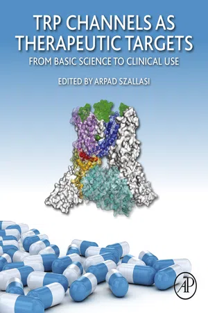 eBook - ePub
TRP Channels as Therapeutic Targets
From Basic Science to Clinical Use
- Arpad Szallasi(Author)
- 2015(Publication Date)
- Academic Press(Publisher)
Chapter 1An Introduction to Transient Receptor Potential Ion Channels and Their Roles in Disease
Michael J. Caterina1 , 2 , 3 , 4 , *1 Department of Neurosurgery, Johns Hopkins School of Medicine, Baltimore, Maryland, USA2 Department of Biological Chemistry, Johns Hopkins School of Medicine, Baltimore, Maryland, USA3 Solomon H. Snyder Department of Neuroscience, Johns Hopkins School of Medicine, Baltimore, Maryland, USA4 Neurosurgery Pain Research Institute, Johns Hopkins School of Medicine, Baltimore, Maryland, USA* Corresponding author: [email protected]Abstract
The transient receptor potential (TRP) cation channel family consists of seven subfamilies that are widely expressed in mammalian tissues. By mediating flux of calcium, sodium, and other cations across cell membranes, in addition to nonionic signaling mechanisms, these channels contribute to many sensory and nonsensory processes throughout the body. Abnormalities in TRP channel function, whether a consequence of mutations in their sequence, alterations in their expression levels, or changes in their myriad regulators, have been associated with numerous disease states ranging from chronic pain to cardiovascular disease, skeletal abnormalities, and cancer. Such prevalent involvement in disease stems not only from the ubiquity of TRP channels but also from their complex pattern of polymodal gating. The connection between TRP channels and disease creates numerous opportunities for therapeutic intervention at these channels, whether through inhibition, activation, or co-opting of their ability to transport cations to alter the course of pathophysiological processes. eBook - PDF
Channels, Carriers, and Pumps
An Introduction to Membrane Transport
- Wilfred D. Stein, Thomas Litman(Authors)
- 2014(Publication Date)
- Academic Press(Publisher)
This often leads to an alteration in protein activity. Certain of these kinases are themselves activated by cAMP or by Ca 2 1 , two substances that act as cellular “second messengers” in the mechanism by which the cell interprets the arrival of a hormonal signal into a change in the metabolic activity of the cell. (See Box 7.7 for an example of a Ion Channels Across Cell Membranes Chapter 3 | 119 “second messenger” affecting the activity of a chloride channel, a process impaired in this case in the hereditable disease, cystic fibrosis.) Ionic chan-nels are, in many cases, targets for the modulatory action of these kinases. The nAChR can be phosphorylated, and this phosphorylation is correlated with the desensitization of the receptor. Calcium ions can directly modulate the activity of ion channels. We saw an example of this when we considered the gap-junction channel in the previous section, but there are numerous other examples. Many cell membranes contain a potassium channel that is opened when the cal-cium concentration of the cell rises from its normal level of less than 1 μ M toward the level of 100 μ M. This calcium-activated potassium channel may play a role in the regulation of cell volume, encouraging osmotically active potassium ions to flow out of the cell, thereby reducing the volume. We might also mention a class of channels that are modulated by the ten-sion at the cell surface. Some of these channels are opened by the stretching of the cell surface; others are closed. These channels may also be involved in the control of the volume of the cell. (We consider volume regulation fur-ther in Section 7.1.) Finally, we must mention a family of membrane-associated proteins, the G proteins, which are vitally concerned in the modulation of cellular processes as a response to incoming signals. They can modulate ionic channels in important ways. One of these G proteins is transducin .
Index pages curate the most relevant extracts from our library of academic textbooks. They’ve been created using an in-house natural language model (NLM), each adding context and meaning to key research topics.