Physics
Magnetic Resonance Imaging
Magnetic Resonance Imaging (MRI) is a non-invasive medical imaging technique that uses strong magnetic fields and radio waves to generate detailed images of the internal structures of the body. It is particularly useful for visualizing soft tissues, such as the brain, muscles, and organs, and is widely used in clinical diagnosis and research.
Written by Perlego with AI-assistance
Related key terms
1 of 5
12 Key excerpts on "Magnetic Resonance Imaging"
 eBook - PDF
Biomagnetics
Principles and Applications of Biomagnetic Stimulation and Imaging
- Shoogo Ueno, Masaki Sekino, Shoogo Ueno, Masaki Sekino(Authors)
- 2018(Publication Date)
- CRC Press(Publisher)
121 C H A P T E R 5 Principles of Magnetic Resonance Imaging Masaki Sekino, Norio Iriguchi, and Shoogo Ueno 5.1 INTRODUCTION Magnetic Resonance Imaging (MRI) is a way of producing cross-sectional images of the human body noninvasively. A proton (or hydrogen nucleus) is spinning with a positive charge, and it is generating a magnetic flux in the direction of the Ampere’s law. Therefore, protons can be regarded as CONTENTS 5.1 Introduction 121 5.2 Magnetic Resonance Signal and Relaxation Times 123 5.2.1 Electromotive Force 123 5.3 Overview of the Spin-Warp Imaging Technique 127 5.3.1 Recognition of Nuclei Distributed in the First Direction 128 5.3.2 Recognition of Nuclei Distributed in the Second Direction 129 5.3.3 Recognition of Nuclei Distributed in the Third Direction 131 5.3.4 k Space 133 5.3.5 Image Contrast 135 5.4 Diversification of MRI Application Techniques 137 5.4.1 Magnetic Resonance Angiography 137 5.4.2 Perfusion and Diffusion Imaging 138 5.4.3 Functional Neuroimaging 140 5.4.4 Magnetic Resonance Spectroscopy 140 5.4.5 MRI of Other Nuclei than Protons 141 5.5 Concluding Remarks 142 References 142 122 ◾ Biomagnetics small, spinning magnets with north poles and south poles in Newtonian physics. When a part of the body is exposed to a static magnetic field, each spinning magnet makes a precession around the axis of the static mag-netic field. If a radiofrequency (rf) wave is then transmitted, some of the magnets begin to make a precession with the same phase of the transmit-ted rf wave. This is the magnetic resonance (MR). In the resonating state, a large number of magnets establish a large magnetic flux rotating around the axis of the static magnetic field at the frequency of the transmitted rf wave. The rf wave is then turned off, and subsequently an electrical coil tuned at the proper rf frequency picks up the electromotive force (EMF) before the rotating magnetic flux decays in amplitude. No longer available |Learn more
- (Author)
- 2014(Publication Date)
- Learning Press(Publisher)
By comparison, the first human X-ray image was taken in 1895. Magnetic Resonance Imaging is a development of nuclear magnetic resonance. Originally, the technique was referred to as nuclear Magnetic Resonance Imaging (NMRI). However, because the word nuclear was associated in the public mind with ionizing radiation exposure, it is generally now referred to simply as MRI. Scientists still use the term NMRI when discussing non-medical devices operating on the same principles. The term magnetic resonance tomography (MRT) is also sometimes used. ___________________________ WORLD TECHNOLOGIES ___________________________ How MRI works The body is largely composed of water molecules. Each water molecule has two hydrogen nuclei or protons. When a person goes inside the powerful magnetic field of the scanner, the magnetic moments of some of these protons changes, and aligns with the direction of the field. In an MRI machine a radio frequency transmitter is briefly turned on, producing an electromagnetic field. The photons of this field have just the right energy, known as the resonance frequency, to flip the spin of the aligned protons in the body. As the intensity and duration of application of the field increase, more aligned spins are affected. After the field is turned off, the protons decay to the original spin-down state and the difference in energy between the two states is released as a photon. It is these photons that produce the electromagnetic signal that the scanner detects. The frequency the protons resonate at depends on the strength of the magnetic field. As a result of conservation of energy, this also dictates the frequency of the released photons. The photons released when the field is removed have an energy — and therefore a frequency — due to the amount of energy the protons absorbed while the field was active. It is this relationship between field-strength and frequency that allows the use of nuclear magnetic resonance for imaging. No longer available |Learn more
- (Author)
- 2014(Publication Date)
- Orange Apple(Publisher)
________________________ WORLD TECHNOLOGIES ________________________ Chapter- 5 Magnetic Resonance Imaging Para-sagittal MRI of the head, with aliasing artifacts (nose and forehead appear at the back of the head) Magnetic Resonance Imaging ( MRI ), nuclear Magnetic Resonance Imaging ( NMRI ), or magnetic resonance tomography ( MRT ) is a medical imaging technique used in radiology to visualize detailed internal structures. MRI makes use of the property of Nuclear magnetic resonance (NMR) to image nuclei of atoms inside the body. An MRI machine uses a powerful magnetic field to align the magnetization of some atoms in the body, and radio frequency fields to systematically alter the alignment of this magnetization. This causes the nuclei to produce a rotating magnetic field detectable by the scanner—and this information is recorded to construct an image of the scanned area of the body. Strong magnetic field gradients cause nuclei at different locations to rotate at different speeds. 3-D spatial information can be obtained by providing gradients in each direction. MRI provides good contrast between the different soft tissues of the body, which make it especially useful in imaging the brain, muscles, the heart, and cancers compared with other medical imaging techniques such as computed tomography (CT) or X-rays. Unlike CT scans or traditional X-rays MRI uses no ionizing radiation. ________________________ WORLD TECHNOLOGIES ________________________ How MRI works The body is largely composed of water molecules. Each water molecule has two hy-drogen nuclei or protons. When a person goes inside the powerful magnetic field of the scanner, the magnetic moments of some of these protons changes, and aligns with the direction of the field. In an MRI machine a radio frequency transmitter is briefly turned on, producing an electromagnetic field. eBook - PDF
- K. Kirk Shung, Michael Smith, Benjamin M.W. Tsui(Authors)
- 2012(Publication Date)
- Academic Press(Publisher)
C H A P T E R 4 Magnetic Resonant Imaging = The phenomenon of nuclear magnetic resonance (NMR) discovered by F. Block and E. Purcell in 1946 has become a standard spectroscopic technique in chem-istry and physics. For this discovery, Block and Purcell were awarded the Nobel Prize in 1952. More recently NMR has been applied as an imaging technique pio-neered by P. Lauterbur (1973), P. Mansfield (1973), and R. Damadian (1971). Nuclear Magnetic Resonance Imaging, abbreviated MRI in the clinical field, has the advantages of being able to penetrate bony and air-filled structures with negli-gible attenuation and artifact. The modality uses non-ionizing radiation and is minimally invasive. It is capable of providing excellent soft-tissue contrast with imaging in any arbitrary plane. Although formerly used primarily for examination of the brain and spinal cord, the development of rapid imaging techniques has ex-tended the role of NMR imaging to the chest and abdomen where motion had previously been a limitation. The recent development of flow imaging provides the possibility of performing NMR angiography. Moreover, with the use of spec-troscopy and functional imaging it is capable of yielding information about the physiological state of the tissue. I. Fu a e tal s o f Nuclea r Ma eti c Reso a ce Like many forms of spectroscopy NMR utilizes electromagnetic radiation to probe the fundamental properties of matter. In NMR the radiation employed is in the radio-frequency (RF) portion of the electromagnetic spectrum, and the prop-erty studied is the interaction of this radiation with the nuclear magnetic moment. An understanding of the principles of nuclear Magnetic Resonance Imaging is founded on principles of quantum and classical mechanics. 213 214 C H A P T E R 4 Magnetic Resonance Imaging A. A ula r Mo e tu An object with a mass, m orbiting about an axis with velocity of v, as shown in Fig. No longer available |Learn more
- (Author)
- 2014(Publication Date)
- College Publishing House(Publisher)
________________________ WORLD TECHNOLOGIES ________________________ Chapter 7 Magnetic Resonance Imaging Sagittal MR image of the knee ________________________ WORLD TECHNOLOGIES ________________________ Para-sagittal MRI of the head, with aliasing artifacts (nose and forehead appear at the back of the head) Magnetic Resonance Imaging ( MRI ), nuclear Magnetic Resonance Imaging ( NMRI ), or magnetic resonance tomography ( MRT ) is a medical imaging technique used in radiology to visualize detailed internal structures. MRI makes use of the property of Nuclear magnetic resonance (NMR) to image nuclei of atoms inside the body. An MRI machine uses a powerful magnetic field to align the magnetization of some atoms in the body, and radio frequency fields to systematically alter the alignment of this magnetization. This causes the nuclei to produce a rotating magnetic field detectable by the scanner—and this information is recorded to construct an image of the scanned area of the body. Strong magnetic field gradients cause nuclei at different locations to rotate at different speeds. 3-D spatial information can be obtained by providing gradients in each direction. MRI provides good contrast between the different soft tissues of the body, which make it especially useful in imaging the brain, muscles, the heart, and cancers compared with other medical imaging techniques such as computed tomography (CT) or X-rays. Unlike CT scans or traditional X-rays MRI uses no ionizing radiation. How MRI works The body is largely composed of water molecules. Each water molecule has two hydro-gen nuclei or protons. When a person goes inside the powerful magnetic field of the scanner, the magnetic moments of some of these protons changes, and aligns with the direction of the field. In an MRI machine a radio frequency transmitter is briefly turned on, producing an electromagnetic field. eBook - PDF
- Andrew Webb(Author)
- 2022(Publication Date)
- Wiley-IEEE Press(Publisher)
189 5 Magnetic Resonance Imaging 5.1 General Principles of MRI Acquisition and Hardware Magnetic Resonance Imaging (MRI) is a modality which produces a spatial map of the hydrogen nuclei in the particular body part being scanned. These hydrogen nuclei occur primarily in tissue water (∼70% of body weight) and lipid. The sig- nal intensity in each voxel in the MR image depends on many different physical characteristics of the tissue, including the number of hydrogen nuclei within the voxel, the tissue viscosity, how fast water diffuses in the tissue, and whether there is blood flow or perfusion. This complicated signal intensity dependence has both advantages and disadvantages. On the one hand, it means that there is a wealth of information on tissue integrity and health that can be analyzed. On the other hand, it means that a single contrast image is difficult to interpret on its own, and therefore a typical MRI scanning protocol might acquire between four and eight different contrasts, which makes the entire scanning session last on average ∼30 min. The major uses of MRI are in the areas of neurological disease, spinal disorders, and musculoskeletal damage. The main strengths of MRI as a clinical imaging modality include: (i) It uses electromagnetic waves which are nonionizing, (ii) Images can be acquired in any three-dimensional orientation (including oblique scans), (iii) Soft tissue contrast is very high, (iv) High spatial resolution images (<1 mm) can be acquired, and (v) Image resolution and signal intensity do not depend on the depth of the tissue within the body, i.e. there are no penetration effects. Introduction to Biomedical Imaging, Second Edition. Andrew Webb. © 2023 The Institute of Electrical and Electronics Engineers, Inc. Published 2023 by John Wiley & Sons, Inc. Companion website: www.wiley.com/go/webb2e No longer available |Learn more
- (Author)
- 2014(Publication Date)
- Research World(Publisher)
________________________ WORLD TECHNOLOGIES ________________________ Chapter- 10 Magnetic Resonance Imaging Sagittal MR image of the knee ________________________ WORLD TECHNOLOGIES ________________________ Para-sagittal MRI of the head, with aliasing artifacts (nose and forehead appear at the back of the head) Magnetic Resonance Imaging ( MRI ), nuclear Magnetic Resonance Imaging ( NMRI ), or magnetic resonance tomography ( MRT ) is a medical imaging technique used in radiology to visualize detailed internal structures. MRI makes use of the property of Nuclear magnetic resonance (NMR) to image nuclei of atoms inside the body. An MRI machine uses a powerful magnetic field to align the magnetization of some atoms in the body, and radio frequency fields to systematically alter the alignment of this magnetization. This causes the nuclei to produce a rotating magnetic field detectable by the scanner—and this information is recorded to construct an image of the scanned area of the body. Strong magnetic field gradients cause nuclei at different locations to rotate at different speeds. 3-D spatial information can be obtained by providing gradients in each direction. MRI provides good contrast between the different soft tissues of the body, which make it especially useful in imaging the brain, muscles, the heart, and cancers compared with other medical imaging techniques such as computed tomography (CT) or X-rays. Unlike CT scans or traditional X-rays MRI uses no ionizing radiation. How MRI works The body is largely composed of water molecules. Each water molecule has two hydrogen nuclei or protons. When a person goes inside the powerful magnetic field of the scanner, the magnetic moments of some of these protons changes, and aligns with the direction of the field. In an MRI machine a radio frequency transmitter is briefly turned on, producing an electromagnetic field.
- Darin D. Dougherty, Scott L. Rauch, Jerrold F. Rosenbaum, Darin D. Dougherty, Scott L. Rauch, Jerrold F. Rosenbaum(Authors)
- 2008(Publication Date)
- American Psychiatric Association Publishing(Publisher)
21 Magnetic Resonance Imaging Martin A. Goldstein, M.D. Bruce H. Price, M.D. Technical Foundations of Nuclear Magnetic Resonance The phenomenon of nuclear magnetic resonance (NMR) was discovered in the 1940s, setting the stage for the development of Magnetic Resonance Imaging (MRI) for medical diagnostic use beginning in the 1970s (Taber et al. 2002). Extraordinary progress has since been made in expanding MRI’s applications, pro- ducing a revolutionizing force in clinical neuroscience. Although rapidly evolving methodology continues to broaden and deepen MRI’s application to research neuroscience (e.g., functional MRI), here we concen- trate on the principles and utility of MRI as they per- tain to clinical applications. A brief review of the tech- nical foundations of MRI can facilitate the technology’s proper use for optimal clinical advantage. MRI exploits the magnetic properties of the atomic constituents of biological matter to construct a visual representation of tissue. The location of the NMR sig- nal within the electromagnetic spectrum is presented in Table 2–1. Although MRI uses electromagnetic radiation, it does not involve exposure to ionizing radiation, so in general patients can safely have multiple scans without concern about aggregate radiation exposure. Table 2–1. Electromagnetic spectrum Wave type Wavelength (nm) (approximate) Frequency (Hz) (approximate) Gamma 10 –4 10 20 X ray 1 10 18 Ultraviolet 10 2 10 16 Visible 10 3 10 15 Microwave 10 8 10 10 Radio (RF), including NMR 10 10 10 5 Note. NMR = nuclear magnetic resonance; RF = radio frequency. 22 ESSENTIALS OF NEUROIMAGING FOR CLINICAL PRACTICE The degree to which a material responds to an ap- plied magnetic field is called magnetic susceptibility. Whereas most body tissues have similar susceptibili- ties, certain atoms with unpaired electrons, which are said to be paramagnetic or ferromagnetic, have signifi- cantly greater magnetic susceptibilities. No longer available |Learn more
- (Author)
- 2014(Publication Date)
- Learning Press(Publisher)
________________________ WORLD TECHNOLOGIES ________________________ Chapter-7 Magnetic Resonance Imaging (MRI) Sagittal MR image of the knee ________________________ WORLD TECHNOLOGIES ________________________ Para-sagittal MRI of the head, with aliasing artifacts (nose and forehead appear at the back of the head) Magnetic Resonance Imaging ( MRI ), nuclear Magnetic Resonance Imaging ( NMRI ), or magnetic resonance tomography ( MRT ) is a medical imaging technique used in radiology to visualize detailed internal structures. The good contrast it provides between the different soft tissues of the body make it especially useful in brain, muscles, heart, and cancer compared with other medical imaging techniques such as computed tomo-graphy (CT) or X-rays. Unlike CT scans or traditional X-rays MRI uses no ionizing radiation. Instead it uses a powerful magnetic field to align the magnetization of some atoms in the body, then uses radio frequency fields to systematically alter the alignment of this magnetization. This causes the nuclei to produce a rotating magnetic field detectable by the scanner—and this information is recorded to construct an image of the scanned area of the body. Magnetic Resonance Imaging is a relatively new technology. The first MR image was published in 1973 and the first cross-sectional image of a living mouse was published in January 1974. The first studies performed on humans were published in 1977. By com-parison, the first human X-ray image was taken in 1895. How MRI works The body is largely composed of water molecules. Each water molecule has two hydro-gen nuclei or protons. When a person goes inside the powerful magnetic field of the scanner, the magnetic moments of some of these protons changes, and aligns with the direction of the field. In an MRI machine a radio frequency transmitter is briefly turned on, producing an electromagnetic field. No longer available |Learn more
- (Author)
- 2014(Publication Date)
- Research World(Publisher)
________________________ WORLD TECHNOLOGIES ________________________ Chapter- 8 Magnetic Resonance Imaging Sagittal MR image of the knee ________________________ WORLD TECHNOLOGIES ________________________ Para-sagittal MRI of the head, with aliasing artifacts (nose and forehead appear at the back of the head) Magnetic Resonance Imaging ( MRI ), nuclear Magnetic Resonance Imaging ( NMRI ), or magnetic resonance tomography ( MRT ) is a medical imaging technique used in radiology to visualize detailed internal structures. MRI makes use of the property of Nuclear magnetic resonance (NMR) to image nuclei of atoms inside the body. An MRI machine uses a powerful magnetic field to align the magnetization of some atoms in the body, and radio frequency fields to systematically alter the alignment of this magnetization. This causes the nuclei to produce a rotating magnetic field detectable by the scanner—and this information is recorded to construct an image of the scanned area of the body. :36 Strong magnetic field gradients cause nuclei at different locations to rotate at different speeds. 3-D spatial information can be obtained by providing gradients in each direction. MRI provides good contrast between the different soft tissues of the body, which make it especially useful in imaging the brain, muscles, the heart, and cancers compared with other medical imaging techniques such as computed tomography (CT) or X-rays. Unlike CT scans or traditional X-rays MRI uses no ionizing radiation. How MRI works The body is largely composed of water molecules. Each water molecule has two hydrogen nuclei or protons. When a person goes inside the powerful magnetic field of the scanner, the magnetic moments of some of these protons changes, and aligns with the direction of the field. In an MRI machine a radio frequency transmitter is briefly turned on, producing an electromagnetic field. The photons of this field have just the right energy, known as the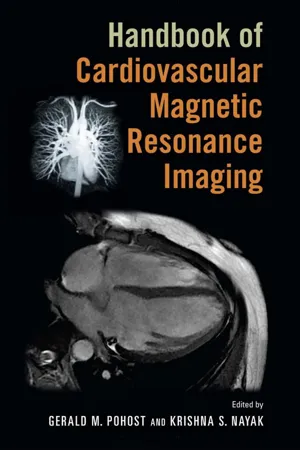
- Gerald M. Pohost, Krishna S. Nayak, Gerald M. Pohost, Krishna S. Nayak(Authors)
- 2006(Publication Date)
- CRC Press(Publisher)
1 Physical Principles of Magnetic Resonance Imaging Mark Doyle Division of Cardiology, Cardiovascular MRI Laboratory, Allegheny General Hospital, Pittsburgh, Pennsylvania, U.S.A. ORIGIN OF THE MRI SIGNAL Of all major imaging modalities, the physical origin of the magnetic reso-nance (MR) signal is quite possibly the least intuitive to comprehend. In brief, Magnetic Resonance Imaging (MRI) forms a ‘‘map’’ or ‘‘image’’ of the body’s water content. Fortunately for the modality, the body is com-posed of approximately 70% water, thus the ‘‘raw material’’ of MRI is naturally abundant. However, the body does not spontaneously generate an MRI signal, and an elaborate and complex configuration of equipment is required to access this rich signal source. In this chapter, the physical prin-ciples of MRI will be addressed in three distinct sections: the signal source, image formation, cardiovascular imaging. SIGNAL SOURCE The signal source for MRI has its origin in the discipline of nuclear magnetic resonance (NMR). The phenomenon of NMR was discovered more than 60 years ago, at which time it received widespread attention from chemists due 1 to its ability to probe the structure of molecules (1,2). Bloch and Purcell received the Nobel Prize for Physics in 1952 for their discovery of the NMR phenomenon. Physically, the NMR procedure involves placing a sample (usually in the form of a liquid contained in a test tube) in a strong uniform magnetic field and irradiating it with radiofrequency (RF) electro-magnetic energy. The atomic nuclei of the sample absorb the RF irradiation and re-emit it, forming the NMR signal. This re-emitted signal has encoded in it unique sample-specific information that reveals information concerning the molecular composition and structure of the sample. The information is encoded in the frequency and amplitude of the re-emitted RF electromag-netic signal.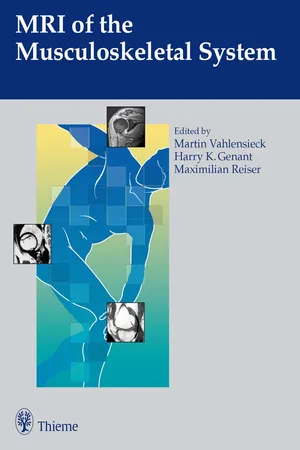 eBook - PDF
- Martin Vahlensieck, Harry K. Genant(Authors)
- 2011(Publication Date)
- Thieme(Publisher)
1 1 Technology of Magnetic Resonance Imaging M. Vahlensieck, F. Träber and J. Gieseke Introduction This chapter will present the fundamental physics un-derlying the techniques used for Magnetic Resonance Imaging (MRI) as applied to the musculoskeletal sys-tem. The discussion will be clinically oriented and fo-cused on how the different techniques affect image con-trast, signal-to-noise ratio, application and practicality. Readers who wish to learn more about the physics and techniques of imaging with magnetic resonance should refer to the appropriate literature. To produce a magnetic resonance signal, the patient is placed in a strong external magnetic field (B o ). This outer magnetic field aligns the tissue’s protons, which can be pictured as small magnets with their mag-netic fields randomly oriented, parallel to the strong field (longitudinal magnetization M z ). When a suitable high frequency pulse (90 degree pulse) is applied, the magnetic fields of the protons change their orientation, with the change measurable as so-called transverse magnetization (M xy ). After the high frequency (radio frequency [RF]) pulse is turned off, the magnetic fields of the protons return to their original orientation paral-lel to the outer magnetic field. The transverse magneti-zation rapidly decays (free induction decay, FID) at a rate dependent on the homogeneity of the external magnetic field and on the tissue itself. This time for decay is given by the constant T 2 * (star) known as the ef-fective T 2 time. Because of the rapid decay, measure-ment of the signal can only be made for a short time. To resolve this, further suitable RF pulses (180 degree pulses) can be applied producing additional signals. These are known as spin echoes (SE) and can be thought of as echoes of the original signal. They gradually decrease in intensity with time and the time constant for this decay is called the spin–spin or T 2 relaxation time.
Index pages curate the most relevant extracts from our library of academic textbooks. They’ve been created using an in-house natural language model (NLM), each adding context and meaning to key research topics.