Physics
X-Ray Imaging
X-ray imaging is a medical imaging technique that uses electromagnetic radiation to create detailed images of the inside of the body. It is commonly used to diagnose and monitor conditions such as broken bones, dental issues, and certain diseases. X-rays are able to penetrate soft tissues and produce images of dense structures like bones, making them a valuable tool in medical diagnostics.
Written by Perlego with AI-assistance
Related key terms
1 of 5
10 Key excerpts on "X-Ray Imaging"
 eBook - PDF
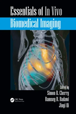
- Simon R. Cherry, Ramsey D. Badawi, Jinyi Qi, Simon R. Cherry, Ramsey D. Badawi, Jinyi Qi(Authors)
- 2016(Publication Date)
- CRC Press(Publisher)
2.2 X-Ray Imaging Basics 11 2.1 INTRODUCTION X-rays are a form of “ionizing radiation” because x-rays are energetic enough to ionize atoms and molecules during interactions. With about 10,000 times more energy than visible light photons, x-ray photons can penetrate objects including the human body. Since the first x-ray image taken in 1895 by Roentgen, X-Ray Imaging has become one of the most common diag-nostic procedures performed in medicine. The development of modern x-ray tubes and detec-tors has enabled a wide range of medical imaging applications, including x-ray radiography, x-ray fluoroscopy, and x-ray computed tomography (CT). With the capability to produce cross-sectional images, x-ray CT revolutionized traditional X-Ray Imaging and provided an invaluable diagnostic tool. The usage of CT has rapidly increased over the past two decades. In 2011, 85.3 million x-ray CT scans were performed in the United States. In addition to their role in diagnostic medicine, x-ray methods are widely used for clinical research across a broad spectrum of disease states. X-ray projection imaging and micro-CT (high-resolution x-ray CT imaging of small volumes) have also become important tools in biomedical research studies of animal models and tissue specimens. This chapter focuses on the fundamentals of X-Ray Imaging and the two major classes of X-Ray Imaging: x-ray projection imaging and x-ray CT. 2.2 X-Ray Imaging BASICS 2.2.1 X-R AY P RODUCTION AND X-R AY S PECTRUM 2.2.1.1 X-Ray Production X-ray photons used for biomedical imaging are produced from a relatively complex device, the x-ray tube. The core of an x-ray tube, called the x-ray tube insert ( Figure 2.1 ), is a vac-uum sealed by a glass or metal enclosure. Within the vacuum insert, a heated filament, the cathode , emits electrons in a process called thermionic emission. eBook - ePub
Methods and Applications of Statistics in Clinical Trials, Volume 2
Planning, Analysis, and Inferential Methods
- Narayanaswamy Balakrishnan, N. Balakrishnan(Authors)
- 2014(Publication Date)
- Wiley(Publisher)
Chapter 14 Imaging Science in Medicine, II: Basics of X-Ray Imaging Anthony B. Wolbarst, Patrizio Capasso and Andrew R. Wyant 14.1 Introduction to Medical Imaging: Different Ways of Creating Visible Contrast Among Tissues The principal job of a medical imaging modality is to provide clear maps of anatomy, or to make it possible to recognize irregularities in physiology, or both— ultimately, that is, to help find and identify pathologies. It does so by creating contrast among tissues, and the various modalities do this in biophysically diverse ways. This chapter and Chapter 15 provide extremely brief sketches of the imaging technologies that involve X rays and are employed routinely in modern clinics to examine the structure and functioning of the body. This chapter covers the modalities that were slowly developed over the first three quarters of the twentieth century, like screen-film radiography and mammography, and image-intensifier tube fluoroscopy. Chapter 15 deals with X-ray technologies that have flourished only with the advent of high-speed but affordable computers—digital planar imaging like computed radiography (CR), digital radiography (DR), digital subtraction angiography (DSA), and computed tomography (CT), culminating in helical, multi-detector ring CT. A much expanded and more in-depth version of this material may be found in the book, Medical Imaging—Essentials for Physicians, by the authors, also published by Wiley (2013). The book also discusses modalities based on gamma rays rather than X rays, including single-photon emission computed tomography (SPECT) and positron emission tomography (PET); on ultrasound waves, namely, advanced forms of B-mode and Doppler US; and on the radio waves and magnetic fields of magnetic resonance imaging (MRI) in all its glory, with T1-, T2-, and proton density-weighted imaging, functional MRI (fMRI), MR angiography (MRA), diffusion tensor imaging (DTI), and other variants eBook - PDF
- Anil Bharath(Author)
- 2022(Publication Date)
- Springer(Publisher)
3 C H A P T E R 2 Diagnostic X-Ray Imaging The contents of this chapter are, essentially, the following: • Basic principles of X-Ray Imaging: Introduction to terminology and mode of operation. • X-Ray physics: Quantum nature; X- Ray interactions with matter; X-Ray spectra; energy range for diagnostic use. • X-Ray image quality parameters: general requirements on physical parameters; image Sig- nal/Noise (S/N); image contrast. • X-Ray production: functional design requirements;tube construction; target selection. • Image receptors: functional design requirements; film-based receptors; image intensifiers. • Patient dosage: trade-offs with image quality; noise and dosage; effective dose equivalent. 2.1 BASIC PRINCIPLES OF X-Ray Imaging In the simplest case, an X-Ray Imaging system requires • X-Ray Source • A patient to image • Film (image receptor) • Radiologist/Diagnostician 2.1.1 IDEAL DESCRIPTION OF IMAGING PROCESS X-Rays are generated within the tube, and they are directed towards the patient. As the x-ray photons pass through the patient, some are absorbed, others scattered, and some pass through the patient with no interaction. The transmitted photons i.e., those which do not interact with the patient) are detected (received) by the photon receptor, usually based around film. The formation of an image on the film is dependent on the number of photons which are captured (detected) by the receptor: areas of the film which are dark have received a large number of photons; brighter areas have received fewer. The distribution of the light and dark areas on film is approximately a projection onto a two- dimensional map of the three-dimensional distribution of attenuating structures within the patient. As we shall see, there are many aspects which complicate the simplistic, ideal situation: 4 CHAPTER 2. DIAGNOSTIC X-Ray Imaging • Statistical arrival of photons (Poisson process). • Photon scatter. • Lines of projection are not parallel i.e., one has beam divergence). eBook - PDF
- Andrew Webb(Author)
- 2022(Publication Date)
- Wiley-IEEE Press(Publisher)
23 2 X-Ray Imaging and Computed Tomography 2.1 General Principles of Imaging with X-rays X-Ray Imaging is a transmission-based technique in which X-rays generated by a source pass through the patient and strike a flat panel detector (FPD) placed underneath the patient, as shown in Figure 2.1a. Contrast in the image arises from differential attenuation of X-rays as they pass through different tissues. For example, X-rays are very efficiently attenuated in bone but much less so in soft-tissue, as shown in Figure 2.1b. In planar X-ray radiography, the image produced is a simple two-dimensional projection of the tissues lying between the X-ray source and the detector. Planar X-ray radiography is used for a number of different purposes including the assessment of possible bone fractures, chest radiography for diseases of the lung, intravenous pyelography (IVP) to detect diseases of the genitourinary tract including kidney stones, and X-ray fluoroscopy (in which images are acquired continuously over a period of several minutes) for image-guided interventional surgeries such as pacemaker placement. For many clinical diagnoses, three-dimensional volumetric imaging of thin slices with good soft tissue contrast is required. In these cases, X-ray computed tomography (CT) is used. The basic principles of CT are shown in Figure 2.2a. The X-ray source and detectors together rotate around the patient, producing a series of one-dimensional transmission projections. The patient bed continuously slides through the rotating source/detector plane to give a three-dimensional data set through the body in only a few seconds. These data are reconstructed to give a series of two dimensional images, which can also be visualized as a 3D surface, as shown in Figure 2.2b. The strengths of CT as an imaging modality include: (i) very high spatial resolution (<1 mm), (ii) very fast acquisition times (brain <1 s, whole body ∼10 s) Introduction to Biomedical Imaging, Second Edition. eBook - PDF
Modern Diagnostic X-Ray Sources
Technology, Manufacturing, Reliability
- Rolf Behling(Author)
- 2015(Publication Date)
- CRC Press(Publisher)
The Interaction of X-Rays with Matter 84 Medical imaging with X-rays is based on the generation of contrast by sending a “probe” of photons into a volume, capturing and analyzing the outgoing X-radiation. This analysis may comprise the simple recording of patterns of attenuation and also other characteristics such as distribu-tion of scattered radiation, spectral modulation, and phase shift. In any case, the result is spatially resolved information about the object. It is convenient to consider the X-ray probe as a spatially delimited shower of photons; a beam. The absence of proper lenses makes this assumption suspi-cious, however. As discussed below, there are no suitable lenses available for human imaging. Hence, an X-ray “beam” for human imaging is always divergent. It travels rectilinearly in air and nearly on straight paths in human tissue. Under the assumption that the dimensions of the source are small with respect to the distance r , the intensity I ( r ) obeys a square distance law, as shown in Figure 3.1, where I ( r → 0) denotes the intensity in an infinitesimal distance to a realistic source: ) ( = A I r I r ( 0) 2 . (3.1) However, the X-ray source is often extended, and the above law may deliver false expectations. When illuminating a patient with X-rays scattered radiation may emerge from wide areas close to the skin. Theoretically, an infinitely large planar source would cause constant intensity independent of the distance. Scattered radiation from a patient, which is illuminated with a delimited beam, decreases less rapidly with distance compared with the point source of Equation 3.1.
- L. Nokes, D. Jennings, T. Flint, B. Turton(Authors)
- 1995(Publication Date)
- Butterworth-Heinemann(Publisher)
6 ImagingTechnology The purpose of this chapter is to introduce a number of the currently used methods of obtaining medical images. The list is far from exhaustive, but is intended to provide a basis of the techniques used. We intend to show from the range of these techniques that although none is a panacea, that each provides characteristic information which can assist a clinician in certain types of diagnosis. Examination for certain conditions may well require the application of more than one method. Sometimes it may be of use to attempt to construct single images derived from a combination of different modalities in order to build up a clearly recognisable picture of a particular pathology. All the techniques outlined in this chapter are progressively being refined by the computer systems which are integrated with them. Manufacturers are now offering both standard network interfaces to enable remote access to the image data and optical disc storage to permit cost effective long term storage of digital images. 6.1. Projection X Radiography The simplest, earliest developed and most frequently used form of medical imaging is by the use of projection X radiography. X rays are shone through the area of the patient under study, taking care to avoid exposure beyond the bounds of interest and to use the energy of X radiation which best shows the aspects of the patient being studied. In the simplest routine forms of processing, a latent image is formed on an X ray film which is subsequently developed and fixed to make the image visible. We outline the technique in Section 6.1.1 together with its technological descendant which uses digital image acquisition to enable images to be processed. Projection X radiographs are either made directly by effectively comparing the absorption of tissues, or may be enhanced by the administration of a radiopaque dye which is localised in either tissues or fluids. Plate 1 (p. 133) shows the sort of apparatus used to obtain these images. eBook - PDF
- Po-Zen Wong(Author)
- 1999(Publication Date)
- Academic Press(Publisher)
This chapter discusses the use of x rays to characterize porous media systems and to quantify fluid distributions within these systems. The process of x-ray attenuation within sample materials, which is the basis of all X-Ray Imaging, is presented. This is followed with a description of one-dimensional x-ray profile measurement and two-dimensional digital radiographic imag-ing. Computed tomography ( CT ) scanning which provides three dimen-sional information is also presented. Specific examples of X-Ray Imaging and applications are presented. 301 EXPERIMENTAL METHODS IN THE PHYSICAL SCIENCES Copyright 1999 by Academic Press Vol. 35 All rights of reproduction in any form reserved. ISBN 0-12-475982-3 ISSN 1079-4042/99 $30.00 F ig . 1. X-ray attenuation measurement. During the X-Ray Imaging process, the attenuation of an x-ray beam is measured as it passes through a sample material. 8.2 Nature and Attenuation of X Rays As illustrated in Fig. 1, during the X-Ray Imaging process, the attenuation of an x-ray beam is measured as it passes through a sample material. The detector may be a single element, a linear array, a two-dimensional array, or a charge-coupled device ( CCD ) with an image intensifier, depending on the specific imaging system and application. X rays are a specific form of electromagnetic radiation as are radio waves; microwaves; and infrared, visible light, ultraviolet, and gamma rays [1, 30]. In the energy range typical of the x rays utilized in most X-Ray Imaging systems ( generally 50 — 150 keV ) , there are two mechanisms by which x rays interact with and are attenuated by matter. These mechanisms are the photoelectric effect and Compton scattering [1, 30]. In photoelectric scattering, if the binding energy ( energy that holds the electron to the atom ) of the photon is greater than the binding energy of the electron, the photon may interact with the electron, giving up all of its energy. eBook - PDF
Fundamentals Of Imaging, The: From Particles To Galaxies
From Particles to Galaxies
- Michael Mark Woolfson(Author)
- 2011(Publication Date)
- ICP(Publisher)
The incom-ing x-radiation is converted into electrical impulses by one of the two techniques illustrated in Fig. 14.6. In Fig. 14.6(a) the x-ray pho-tons fall on a layer of scintillating material and generate pulses of visible light when they are absorbed. This light is received by the 282 The Fundamentals of Imaging: From Particles to Galaxies x-ray photon Light flash Amorphous silicon element Glass substrate Contacts x-ray photon Electron Semiconductor Scintillator (a) (b) Figure 14.6 X-Ray Imaging using amorphous silicon arrays with (a) a scintillator and (b) a semiconductor. tiny amorphous silicon element that converts it into an electrical sig-nal that is sent to a computer and stored. The problem with this system is that the light pulse can influence more than one element so that the resolution is somewhat reduced. Having a very thin layer of scintillator can minimize the reduction of resolution, but this is at the expense of reducing the conversion efficiency of x-rays to light. In practice a compromise is reached between the requirements of efficiency and resolution. The resolution problem can be resolved by replacing the scin-tillator with a layer of a heavy-atom semiconductor, coated with a conductor on the input surface (Fig. 14.6(b)). X-rays are absorbed by the semiconductor and generate electrons that are accelerated directly towards the silicon elements by an applied potential differ-ence. As for the previous method, the generated electrical signal is passed to a computer. 14.4. Computed Tomography — CT Scans The word tomography is derived from two Greek words that together mean ‘writing a slice’ and, in medicine, is concerned with producing a representation of the structure within a plane section of a body. An Italian radiologist, Alessandro Vallebona (1899–1987), first proposed a way to do this in the 1920s and it was a technique widely used until about 1970, when it was largely replaced by a superior procedure, computed tomography . eBook - PDF
- William R. Hendee, E. Russell Ritenour(Authors)
- 2003(Publication Date)
- Wiley-Liss(Publisher)
INFORMATION MANAGEMENT AND COMMUNICATION ❘ 471 The third challenge listed above, if met and solved, raises the possibility of a new technique for functional imaging at the molecular level. It may be possible to tag a substance with a “marker” that emits visible light only when the substance undergoes a specific chemical reaction in the body. Thus, levels of activity involving, for example, minute amounts of specific enzymes could be detected. PHASE-CONTRAST X-Ray Imaging Conventional x-ray images exhibit contrast that depicts differences in x-ray absorp- tion among various tissue constituents in the path of the x-ray beam. These images provide excellent visualization of tissues with significantly different absorption char- acteristics resulting from differences in physical density and atomic number. When these differences are slight, however, conventional X-Ray Imaging methods are lim- ited. Mammography is one example where conventional X-Ray Imaging methods are challenged. In addition to absorption differences, x-rays also experience phase shifts during transmission through materials. At x-ray energies employed in mammography, the phase shifts may exceed the absorption differential by as much as 1000 times. Hence, it is possible to observe phase contrast in the image when absorption contrast is undetectable. Three methods to detect phase differences are conceivable. 7 They are (1) x-ray interferometry, (2) diffraction-enhanced imaging, and (3) phase-contrast radiography. For phase-contrast X-Ray Imaging, a spatially coherent source of x rays is re- quired. To date, synchrotron radiation sources have been employed. These sources are impractical for routine clinical use. Several efforts are underway to develop small x-ray tubes with a microfocal x-ray source to provide spatial coherence, and with enough x-ray intensity to achieve reasonable exposure times. eBook - ePub
Biomedical Imaging
Principles and Applications
- Reiner Salzer(Author)
- 2012(Publication Date)
- Wiley(Publisher)
Chapter 3 X-Ray Imaging Volker Hietschold and “Carl Gustav Carus” Department of Radiology, University Hospital, Dresden, Germany3.1 Basics
3.1.1 History
Wilhelm Conrad Röntgen was born in Lennep (nowadays a part of Remscheid/Germany) in 1845 and grew up in Apeldoorn (Holland). From 1865 to 1868 he studied mechanical engineering in Zürich, and in 1870 moved to Würzburg. After employment in Straßburg, Hohenheim, and Gießen, he was offered a professorship in Würzburg in 1888, and in 1893 he was elected as the rector of the University of Würzburg. His primary experiments were with cathode rays, which he had studied since 1894. On November 8, 1895, late in the evening, he happened to notice that a barium-platine-cyanuere coated screen fluoresced each time he switched on the cathode ray tube (a Hittorf-Crooke tube). This fortunate observation allowed him to reach the conclusion that the radiation responsible for the fluorescence must be able to penetrate opaque materials. About six weeks later, on December 22, he took the famous X-ray of his wife Bertha's hand (Leicht, 1994; Schedel, 1995). Röntgen termed this “unknown radiation” he had discovered “X-Strahlen” (X-rays). Although this terminology was kept unchanged by the English-speaking world, the radiation is called in German and (rentgenovskoe izluenie) in Russian, to honor its discoverer.3.1.2 Basic Physics
X-rays are part of the electromagnetic spectrum. By definition, X-rays are photon radiation generated either by the rapid acceleration (or deceleration) of charged particles (“Bremsstrahlung,” from the German bremsen = to brake and Strahlung = radiation) or as a result of high energy transitions between the electron shells of atoms or molecules.For diagnostic applications, X-rays usually are produced in X-ray tubes, in which electrons, accelerated to a certain kinetic energy using a high voltage, are shot onto a metallic target. They are decelerated mainly by the Coulomb interaction with the electron shell of the target material, and the difference in kinetic energy is emitted as electromagnetic radiation (bremsstrahlung). The intensity of bremsstrahlung is continuously distributed with a linear decrease down to the initial kinetic energy of the electrons. If the kinetic energy is sufficient to strip an electron from the inner shell of the target, an electron from the target's outer energy level may transition to this unoccupied energy level. The energy difference between the initial and final energy levels of the transitioning electron is emitted from the target's electron shell in the form of a photon. This emitted photon will have an energy characteristic of the electron shell of the target material. The contribution of this process to the energy distribution obtained from an X-ray tube (Fig. 3.1
Index pages curate the most relevant extracts from our library of academic textbooks. They’ve been created using an in-house natural language model (NLM), each adding context and meaning to key research topics.