Physics
CT Scanners
CT scanners, short for computed tomography scanners, are medical imaging devices that use X-rays to create detailed cross-sectional images of the body. They are based on the principles of X-ray absorption and computer processing to generate detailed images of internal structures. CT scanners are widely used in medical diagnosis and treatment planning due to their ability to provide clear and precise images of the body's internal anatomy.
Written by Perlego with AI-assistance
Related key terms
1 of 5
12 Key excerpts on "CT Scanners"
 No longer available |Learn more
- (Author)
- 2014(Publication Date)
- Learning Press(Publisher)
________________________ WORLD TECHNOLOGIES ________________________ Chapter-5 X-ray Computed Tomography A patient is receiving a CT scan for cancer. Outside of the scanning room is an imaging computer that reveals a 3D image of the body's interior. Computed tomography (CT) is a medical imaging method employing tomography created by computer processing. Digital geometry processing is used to generate a three-dimensional image of the inside of an object from a large series of two-dimensional X-ray images taken around a single axis of rotation. CT produces a volume of data which can be manipulated, through a process known as windowing, in order to demonstrate various bodily structures based on their ability to block the X-ray beam. Although historically the images generated were in the axial or transverse plane, orthogonal to the long axis of the body, modern scanners allow this volume of data to be reformatted in various planes or even as volumetric (3D) repre-sentations of structures. Although most common in medicine, CT is also used in other fields, such as nondestructive materials testing. Another example is archaeological uses such as imaging the contents of sarcophagi or the DigiMorph project at the University of ________________________ WORLD TECHNOLOGIES ________________________ Texas at Austin which uses a CT scanner to study biological and paleontological specimens. Usage of CT has increased dramatically over the last two decades. An estimated 72 million scans were performed in the United States in 2007. Terminology The word tomography is derived from the Greek tomos (slice) and graphein (to write). Computed tomography was originally known as the EMI scan as it was developed at a research branch of EMI, a company best known today for its music and recording business. It was later known as computed axial tomography (CAT or CT scan) and body section röntgenography . No longer available |Learn more
- (Author)
- 2014(Publication Date)
- Research World(Publisher)
________________________ WORLD TECHNOLOGIES ________________________ Chapter- 7 X-ray Computed Tomography A patient is receiving a CT scan for cancer. Outside of the scanning room is an imaging computer that reveals a 3D image of the body's interior. X-ray computed tomography (CT) is a medical imaging method employing tomography created by computer processing. Digital geometry processing is used to generate a three-dimensional image of the inside of an object from a large series of two-dimensional X-ray images taken around a single axis of rotation. CT produces a volume of data which can be manipulated, through a process known as windowing, in order to demonstrate various bodily structures based on their ability to block the X-ray beam. Although historically the images generated were in the axial or transverse plane, orthogonal to the long axis of the body, modern scanners allow this volume of data to be reformatted in various planes or even as volumetric (3D) representations of structures. Although most common in medicine, CT is also used in ________________________ WORLD TECHNOLOGIES ________________________ other fields, such as nondestructive materials testing. Another example is archaeological uses such as imaging the contents of sarcophagi or the DigiMorph project at the University of Texas at Austin which uses a CT scanner to study biological and paleontological specimens. Usage of CT has increased dramatically over the last two decades in many countries. An estimated 72 million scans were performed in the United States in 2007. It is estimated that 0.4% of current cancers in the United States are due to CTs performed in the past and that this may increase to as high as 1.5-2% with 2007 rates of CT usage. Terminology The word tomography is derived from the Greek tomos (slice) and graphein (to write). No longer available |Learn more
- (Author)
- 2014(Publication Date)
- Learning Press(Publisher)
___________________________ WORLD TECHNOLOGIES ___________________________ Chapter- 5 X-ray Computed Tomography A patient is receiving a CT scan for cancer. Outside the scanning room is the imaging computers that reveal a 3D image of inside the body. Computed tomography (CT) is a medical imaging method employing tomography created by computer processing. Digital geometry processing is used to generate a three-dimensional image of the inside of an object from a large series of two-dimensional X-ray images taken around a single axis of rotation. CT produces a volume of data which can be manipulated, through a process known as windowing, in order to demonstrate various bodily structures based on their ability to block the X-ray beam. Although historically the images generated were in the axial or transverse plane, orthogonal to the long axis of the body, modern scanners allow this volume of data to be reformatted in various planes or even as volumetric (3D) rep-resentations of structures. Although most common in medicine, CT is also used in other fields, such as nondestructive materials testing. Another example is archaeological uses such as imaging the contents of sarcophagi or the DigiMorph project at the University of Texas at Austin which uses a CT scanner to study biological and paleontological specimens. ___________________________ WORLD TECHNOLOGIES ___________________________ Usage of CT has increased dramatically over the last two decades. An estimated 72 million scans were performed in the United States in 2007. Terminology The word tomography is derived from the Greek tomos (slice) and graphein (to write). Computed tomography was originally known as the EMI scan as it was developed at a research branch of EMI, a company best known today for its music and recording business. It was later known as computed axial tomography (CAT or CT scan) and body section röntgenography . No longer available |Learn more
- (Author)
- 2014(Publication Date)
- College Publishing House(Publisher)
________________________ WORLD TECHNOLOGIES ________________________ Chapter 14 X-ray Computed Tomography A patient is receiving a CT scan for cancer. Outside of the scanning room is an imaging computer that reveals a 3D image of the body's interior. X-ray computed tomography (CT) is a medical imaging method employing tomo-graphy created by computer processing. Digital geometry processing is used to generate a three-dimensional image of the inside of an object from a large series of two-dimensional X-ray images taken around a single axis of rotation. CT produces a volume of data which can be manipulated, through a process known as windowing, in order to demonstrate various bodily structures based on their ability to block the X-ray beam. Although historically the images generated were in the axial or transverse plane, orthogonal to the long axis of the body, modern scanners allow this volume of data to be reformatted in various planes or even as volumetric (3D) repr-esentations of structures. Although most common in medicine, CT is also used in other fields, such as nondestructive materials testing. Another example is archaeological uses such as imaging the contents of sarcophagi or the DigiMorph project at the University of Texas at Austin which uses a CT scanner to study biological and paleontological specimens. ________________________ WORLD TECHNOLOGIES ________________________ Usage of CT has increased dramatically over the last two decades in many countries. An estimated 72 million scans were performed in the United States in 2007. It is estimated that 0.4% of current cancers in the United States are due to CTs performed in the past and that this may increase to as high as 1.5-2% with 2007 rates of CT usage. Terminology The word tomography is derived from the Greek tomos (slice) and graphein (to write). No longer available |Learn more
- (Author)
- 2014(Publication Date)
- College Publishing House(Publisher)
________________________ WORLD TECHNOLOGIES ________________________ Chapter 10 X-ray Computed Tomography A patient is receiving a CT scan for cancer. Outside of the scanning room is an imaging computer that reveals a 3D image of the body's interior. X-ray computed tomography (CT) is a medical imaging method employing tomo-graphy created by computer processing. Digital geometry processing is used to generate a three-dimensional image of the inside of an object from a large series of two-dimensional X-ray images taken around a single axis of rotation. CT produces a volume of data which can be manipulated, through a process known as windowing, in order to demonstrate various bodily structures based on their ability to block the X-ray beam. Although historically the images generated were in the axial or transverse plane, orthogonal to the long axis of the body, modern scanners allow this volume of data to be reformatted in various planes or even as volumetric (3D) repre-sentations of structures. Although most common in medicine, CT is also used in other fields, such as nondestructive materials testing. Another example is archaeological uses such as imaging the contents of sarcophagi or the DigiMorph project at the University of ________________________ WORLD TECHNOLOGIES ________________________ Texas at Austin which uses a CT scanner to study biological and paleontological specimens. Usage of CT has increased dramatically over the last two decades in many countries. An estimated 72 million scans were performed in the United States in 2007. It is estimated that 0.4% of current cancers in the United States are due to CTs performed in the past and that this may increase to as high as 1.5-2% with 2007 rates of CT usage. Terminology The word tomography is derived from the Greek tomos (slice) and graphein (to write). No longer available |Learn more
- (Author)
- 2014(Publication Date)
- Orange Apple(Publisher)
________________________ WORLD TECHNOLOGIES ________________________ Chapter- 6 X-ray Computed Tomography A patient is receiving a CT scan for cancer. Outside of the scanning room is an imaging computer that reveals a 3D image of the body's interior. X-ray computed tomography (CT) is a medical imaging method employing to-mography created by computer processing. Digital geometry processing is used to generate a three-dimensional image of the inside of an object from a large series of two-dimensional X-ray images taken around a single axis of rotation. CT produces a volume of data which can be manipulated, through a process known as windowing, in order to demonstrate various bodily structures based on their ability to block the X-ray beam. Although historically the images generated were in the axial or transverse plane, orthogonal to the long axis of the body, modern scanners allow this volume of data to be reformatted in various planes or even as volumetric (3D) rep-resentations of structures. Although most common in medicine, CT is also used in other fields, such as nondestructive materials testing. Another example is archaeological uses such as imaging the contents of sarcophagi or the DigiMorph project at the University of ________________________ WORLD TECHNOLOGIES ________________________ Texas at Austin which uses a CT scanner to study biological and paleontological specimens. Usage of CT has increased dramatically over the last two decades in many countries. An estimated 72 million scans were performed in the United States in 2007. It is estimated that 0.4% of current cancers in the United States are due to CTs performed in the past and that this may increase to as high as 1.5-2% with 2007 rates of CT usage. Terminology The word tomography is derived from the Greek tomos (slice) and graphein (to write).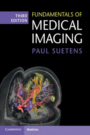 eBook - PDF
- Paul Suetens(Author)
- 2017(Publication Date)
- Cambridge University Press(Publisher)
Chapter 3 X-ray Computed Tomography 3.1 Introduction X-ray computed tomography or CT (Figure 3.1) is an imaging modality that produces cross-sectional images representing the X-ray attenuation proper- ties of the body. The word tomography originates from the Greek words τ oμoς (slice) and γραφιν (to write). Image formation of a cross-section is based on the following procedure. X-rays are produced by an X-ray tube, attenuated by the patient, and mea- sured by an X-ray detector. By densely sampling a wide X-ray beam, a set of lines is scanned cover- ing the entire field of view (Figure 3.2(a) shows a parallel-beam geometry and Figure 3.2(b) shows a fan-beam geometry). This process is repeated for a large number of angles (Figures 3.2(c) and (d)), yielding line attenuation measurements for all pos- sible angles and for all possible distances from the center. Based on all these measurements, the actual attenuation at each point of the scanned slice can be reconstructed. Although the imaging modal- ities of Chapters 4 and 5 (MR, PET, and SPECT) also represent a kind of computed tomography, the term CT (originally CAT) is allocated for X-ray comput(eriz)ed (axial) tomography. The physics of X-rays, their production, and interactions with tissue have already been discussed in Chapter 2. The history of CT began in 1895, when Wilhelm Konrad Röntgen reported the discovery of what he called “a new kind of rays.” Röntgen received the first Nobel Prize in Physics in 1901. Reconstruction of a function from its projections was first formulated by Johann Radon in 1917. Before the invention of CT, other kinds of tomography existed. Linear tomography (Figure 3.3(a)) The X-ray source and film move at constant speed in opposite directions. Under these circumstances, one section of the patient (plane P 1 − P 2 ) is always projected at the same position on the film, whereas the rest of the body is averaged out. eBook - PDF
- Joseph D. Bronzino, Donald R. Peterson, Joseph D. Bronzino, Donald R. Peterson(Authors)
- 2014(Publication Date)
- CRC Press(Publisher)
In practice, this plane (also called a slice) has a selectable thickness between 1.0 and 10 mm centered on the image plane. Pencil beam: A narrow, well-collimated beam of x-rays. Projection data: The set of transmission measurements used to reconstruct the image. Reconstruct: The mathematical operation of generating the tomographic image from the projection data. Scan time: The time required to acquire the projection data for one image, typically 1.0 sec. Scattered radiation: Radiation that is removed from the primary beam by a scattering process. This radiation is not absorbed but continues along a path in an altered direction. Slice: See Image plane. Spiral scanning: See Helical scanning. Three-dimensional imaging: See Helical scanning. Tomography: A technique of imaging a cross-sectional slice. 9 -12 Medical Imaging Volume CT: See Helical scanning. X-ray detector: A device that absorbs radiation and converts some or all of the absorbed energy into a small electrical signal. X-ray linear attenuation coefficient m : Expresses the relative rate of attenuation of a radiation beam as it passes through a material. The value of m depends on the density and atomic number of the material and on the x-ray energy. The units of m are cm − 1 . X-ray source: The device that generates the x-ray beam. All CT Scanners are rotating-anode bremsstrah-lung x-ray tubes except one-fifth generation system, which uses a unique scanned electron beam and a strip anode. X-ray transmission: The fraction of the x-ray beam intensity that is transmitted through the patient without being scattered or absorbed. It is equal to I t / I o in Equation 9.2, can be determined by measuring the beam intensity both with ( I t ) and without ( I o ) the patient present, and is expressed as a fraction. As a rule of thumb, n 2 independent transmission measurements are required to reconstruct an image with an n × n sized pixel matrix. eBook - PDF
Introduction to Medical Imaging
Physics, Engineering and Clinical Applications
- Nadine Barrie Smith, Andrew Webb(Authors)
- 2010(Publication Date)
- Cambridge University Press(Publisher)
A bank of solid-state detectors is situated opposite the X-ray tube and together they record a one-dimensional projection of the patient. The X-ray source and detectors are rotated through one complete revolution around the patient, with data being acquired essentially continuously. Most commercial scanners are so-called ‘third generation scanners’ which use a wide X-ray fan-beam and between 512 and 768 detectors. Two separate collimators are used in front of the source. The first collimator restricts the beam to an angular width of 45–60 ° . The second collimator, placed perpendicular to the first, restricts the beam to the desired slice thickness, typically 1–5 mm, in the patient head/foot direction. The scanner usually operates at a kVp of ~140 kV, with filtration giving an effective X-ray energy of 70–80 keV, and a tube current between 70 and 320 mA, although much higher values can be used for large patients. The focal spot size is between 0.6 and 1.6 mm. Typical operating conditions are a rotation speed of once per second, a data matrix filtered backprojection source detectors Figure 2.27 (left) The physical principle of computed tomography involves synchronous rotation of the X-ray tube and multiple detectors to record a series of one-dimensional projections. The CT image (right) is produced by the process of filtered backprojection. 66 X-ray planar radiography and computed tomography of the reconstructed image of either 512 3 512 or 1024 3 1024, and a spatial resolution of ~0.35 mm. 2.12.1 Spiral / helical CT Acquiring a single axial slice through a particular organ is of very limited diagnostic use, and so a volume consisting of multiple adjacent slices is always acquired. One way to do this is for the X-ray source to rotate once around the patient, then the patient table to be electronically moved a small distance in the head/foot direction, the X-ray source to be rotated back to its starting position, and another slice acquired. eBook - PDF
- Anthony B. Wolbarst, Robert G. Zamenhof, and William R. Hendee(Authors)
- 2006(Publication Date)
- Medical Physics Publishing(Publisher)
CHAPTER 3 Computed Tomography Thomas G. Flohr, Dianna D. Cody, and Cynthia H. McCollough* 59 3.1 History and Significance of X-Ray CT 60 3.2 Basics of Image Formation 61 3.3 Five Generations of CT Scanner Design 61 3.4 Spiral CT 64 3.4.1 Spiral Pitch 65 3.4.2 Spiral Interpolation Techniques 65 3.5. Multiple Detector-Row CT (MDCT) 67 3.5.1 MDCT Detector Designs and Evolution of Detectors 69 3.5.2 The Cone Beam Problem 71 3.5.3 Sequential Data Acquisition Has a Few Specialized Applications 72 3.5.4 Spiral MDCT Data Acquisition 73 3.5.5 Reconstruction Algorithms Addressing the Cone Beam Problem 77 3.5.6 Double z-Sampling 81 3.6 CT System Performance 81 3.6.1 Scan Acquisition Parameters and their Effects on Image Quality 82 3.6.2 CT Artifacts 82 3.7 Radiation Dose 85 3.7.1 Effective Dose 87 3.7.2 Risk 87 3.7.3 Dose Reduction Strategies 88 3.8 Cardiac CT 90 3.8.1 ECG-Triggered Prospective Sequential Scanning 91 3.8.2 ECG-Gated Retrospective Spiral Scanning 91 3.9 Future Direction of Multi-Detector CT 93 3.10 References 96 *Corresponding author. 3.1 History and Significance of X-Ray CT From Roentgen’s discovery of x-rays until the late 1960s, radiographic imaging was performed solely with large-area detectors such as radiographic film or image intensifier tubes. These systems permitted reasonably good visualization of high-contrast objects, but they could not record and display small differences in transmitted x-ray signal, such as those that occur among soft tissues. Several factors contributed to their inability to resolve low-contrast signals. Large-area detectors record a large amount of scattered radiation, making it difficult to resolve small differences in the transmitted patterns of primary, unscattered x-rays. Further, the superposition of the shadow of three-dimensional (3-D) objects onto a two-dimensional (2-D) detector obscures most low-contrast information. eBook - PDF
- Simon R. Cherry, Ramsey D. Badawi, Jinyi Qi, Simon R. Cherry, Ramsey D. Badawi, Jinyi Qi(Authors)
- 2016(Publication Date)
- CRC Press(Publisher)
2.2 X-Ray Imaging Basics 11 2.1 INTRODUCTION X-rays are a form of “ionizing radiation” because x-rays are energetic enough to ionize atoms and molecules during interactions. With about 10,000 times more energy than visible light photons, x-ray photons can penetrate objects including the human body. Since the first x-ray image taken in 1895 by Roentgen, x-ray imaging has become one of the most common diag-nostic procedures performed in medicine. The development of modern x-ray tubes and detec-tors has enabled a wide range of medical imaging applications, including x-ray radiography, x-ray fluoroscopy, and x-ray computed tomography (CT). With the capability to produce cross-sectional images, x-ray CT revolutionized traditional x-ray imaging and provided an invaluable diagnostic tool. The usage of CT has rapidly increased over the past two decades. In 2011, 85.3 million x-ray CT scans were performed in the United States. In addition to their role in diagnostic medicine, x-ray methods are widely used for clinical research across a broad spectrum of disease states. X-ray projection imaging and micro-CT (high-resolution x-ray CT imaging of small volumes) have also become important tools in biomedical research studies of animal models and tissue specimens. This chapter focuses on the fundamentals of x-ray imaging and the two major classes of x-ray imaging: x-ray projection imaging and x-ray CT. 2.2 X-RAY IMAGING BASICS 2.2.1 X-R AY P RODUCTION AND X-R AY S PECTRUM 2.2.1.1 X-Ray Production X-ray photons used for biomedical imaging are produced from a relatively complex device, the x-ray tube. The core of an x-ray tube, called the x-ray tube insert ( Figure 2.1 ), is a vac-uum sealed by a glass or metal enclosure. Within the vacuum insert, a heated filament, the cathode , emits electrons in a process called thermionic emission. eBook - PDF
- Gilbert R Thompson(Author)
- 2011(Publication Date)
- ICP(Publisher)
This has greatly facilitated the monitoring of the effects of medical treatments in clinical trials. As the CT scan is non-invasive the threshold for performing a radiological examination is reduced, since doctors are understandably reluctant to perform potentially haz-ardous examinations without a very strong clinical indication. The introduction of the CT scanner has lowered the threshold for per-forming radiological investigations, and whilst this has had the effect of considerably increasing the work of X-ray departments it has also meant that abnormalities may be diagnosed at an earlier stage in their natural history, and earlier diagnosis means that treatment is facili-tated. The CT scanner can also be used to guide the radiologist in interventional radiology and facilitate the biopsy of tumours, the drainage of fluid collections and radiofrequency ablation. When the team at EMI were developing the whole body scanner it became apparent that a cross-section of the body would have 166 A. M. K. Thomas significant utility in planning radiotherapy treatment. Prior to the introduction of CT scanning radiotherapy treatment planning was imprecise and time consuming. There was an imbalance between the accuracy of the treatment that could be delivered by the linear accel-erator and the then available treatment plans. The CT scan allowed computer programs to guide the treatment beam in a process requir-ing only a few minutes. The radiotherapy planning system is linked to the CT diagnostic display console and the radiation isodose distribu-tion curves can be overlaid onto the CT image. The CT density numbers could be used to calculate the effect of inhomogeneities in the tissues in the path of the radiation beam. This was the very prob-lem that Allan Cormack had been considering back in 1956. The areas to be irradiated at therapy could be marked as could sensitive areas to be avoided, which could be located precisely.
Index pages curate the most relevant extracts from our library of academic textbooks. They’ve been created using an in-house natural language model (NLM), each adding context and meaning to key research topics.