Biological Sciences
Biological Imaging
Biological imaging refers to the use of various techniques to visualize and study biological structures and processes at the cellular, tissue, and organismal levels. This includes methods such as microscopy, magnetic resonance imaging (MRI), and positron emission tomography (PET) to observe and analyze living organisms and their components. These techniques are essential for understanding biological functions and disease processes.
Written by Perlego with AI-assistance
Related key terms
1 of 5
9 Key excerpts on "Biological Imaging"
 eBook - ePub
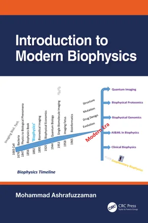
- Mohammad Ashrafuzzaman(Author)
- 2023(Publication Date)
- CRC Press(Publisher)
Table 12.1 presents a comparison of these imaging technologies. Each imaging technique is profiled with its respective underlying principle, a description of selected current applications, and a discussion of advantages and known limitations.Table 12.1Comparison of Imaging Technology for Systems BiologyImaging technique Spatial resolution Scan time Contrast agents and molecular probes Key use Multi-photon microscopy 15 – 1000 nm Secs Fluorescent proteins, dyes, rhodamine amide, quantum dots Visualization of cell structures Atomic force microscopy 10 – 20 nm Mins Intermolecular forces Mapping cell surface Electron microscopy ~5 nm Secs Cyrofixation Discerning protein structure Ultrasound 50 μm Secs Microbubbles, nanoparticles Vascular imaging CT/microCT 12 – 50 μm Mins Iodine Lung and bone tumor imaging MRI/microMRI 4 – 100 μm Mins – Hrs Gadolinium, dysprosium, iron oxide particles Anatomical imaging fMRI ~1 mm Secs – Mins Oxygenated hemoglobin (HbO2 ) deoxygenated hemoglobin (Hb)Functional imaging of brain activity MRS ~2 mm Secs N-acetylaspartate (NAA), creatine, choline, citrate Detection of metabolites PET/microPET 1 – 2 mm Mins Fluorodeoxyglucose (FDG), 18 F, 11 C, 15 OMetabolic imaging Source: Kherlopian et al. (2008 ).Systems biology approaches involve determining the structure of biological circuits using genomewide analyses. Imaging is known to offer the unique advantage of monitoring biological circuits function over time at single-cell resolution in the intact animal. The power of integrating imaging tools with more conventional genomic approaches has been detailed to analyze the biological circuits of microorganisms, plants, and animals (Megason and Fraser, 2007 ). Conventional genomic approaches excel at the first goal of systems biology: characterizing the structure of biological networks. The systems-level analysis works best when all the components and interactions associated with the biological circuitry are defined clearly. For this purpose, comprehensive approaches including sequencing, microarrays, and interactomic approaches are found ideal. As the network structure has been elucidated, how it functions as a circuit may be investigated. There are four important considerations about how biological circuits function that illustrate the advantages of imaging for systems biology (see Figure 12.10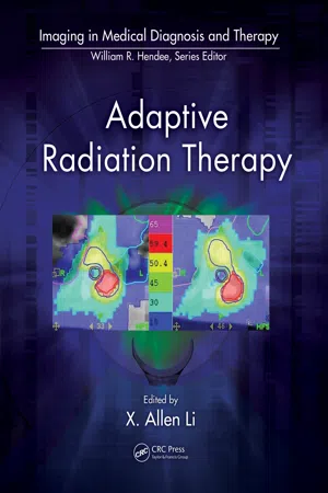 eBook - PDF
- X. Allen Li(Author)
- 2011(Publication Date)
- CRC Press(Publisher)
Many more studies like this are needed to finally uncover the complexity of treatment response and get closer to the goal of personalized approach to treatment. 3.4 Conclusions Biological Imaging is quickly becoming an integral part of radiation oncology practice. As the flagship Biological Imaging method, FDG-PET has proven indispensable in tumor diagnosis and staging. Although it will continue to play the central role in cancer management, other Biological Imaging methods that target general tumor phenotypes like hypoxia, cellular prolif-eration, angiogenesis, or even more specific cellular processes will come to play increasingly important roles in future. Their roles will be multifaceted—to design the most optimal treatment strategy, better define the target volume, or estimate the expected treatment outcome. As the complexity of treatment manage-ment, which often includes chemotherapy or molecular targeted therapies in addition to RT, increases, the appropriate selection and use of Biological Imaging will become essential. The path to optimal selection is not straightforward, as it requires a compre-hensive clinical validation of the particular imaging modality in addition to its biological validation. Because tumor biology is incredibly complex and Biological Imaging surrogates are imperfect, it is unreasonable to expect that such validations will be perfect. One of the most detrimental aspects of surrogacy is the limited quantitative accuracy of Biological Imaging modali-ties. Understanding these limitations is of paramount impor-tance, although they are often neglected in the desire to push the field forward. Target definition is at the heart of the RT process and will benefit significantly from expanded incorporation of biologi-cal imaging information. The efforts to incorporate biological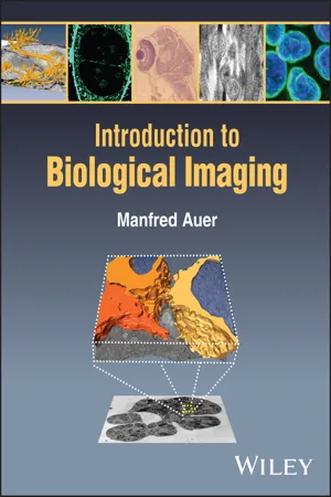 eBook - PDF
- Manfred Auer(Author)
- 2024(Publication Date)
- Wiley(Publisher)
Moreover, life happens at multiple scales, often spanning at least six orders of magnitude, which in terms of imaging requires the utilization of a large variety of imaging tech- niques, whose information content needs to be correlated and integrated. I will discuss at the end what a biological information system (BIS) – the biological equivalent of a geospatial information system (GIS) – could look like and how it would revolutionize biology as a discipline and making biological research even more powerful and useful than it already is. I will attempt to make some predictions about the future of bioimaging, fully knowing (to quote the “baseball-playing philosopher” Yogi Berra) that “it is tough to make predictions, particularly about the future.” 12.1 Future of Macromolecular Imaging Since its inception in the early to the mid 20th century, X-ray crystallography was the dominant player and the gold standard in the structure determination of biological 12 The Future of Bioimaging 12 The Future of Bioimaging 302 macromolecules, particularly proteins, multi-protein com- plexes as well as nucleic acid complexes. After its biggest theoretical problem of phasing was overcome, its only practical drawback was the need to grow large diffraction quality crystals. For a couple of decades, nuclear magnetic resonance (NMR) spectroscopy emerged as a rival tech- nique for structure determination, but it remained the domain of smaller proteins and their study in solution. Cryo-electron microscopy slowly but steadily emerged as an alternative to structure determination, particularly when 2D crystalline and helical or icosahedral symmetry could be exploited, as has been frequently the case for membrane proteins and cytoskeletal protein assemblies or viruses, respectively.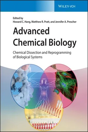 eBook - PDF
Advanced Chemical Biology
Chemical Dissection and Reprogramming of Biological Systems
- Howard C. Hang, Matthew R. Pratt, Jennifer A. Prescher, Howard C. Hang, Matthew R. Pratt, Jennifer A. Prescher(Authors)
- 2023(Publication Date)
- Wiley-VCH(Publisher)
Imaging in such opaque animals requires special considerations beyond those for cultured cells or transparent organ- isms. We also focus on noninvasive techniques, versus those requiring surgical manipulation or exposed tissue. We first introduce the general principles of molecular imaging in live organisms, along with the common probe sets employed. We then highlight the individual modalities used to illuminate cellular and molecular fea- tures in vivo. This latter category is dominated by optical methods, and several examples are showcased. Imaging Advanced Chemical Biology: Chemical Dissection and Reprogramming of Biological Systems, First Edition. Edited by Howard C. Hang, Matthew R. Pratt, and Jennifer A. Prescher. © 2023 WILEY-VCH GmbH. Published 2023 by WILEY-VCH GmbH. Companion website: www.wiley.com/go/hang 436 18 In Vivo Imaging discoveries go hand in hand with probe development; thus, the chemical innovations underlying visualization in whole organisms are discussed throughout. 18.2 Basic Concepts for Imaging In Vivo In vivo imaging modalities rely on distinct types of energy to interrogate living subjects and provide visual readouts. The relevant energies span the electromag- netic spectrum (Figure 18.1), and they can be deployed to report on a variety of features – from anatomical struc- tures to physiological functions to molecular activities. The signal outputs must penetrate skin, tissues, and other biological barriers to reach external detectors. The collected signals are then used to generate pictures of the targeted structures or processes. Serial imaging is also possible in most cases. In human patients, continuous monitoring enables disease progression to be tracked without the need for biopsy. Such approaches are critical for early disease detection, as molecular changes typ- ically arise long before any morphological differences appear. eBook - PDF
- Robert Splinter, Brett A. Hooper(Authors)
- 2006(Publication Date)
- CRC Press(Publisher)
551 16 Diagnostic Methods Using Light: Photobiological This chapter focuses on the biological information that can be obtained by optical means. The list of items is only a selection; new applications are added daily. Some of the examples of photobiological diagnostics are spec-troscopic imaging and immunofluorescence, various biosensors, and possibly the oldest of all diagnostic methods, i.e., blood oxygenation determination by attenuation spectroscopy. Another relatively new method using quantum dots for labeling is also described. Although fluorescence was described in Chapter 15, the particular diagnostic application discussed in this chapter is definitely of biological interest. The first method describes the uses of fluorescence for functional imaging, thus combining the photophysical and photochemical effects to provide bio-logical information. 16.1 Immunostaining (“Functional Imaging”) General immunostaining with antibodies is possible and the cells can then be viewed using a compound microscope. A primary antibody is incubated within the cells upon staining. Once the antibodies bind to their specific pro-teins on or in the cells, a secondary antibody is incubated with the sample. The secondary antibody is specific for the primary antibody and is conju-gated to an enzyme such as horseradish peroxidase (HRP). A substrate is then added that is cleaved by the HRP, which results in a color change, and the specimen is observed with a compound microscope. The stained por-tions of the cells appear darker than unstained portions. Immunofluorescence is another technique used to examine cell-surface molecules as well as other molecules. 551 16.2 Immunofluorescence Immunofluoresence is the use of antibodies to label a specific protein within a sectioned tissue or a population of cells. Prior to describing the process of antibody labeling a brief explanation is presented about the immunology that led scientists to develop this type of assay. eBook - PDF
- Young-Tae Chang, Nam-Young Kang(Authors)
- 2023(Publication Date)
- Wiley-VCH(Publisher)
1 1 Introduction to Bioimaging Bioimaging can be defined as visualization of a biological object. The most basic bioimaging may be just “seeing” the living object using our own eyes. This function is called “vision” and the procedure is mediated by visible light. The visible light is a part of electromagnetic wave in the wavelength range of 400–700 nm, and the image information is generated by the interaction between light and object, such as reflection, scattering, and diffraction. The generated information-rich light package travels and reaches our eyes. The focused light through lens would be projected to the screen as in a camera. The retina in our eye is the screen of the image, which is composed of the two-dimensional array of optic nerves. The photon in light signal (containing the information of the object) reaches the retina and activates optical neurons, and the signal is transferred to the brain and is reconstructed into the image of the object by neuronal processing. Even though the screen is two-dimensional, the processed images via two retinas provide three-dimensional information about the shape and distance of the object. Visible light travels at a so-called speed of light (3 × 10 8 m/s), so the information transfer in the vision could be almost instantaneous. If there is a possible delay, it may be from the signal transition step from the optical nerve to the brain and the information processing time in the brain. Bats live in the dark environment without enough environmental light for vision. Instead, they use ultrasound for bioimaging platform. If other conditions are same, the light vision could be million times faster than ultrasonic sensing (340 m/s) (Figure 1.1). Among all the sensors, light vision is the fastest and most information-rich system. Therefore, the invention of eye (in more general term, photoreceptor) is one of the most dramatic events in the evolution of life. eBook - PDF
- Shayne C. Gad, Shayne C. Gad, Shayne C. Gad, Shayne C. Gad, Shayne C. Gad(Authors)
- 2016(Publication Date)
- CRC Press(Publisher)
physical.phenomena.(see.Figure.14 .1), .such.as.magnetic.field/radiofrequency.(magnetic.resonance. imaging/MRI),. x-ray. (computed. tomography/CT),. high. frequency. sound. waves. (ultrasound/US),. optical.(bioluminescence/fluorescence),.gamma.rays.(single.photon.emission.computed.tomogra-phy/SPECT),. and. annihilation. twin. photons. from. beta. emission. (positron-emission. tomography/ PET). .These.modern.imaging.modalities.have.been.re-engineered.for.use.with.laboratory.animals. by.pushing.the.resolution.and.sensitivity.of.each.modality.to.the.physical.limit . .The.improved.high. resolution.and.extreme.sensitivity.make.the.imaging.technology.highly.translational.in.the.preclini-cal.drug.development.process . MRI and MRS Optical (fluorescence and bioluminescence) microSPECT Ultrasound microCT microPET Figure 14.1 Multimodality imaging instrumentations. The modern molecular imaging equipment includes magnetic field/radiofrequency (magnetic resonance imaging/MRI), x-ray (computed tomogra-phy/CT), high frequency sound waves (ultrasound/US), optical (bioluminescence/fluorescence), gamma rays (single photon emission computed tomography/SPECT), and annihilation twin pho-tons from beta emission (positron-emission tomography/PET). (Images provided by LoMIN of NIBIB at National Institutes of Health, Bethesda, MD.) 897 MOLECULAR IMAGING TECHNIQUES IN LABORATORY ANIMALS The. cutting. edge. non-invasive. imaging. technology. is. able. to. provide. evidence. of. in vivo. biodistribution.of.imaging.probes,.confirm.on-target.biological.activity,.illustrate.disease.mecha-nism,.and.validate.efficacy.of.drug.treatment . .Maximizing.collection.of.in vivo.biological.infor-mation.during.the.preclinical.development.can.be.highly.valuable.during.the.critical.period.of. lead.selection/optimization.of.promising.drug.candidates . eBook - PDF
- Joseph D. Bronzino, Donald R. Peterson(Authors)
- 2018(Publication Date)
- CRC Press(Publisher)
63 -1 63.1 Introduction Readers of this book are well aware that tissue engineering is a vibrant field that aims to develop bio-logical substitutes that can replace, repair, or enhance lost tissue or organ function. As this field comes to the forefront of medicine, it becomes critical for basic and clinical researchers to understand some of the methods that can be employed to study engineered constructs and tissues under development in vitro , or while functioning in vivo . Methods designed to image tissues can greatly aid in the advance-ment of tissue engineering. Today’s imaging techniques, such as x-ray, computed tomography (CT), ultrasound, positron emission tomography (PET), single photon emission computed tomography, and magnetic resonance imaging (MRI), can allow for more than just mere pictures of the tissues of inter-est. Indeed, even optical techniques have progressed significantly, and can generate images of exquisite detail and clarity. These modern biomedical imaging techniques can see into objects without physically peeling through the interposing layers. They also have the desired ability to yield information related to many physical and physiological characteristics important to the study of engineered tissues. Among these characteristics are the structural integrity and physical attributes of the scaffolding; the perfusion/ diffusion of blood and nutrients into the tissue; the distribution of oxygen within the tissue; and changes in the cellular function and remodeling of engineered tissue over time. These critical data can be col-lected and used to optimize design, monitor function, and observe, predict and possibly also prevent failure of engineered tissues. The subject of biomedical imaging is far too vast to be comprehensive in this short chapter. Therefore, it is the purpose of this chapter to briefly touch upon some of the important imaging techniques that are appropriate for observing engineered tissues.
- Nick Van Bruggen, Timothy P.L. Roberts, Nick Van Bruggen, Timothy P.L. Roberts(Authors)
- 2002(Publication Date)
- CRC Press(Publisher)
323 0-8493-0122-X/03/$0.00+$1.50 ' 2003 by CRC Press LLC The Future for Biomedical Imaging: Emerging and Alternative Technologies Nick van Bruggen and Timothy P.L. Roberts CONTENTS 11.1 Introduction: The Importance of Noninvasive Imaging .............................. 323 11.2 Molecular Imaging Using Novel MRI Contrast Agents ............................. 325 11.3 Optical Imaging ............................................................................................ 326 11.3.1 Brain Activity Monitoring with Intrinsic Optical Signals ............... 327 11.3.2 Bioluminescence .............................................................................. 328 11.3.3 Fluorescent Probes ........................................................................... 329 11.3.4 Imaging in Near-Infrared Range ...................................................... 330 11.4 Near-Infrared Spectroscopy ......................................................................... 331 11.5 Magnetoencephalography ............................................................................ 333 11.6 Conclusions .................................................................................................. 333 References .............................................................................................................. 335 11.1 INTRODUCTION: THE IMPORTANCE OF NONINVASIVE IMAGING Studies of natural development, disease progression, response to external stimuli, and in ß uences of new therapies and interventions share a common demand — the ability to characterize or determine some aspect of brain physiology at successive time points. Such studies are facilitated by serial or longitudinal examinations. If the nature of an examination (e.g., brain sectioning or immunohistologic staining) precludes repeated performance, alternative strategies involving large cohorts of similar preparations are required.
Index pages curate the most relevant extracts from our library of academic textbooks. They’ve been created using an in-house natural language model (NLM), each adding context and meaning to key research topics.