Biological Sciences
Microscopes
Microscopes are essential tools used to observe and study biological specimens at the cellular and molecular levels. They work by magnifying the image of tiny objects, allowing scientists to see details that are not visible to the naked eye. There are different types of microscopes, such as light microscopes and electron microscopes, each with its own specific applications and advantages.
Written by Perlego with AI-assistance
Related key terms
1 of 5
10 Key excerpts on "Microscopes"
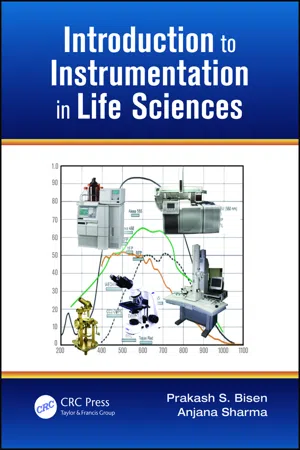
- Prakash Singh Bisen, Anjana Sharma(Authors)
- 2012(Publication Date)
- CRC Press(Publisher)
1 1 Microscopy 1.1 INTRODUCTION A microscope is defined as an instrument that magnifies objects by means of lenses to reveal details that are invisible to the naked eye. It was only after the discovery of the first microscope around 1590 by a Dutch spectacle maker, Zacharias Jensen, that it was realized there were certain living organisms so small in size that they were invisible to the naked eye and hence never believed to be in existence. The microscope opened new doors into the living world, bringing forth a realm of microorganisms. This gave concrete evidence to Louis Pasteur’s germ theory of disease. Thus began the science of microbiology. Through the microscope, the different shapes, sizes, and even colors of microorganisms can be seen. The degree of magnification needed to see a microorganism depends on the size of the microbe. Protozoa, fungi, algae, and bacteria whose sizes range from 1–200 μ m can be viewed with a light microscope, that is, a microscope that uses visible light to illuminate a specimen. It gives a magnification of about 1500 × . Viewing smaller microorganisms like viruses, whose size varies from 0.015 to 2.0 µ m, as well as the internal structure of bacterial cells or eukaryotic cell organ-elles, requires the use of a more specialized electron microscope, which has a higher magnification, that is, 200,000 × (Table 1.1). It may be noted here that the extent of magnification is limited by the capacity of resolution, which we shall discuss in Section 1.2. Without resolution, magnification is called empty magnification and it is of practically no use whatsoever. Today, microbiologists have a variety of Microscopes at their disposal (Table 1.1). Together with the different techniques available for exploration, they can choose and use these Microscopes for study. The choice of a particular microscope depends on the size of the object, degree of detail to be viewed, and purpose of microscopic observation. eBook - PDF
- Smita Nair(Author)
- 2019(Publication Date)
- Delve Publishing(Publisher)
The microscope magnifies the view of the smallest bacteria or infection, which permits the experts to create the inoculations, injections, and treatments with respect to the infectious illnesses in the humans as well as in the animals. Scanning electron Microscopes have the potential to intensify up to several million times to view the smallest areas of molecules, the viruses, and the nanoparticles, the Scanning electron Microscopes use the counteractive software to elevate the enlargement and the firmness of the images, the computers support the nano-technologists utilizes the high-powered electron Microscopes to interpretation the objects only a few molecules thick. Microscope and Its Significance in Life Sciences 161 8.2 DIFFERENT TYPES OF MICRO-SCOPES There are several various types of Microscopes which are applied in the light microscopy, and there are four most general kinds of Microscopes are Compound Microscopes, Stereo Microscopes, Digital Microscopes, and the Pocket Microscopes or handheld Microscopes. Some kinds which are best well-matched for the biological uses, where others of the Microscopes are best for teaching space or individual hobby usage. Outside of light microscopy are the exhilarating growths with the help of electron Microscopes and in scanning probe microscopy. Figure 8.3: Various types of Microscopes. Source: https://cdn.pixabay.com/ p h o t o / 2 0 1 8 / 0 7 / 0 6 / 0 4 / 3 8 / t e c h n o l o -gy-3519747_960_720.jpg There is a descriptive introduction of the several different kinds of Microscopes which are avail-able in the present time. Introduction to Life Science 162 8.2.1 The Compound Light Microscope Usually binocular (which are two eyepieces), the compound light microscope is the type of microscope which integrate the power of the lenses and light which helps to magnifies the sample or specimen which is being under the inspection or analysis.
- Khuntia, B K(Authors)
- 2021(Publication Date)
- Daya Publishing House(Publisher)
4 Microscopes Microorganisms are usually not visible to naked human eye. They can be made visible, only when they are magnified under Microscopes. Microscopes are instruments, which can produce enlarged images of very small objects, making it possible to view them, which, otherwise, cannot be seen distinctly by naked human eye. Different types of Microscopes are used by the microbiologists for specific purposes. Microscopes are of the following types ( Figure 4.1 ). Figure 4.1: Types of Microscopes A. Light or Optical Microscopes In light or optical Microscopes, ‘light waves’ are used to produce the enlarged images of very small objects and magnification is obtained by a system of ‘optical lenses’. Ordinarily, microbes do not absorb much light, but This ebook is exclusively for this university only. Cannot be resold/distributed. staining them with dyes greatly increases their light absorbing ability, resulting in greater contrast and color differentiation. Light Microscopes are of four types as described below. (1) Bright-field Microscope In a bright-field microscope, the microscopic field (the circular area visible under microscope) is brightly illuminated and the microbes (or biological specimen) appear darker, as they absorb some of the light passing through them. It is of two types as follows. ( a ) Simple Microscope A simple microscope is used to obtain small magnifications. A single biconvex lens magnifies the size of the object to get an enlarged virtual image. ( b ) Compound Microscope The most commonly used microscope for general purposes is the standard compound microscope. It magnifies the size of the object by a complex system of lens arrangement. It has a series of two lenses; the objective lens and the ocular lens, to magnify the size of the object. 2. Dark-field Microscope In a dark-field microscope, the object is brilliantly illuminated against a dark background. eBook - ePub
Introduction to Experimental Biophysics
Biological Methods for Physical Scientists
- Jay L. Nadeau(Author)
- 2017(Publication Date)
- CRC Press(Publisher)
7Introduction to Biological Light Microscopy Coauthored with Michael W. Davidson7.1 IntroductionLight microscopy is an essential technique for the life sciences. It allows for the identification and counting of cells and the observation of cellular growth, development, communication, and death. These processes can all be imaged down to the single-molecule scale thanks to a revolution in labeling techniques, particularly fluorescent labeling. Several excellent, thorough textbooks have been written on light microscopy, and this chapter is not a substitute for any of them. Neither is it a substitute for a complete knowledge of your own instrument, which should be obtained from the literature of the company that made the microscope. All of the large manufacturers provide detailed documentation in their user manuals and websites, often including introductions to the principles of microscopy. What you will find in this chapter is a review of the physics that applies to all light Microscopes and their key illumination modes: brightfield, darkfield, phase contrast, differential interference contrast (DIC), epifluorescence, and confocal. The structure of a typical inverted biological microscope is discussed with its practical implications for imaging dishes, plates, and living cells. We then discuss the physics of fluorescence and introduce some of the most common biological fluorescent dyes. At the end of this chapter, you should be comfortable preparing a specimen for a particular type of light microscopy and know why it was prepared the way it was—its substrate of growth, whether it is fixed or live, the presence of one or more labels. You should know how it should be illuminated and which parameters will need to be adjusted to obtain a good image.Obtaining those good images is then a matter of training, patience, and art. Biological imaging is a lot like wildlife photography, especially when working with living cells. The observer needs to be in the right place at the right time when the cells decide to cooperate. No longer available |Learn more
- (Author)
- 2014(Publication Date)
- Library Press(Publisher)
________________________ WORLD TECHNOLOGIES ________________________ Chapter- 1 Microscopy Microscopy is the technical field of using Microscopes to view samples and objects that cannot be seen with the unaided eye (objects that are not within the resolution range of the normal eye). There are three well-known branches of microscopy, optical, electron, and scanning probe microscopy. Optical and electron microscopy involve the diffraction, reflection, or refraction of electromagnetic radiation/electron beams interacting with the specimen, and the subsequent collection of this scattered radiation or another signal in order to create an image. This process may be carried out by wide-field irradiation of the sample (for example standard light microscopy and transmission electron microscopy) or by scanning of a fine beam over the sample (for example confocal laser scanning microscopy and scanning electron microscopy). Scanning probe microscopy involves the interaction of a scanning probe with the surface of the object of interest. The development of microscopy revolutionized biology and remains an essential technique in the life and physical sciences. ________________________ WORLD TECHNOLOGIES ________________________ Scanning electron microscope image of pollen ________________________ WORLD TECHNOLOGIES ________________________ Optical microscopy Stereo microscope Optical or light microscopy involves passing visible light transmitted through or reflected from the sample through a single or multiple lenses to allow a magnified view of the sample. The resulting image can be detected directly by the eye, imaged on a pho-tographic plate or captured digitally. The single lens with its attachments, or the system of lenses and imaging equipment, along with the appropriate lighting equipment, sample stage and support, makes up the basic light microscope. The most recent development is the digital microscope, which uses a CCD camera to focus on the exhibit of interest. eBook - PDF
Microbiology
Principles and Explorations
- Jacquelyn G. Black, Laura J. Black(Authors)
- 2018(Publication Date)
- Wiley(Publisher)
This technique is especially important 64 CHAPTER 4 Microscopy and Staining (a) (b) wide area network for distance learning or teleconferenc- ing. Imagine being able to show your cousin in Kansas live images of what’s swimming in your sample of pond water. There are, however, some limitations: Maximum magnification is quite limited, and the price is high. ELECTRON MICROSCOPY The light microscope opened doors to the world of microbes. However, because it could not resolve objects separated by less than 0.2 μm, the view was limited to observations at the level of whole cells and their arrange- ments. Few subcellular structures could be seen; neither could viruses. The advent of the electron microscope (EM) allowed these small structures to be visualized and stud- ied. The EM was developed in 1932 and was in use in many laboratories by the early 1940s. The EM uses a beam of electrons instead of a beam of light, and electromagnets rather than glass lenses are disturbing them, as in examining living biofilms. Time-lapse images can also be collected. Digital Microscopy Have you had frustrating moments in the lab when you just couldn’t get a slide into focus? Then you would like the auto-focus, auto-aperture, auto-light, motor- ized stage and magnification changers of a digital micro- scope (Figure 4.20). Not only that, but these Microscopes also come with a built-in digital camera and preloaded software. All you do is plug in the unit, turn on the power, and use the mouse to view live or stained specimens on a monitor, or in a group situation on a screen through use of a projector. It can also be integrated into a local or FIGURE 4.19 (a) A confocal microscope system manufactured by Olympus. Cell with microtubular fragments shown using (b) standard fluorescent microscopy, and (c) confocal micro- scopy. (a, b, c: Courtesy Olympus Corporation, Scientific Equipment Division) (c) (b) (a) FIGURE 4.20 (a) A digital microscope system manufactured by Nikon Instruments, Inc.
- Robert N. Trigiano, Dennis J. Gray, Robert N. Trigiano, Dennis J. Gray(Authors)
- 2016(Publication Date)
- CRC Press(Publisher)
75 6 Proper Use of Microscopes David T. Webb Although the compound microscope is the most commonly used biological instrument, it is often used improperly. This may not matter with very thin commercial slides at low-to-medium magni-fications. However, proper alignment of the illumination system is essential for viewing thick sec-tions, whole mounts, and, for highly magnified samples of cells, fungi and bacteria. It is also crucial for studying unstained specimens and for photomicroscopy. This chapter was written as if you knew nothing about using a microscope. In some cases you may have learned bad practices that need to be corrected. It is also written for Microscopes that have a field diaphragm, and a condenser that can be centered and focused to achieve Kohler illumination (Kohler, 1893). Many student scopes do not have these features because their condensers and field diaphragms are rudimentary. In the course of your careers you will encounter Microscopes that have the ability to achieve Kohler illumination. At that point this article will be even more useful. You will be using Microscopes throughout your career. If you learn the simple lessons in this chapter, you will do much better work and see the exciting world of microscopy in a new light. The modified procedure we present was developed by the German scientist, August Kohler (1866–1948), and bears his name. Recently, his ideas were used to make the EM 910 Electron Microscope by Carl Zeiss. Thus, this procedure, which was introduced in 1893, has been of lasting value. Microscopes are partly categorized by the number of oculars they contain. The first Microscopes had one ocular and were called Monocular. Binocular scopes have two oculars, while Trinocular scopes have three. The third ocular is typically modified for the use of a camera. THE COMPOUND MICROSCOPE Because the optical units in a microscope are composed of many lenses, the term compound micro-scope is used. eBook - PDF
- Robert Blanchard(Author)
- 2012(Publication Date)
- Academic Press(Publisher)
10. Body—Movable component enclosing lens systems. 11. Objective lens—Lens system which supplies primary magnification of specimen. Uses The dissecting microscope is used for low power, three-dimensional magnifi-cation of opaque, transparent, or translucent specimens that are difficult to see with the naked eye. Typical uses might include observations of fungal fruiting structures in culture plates or on living tissues, or observations of disease symp-Fig. 1.2 Typical dissecting microscope. 8 1. The Microscope toms on infected plant parts. Most dissecting Microscopes allow low power magnification of transparent or translucent specimens such as leaves. The fol-lowing procedures should be used when observing specimens: 1. Clean the stage plate, eyepieces, illuminator glasses, and objective lenses with lens paper. 2. Place a specimen or object in the center of the stage. 3. Illuminate the object (reflected light for opaque objects; transmitted light for transparent or translucent objects). 4. Raise the body above the focus point with the focusing knob. Then lower the body to bring the object into sharp focus. 5. Start with the magnification knob in the lowest position and adjust upward as necessary. Sterile Technique I N T R O D U C T I O N Identification of microorganisms that cause tree diseases often requires micro-scopic examination. If a disease-causing microorganism has produced fruiting structures on the surface of diseased parts, observation of these structures and their accompanying spores with the microscope may provide adequate clues for identification. However, the presence of more than one type of fruiting structure may make it difficult to know which is the pathogen, or may make it impossible to obtain a pure sample of the pathogen for examination. Therefore, it is desir-able to make pure cultures of pathogens for identification and subsequent storage for future reference. eBook - PDF
Basic Bioscience Laboratory Techniques
A Pocket Guide
- Philip L.R. Bonner, Alan J. Hargreaves(Authors)
- 2022(Publication Date)
- Wiley-Blackwell(Publisher)
The oil immersion lens is essential for microbiological samples, especially bacteria. • Remember that there are usually crude (outer) and fine (inner) focus controls, the latter being used at higher magnifications. However, the high-power lenses need to be much closer to the specimen; care is therefore needed to prevent contact with the coverslip, which could jam the slide in position. • Remember also that the coverslip should be fixed in position, particularly if the sample is covered by a layer of liquid (e.g. in fluorescence microscopy). MICROSCOPY 33 2.4.6 Data Analysis and Presentation 2.4.6.1 Drawing and Labelling a Microscope Image • Remember to give each image a clear descriptive title. • If drawing the image, draw representative areas of cells rather than every detail. Label the major structures or cell types visible in the field of view. • If drawn by hand, indicate the magnification at which the specimen was viewed or, if using a camera image, include the final magnification (including image enlargement) or a calibrated scale bar to give a clear idea of dimensions and distances. Magnification can be determined by multiplying the magnifying powers of the eyepiece (ocular) and the objective lens used to view the specimen (see Worked Example 2.1). Note that, if making prints from a negative, the enlargement factor of the print from the negative also needs to be taken into account when calculating the final magnification. Many microscope cameras are digital, and the camera software allows the insertion of a scale bar on to the image, which would adjust according to the final image size. 2.4.6.2 Making Size Measurements Using the Light Microscope Microscopes may be fitted with a scale (reticule) in one eyepiece lens, which can be turned and aligned with any focused object to obtain an estimate of its size. eBook - PDF
- Geoffrey Bourne(Author)
- 2012(Publication Date)
- Academic Press(Publisher)
Microscopy R. Barer Department of Human Biology and Anatomy, University of Sheffield, Sheffield, England I. Introduction 91 II. Some General Principles 92 A. Numerical Aperture and Resolution 92 B. Lens Aberrations 93 C. Correction of Aberrations 94 D. The Condenser 95 E. Methods of Illumination 97 III. Special Methods of Microscopy 98 A. Phase Contrast Microscopy 98 B. Interference Microscopy 109 C. Ultraviolet Microscopy 119 D. Fluorescence Microscopy 131 E. Polarizing Microscopy 136 F. Electron Microscopy 140 IV. Conclusion 153 References 154 I. Introduction Few cytological research methods are as satisfactory as direct obser-vation; much can be inferred by indirect physiological and chemical means, but eventually the information must be correlated with cell struc-ture as observed with the microscope. The purpose of this chapter is to present certain fundamental principles which are common to most meth-ods of microscopy and then to discuss a number of methods of special value in cytology. It is not possible to give detailed practical instruc-tions, but it is hoped that the general reader will gain enough infor-mation to be able to select the most suitable methods for studying his own particular problem. 91 92 «R. B A R E R II. Some General Principles There are two main aims in all methods of microscopy. The first is the formation of a magnified image as free as possible from optical de-fects. This is by no means the only requirement, because a technique that gives excellent results with one type of specimen may fail com-pletely with another. Living cells, for example, would be virtually in-visible if examined by methods commonly used for studying stained sections. Mere magnification or resolution is not enough; achievement of contrast, which involves a difference in intensity or color between the object and the background, is equally important.
Index pages curate the most relevant extracts from our library of academic textbooks. They’ve been created using an in-house natural language model (NLM), each adding context and meaning to key research topics.