
eBook - ePub
Essentials of Clinical Anatomy of the Equine Locomotor System
Jean-Marie Denoix
This is a test
Condividi libro
- 296 pagine
- English
- ePUB (disponibile sull'app)
- Disponibile su iOS e Android
eBook - ePub
Essentials of Clinical Anatomy of the Equine Locomotor System
Jean-Marie Denoix
Dettagli del libro
Anteprima del libro
Indice dei contenuti
Citazioni
Informazioni sul libro
Essentials of Clinical Anatomy of the Equine Locomotor System presents a unique photographic record of dissections showing the topographical anatomy of the locomotor system of the horse. Readers of this book will be able to see the position and relationships of the bones, joints, muscles, nerves and blood vessels that make up each region of the forelimb, vertebral column and hindlimb.
Key features:
- Important features of regional and topographical anatomy are presented using full-color photos of detailed dissections
- Anatomy is presented in a clinical context
- Preparations of cross-sectional anatomy facilitate interpretation of diagnostic imaging, such as ultrasonography, MRI images and CT scans
- All dissections are of fresh material, rather than preserved specimens, to demonstrate the appearance of tissues in the living animal, or at post mortem autopsy
This new atlas is essential for anybody involved in detailed anatomical study, complex lameness evaluation or advanced imaging techniques in horses. It will be a useful guide for veterinary students, and a reference for equine vets in practice.
Domande frequenti
Come faccio ad annullare l'abbonamento?
È semplicissimo: basta accedere alla sezione Account nelle Impostazioni e cliccare su "Annulla abbonamento". Dopo la cancellazione, l'abbonamento rimarrà attivo per il periodo rimanente già pagato. Per maggiori informazioni, clicca qui
È possibile scaricare libri? Se sì, come?
Al momento è possibile scaricare tramite l'app tutti i nostri libri ePub mobile-friendly. Anche la maggior parte dei nostri PDF è scaricabile e stiamo lavorando per rendere disponibile quanto prima il download di tutti gli altri file. Per maggiori informazioni, clicca qui
Che differenza c'è tra i piani?
Entrambi i piani ti danno accesso illimitato alla libreria e a tutte le funzionalità di Perlego. Le uniche differenze sono il prezzo e il periodo di abbonamento: con il piano annuale risparmierai circa il 30% rispetto a 12 rate con quello mensile.
Cos'è Perlego?
Perlego è un servizio di abbonamento a testi accademici, che ti permette di accedere a un'intera libreria online a un prezzo inferiore rispetto a quello che pagheresti per acquistare un singolo libro al mese. Con oltre 1 milione di testi suddivisi in più di 1.000 categorie, troverai sicuramente ciò che fa per te! Per maggiori informazioni, clicca qui.
Perlego supporta la sintesi vocale?
Cerca l'icona Sintesi vocale nel prossimo libro che leggerai per verificare se è possibile riprodurre l'audio. Questo strumento permette di leggere il testo a voce alta, evidenziandolo man mano che la lettura procede. Puoi aumentare o diminuire la velocità della sintesi vocale, oppure sospendere la riproduzione. Per maggiori informazioni, clicca qui.
Essentials of Clinical Anatomy of the Equine Locomotor System è disponibile online in formato PDF/ePub?
Sì, puoi accedere a Essentials of Clinical Anatomy of the Equine Locomotor System di Jean-Marie Denoix in formato PDF e/o ePub, così come ad altri libri molto apprezzati nelle sezioni relative a Medicine e Opthalmology & Optometry. Scopri oltre 1 milione di libri disponibili nel nostro catalogo.
Informazioni
Chapter I
THE PELVIS
I.1 Physical aspect

Fig. I.1 Caudolateral aspect of the equine pelvis. (A) Bones; (B) superficial aspect.
1- Sacral tuber; 2- Tuber coxae; 3a- Tuber ischiadicum, 3b- point of the croup; 4a- Greater trochanter, 4b- Hip; 5- Third trochanter; 6- Gluteus medius muscle; 7- Gluteus superficialis muscle; 8- Gluteofemoralis muscle; 9- Biceps femoris muscle; 10- Semitendinosus muscle; 11- Semimembranosus muscle; 12- Tensor fascia latae muscle; 13- Sacrum (median sacral crest); 14a- Caudal vertebrae, 14b- Tail.
I.2 Bones
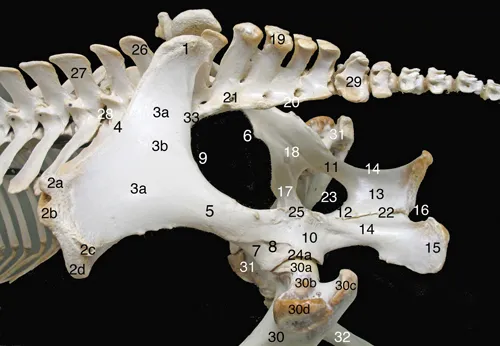
Fig. I.2 Dorsolateral aspect of the equine pelvic bones.
Ilium: 1- Sacral tuber; 2- Tuber coxae, 2a- dorsocranial cuspid, 2b- ventrocranial cuspid, 2c- dorsocaudal cuspid, 2d-ventrocaudal cuspid; 3- Ilium wing, 3a- gluteal face, 3b- accessory gluteal line; 4- Ilium crest, 5- Ilium neck; 6- Psoas minor muscle tubercle; 7- Ilium body; 8- Lateral rectus femoris muscle area; 9- Major sciatic incisura;
Ischium: 10- Ischium body; 11- Lateral (acetabular) ramus; 12- Medial ramus; 13- Ischium table; 14- Minor sciatic incisura; 15- Tuber ischiadicum (ischiatic tuberosity); 16- Ischiatic arch;
Pubis: 17- Cranial ramus; 18- Obturator sulcus;
Sacrum: 19- Median sacral crest (five spinal processes); 20- Lateral sacral crest (fused transverse processes); 21- Dorsal sacral foramen;
Pelvis and connected bones: 22- Pelvic symphysis; 23- Obturator foramen; 24- Acetabulum, 24a- Acetabular margin; 25- Ischiatic spine (sciatic crest); 26- Spinal process of the sixth lumbar vertebra; 27- Fourth lumbar vertebra; 28- Intervertebral foramen; 29- First caudal vertebra; 30- Left femur, 30a- head, 30b- neck, 30c- major trochanter (caudal part); 30d- major trochanter (cranial part); 31- Right femur; 32- Right tibia; 33- Sacroiliac joint.

Fig. I.3 Ventral aspect of the equine pelvic bones.
Ilium: 1- Tuber coxae, 1a- dorsocranial cuspid, 1b- ventrocranial cuspid, 1c- ventrocaudal cuspid; 2- Ilium wing, 2a- iliac face, 2b- sacropelvic face (insertion of the interosseous sacroiliac ligament); 3- Ilium crest; 4- Ilium neck; 5- Arch line; 6- Psoas minor muscle tubercle; 7- Ilium body; 8- Medial rectus femoris muscle area; 9- Major sciatic incisura;
Pubis: 10- Pubis body; 11- Cranial ramus; 12- Caudal ramus, 12a- symphysial face; 13- Pubis pecten; 14- Accessory ligament sulcus;
Ischium: 15- Ischium body; 16- Lateral (acetabular) ramus; 17- Ischium table; 18- Medial ramus, 18a- symphysial face; 19- Tuber ischiadicum (ischiatic tuberosity); 20- Ischiatic arch;
Pelvis: 21- Pelvic symphysis; 22- Obturator foramen; 23- Acetabulum, 23a- acetabular margin, 23b- lunar surface, 23c- acetabular fossa, 23d- acetabular notch;
Sacrum: 24- First sacral vertebra; 25- Fourth sacral vertebra; 26- Sacral wing; 27- First ventral (intervertebral) sacral foramen; 28- Third ventral (intervertebral) sacral foramen; 29- Promontory; 30- Sacroiliac joint;
Lumbar spine: 31- Sixth lumbar vertebra (L6); 32- Transverse process of the fifth lumbar vertebra (L5, fused with L6); 33- Lumbar ventral intervertebral foramen; 34- Third lumbar vertebra (L3, ventral crest); 35- Transverse process of L3; 36- Intertransverse lumbosacral joint;37- Intertransverse synostosis between L5 and L6; 38- Intervertebral symphysis between L3 and L4.

Fig. I.4 Dorsal aspect of the equine pelvic bones.
Ilium: 1- Tuber coxae, 1a- dorsocranial cuspid, 1b- dorsocaudal cuspid, 1c- ventrocaudal cuspid; 2- Sacral tuber; 3- Ilium wing; 4- Ilium crest; 5- Ilium neck; 6- Ilium body; 7- Major sciatic incisura;
Ischium: 8- Ischium body; 9- Lateral (acetabular) ramus; 10- Ischium table; 11- Medial ramus, 11a- symphysial face; 12- Tuber ischiadicum (ischiatic tuberosity); 13- Ischiatic arch;
Pubis: 14- Cranial ramus; 15- Caudal ramus, 15a- symphysial face;
Pelvis: 16- Pelvic symphysis, 16a- cranial (pubic part), 16b- caudal (ischiatic) part; 17- Obturator foramen; 18- Ischiatic spine (sciatic crest); 19- Acetabulum, 19a- acetabular margin;
Sacrum: 20- Median sacral crest; 21- Lateral sacral crest; 22- Transverse process of the first sacral vertebra (sacral wing); 23- Right lumbosacral articular process joint; 24- Sacroiliac joint; 25- Third dorsal (intervertebral) sacral foramen; 26- Sacral canal (caudal opening); 27- First caudal vertebra (vertebral body);
Lumbar spine: 28- Transverse process of the third lumbar vertebra (L3); 29- Transverse process of the fifth lumbar vertebra (L5); 30- Right articular process joint between L3 and L4.

Fig. I.5 Dorsal aspect of the equine sacrum.
1- Spinal process of the first sacral vertebra (S1); 2- Vertebral arch of S1; 3- Left cranial articular process of S1; 4- Transverse process of the first sacral vertebra (sacral wing), 4a- auricular surface (contributing to the sacroiliac joint), 4b- insertion surface of the interosseous sacroiliac ligament; 5- Articular surface of the intertransverse lumbosacral joint seen through the cranial intervertebral incisura; 6- Median sacral crest, 6a- spinal process of the second sacral vertebra, 6b- spinal process of the fifth sacral vertebra; 7- First dorsal (intervertebral) sacral foramen; 8- Second dorsal (intervertebral) sacral foramen; 9- Lateral sacral crest; 10- First caudal vertebra, 10a- vertebral arch, 10b- vertebral body.
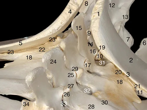
Fig. I.6 Craniolaterodorsal aspect of the lumbosacroiliac area.
Coxal bones: 1- Sacral tuber of the left ilium; 2- Ilium wing of the left ilium; 3- Ilium crest of the left ilium; 4- Sacral tuber of the right ilium; 5- Ilium crest of the right ilium; 6- Left ischium (angle between ilium arch and pelvic symphysis); 7- Right ischium (angle between ilium arch and pelvic symphysis); 8- Right tuber ischiadicum;
Sacrum: 9- Spinal process of the first sacral vertebra (S1); 10- (Cranial) articular process of S1; 11- Left transverse process of S1 (sacral wing); 12- Median sacral crest; 13- Lateral sacral crest; 14- Interarcual space between the last lumbar vertebra (L6) and S1;
Lumbar vertebrae and sacroiliac joints: 15- Spinal process of L6; 16- Caudal articular process of L6; 17- Cranial articular process of L6; 18- Transverse process of L6 (fused with the transverse process of the fifth lumbar vertebra (L5)); 19- Left lumbosacral articular process joint; 20- Left lumbosacral intertransverse joint; 21- Left sacroiliac joint; 22- Right lumbosacral intertransverse joint; 23- Right sacroiliac joint; 24- Spinal process of the fourth lumbar vertebra (L4); 25- Caudal articular process of L4; 26- Cranial articular process of L4; 27- Mammillary process; 28- Transverse process of L4; 29- Left articular process joint between L3 and L4; 30- Left intertransverse joint between L4 and L5; 31- Dorsal lumbosacral intertransverse foramen; 32- Dorsal intertransverse foramen between L4 and L5; 33- Left intervertebral foramen between L3 and L4; 34- Right cranial articular process of the third lumbar vertebra.
I.3 Dissected specimen
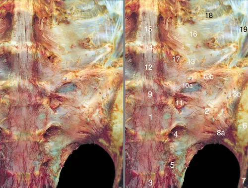
Fig. I.7 Ventral aspect of the roof of the pelvis: bones and ligaments.
1- Body of the first sacral vertebra (S1); 2- Transverse process of S1 (sacral wing); 3- Body of the third sacral vertebra (S3); 4- First ventral (intervertebral) sacral foramen; 5- Second ventral (intervertebral) sacral foramen; 6- Ilium wing; 7- Ilium neck; 8- Sacroiliac joint covered by the ventral sacroiliac ligament, 8a- caudomedial part, 8b- craniolateral part (covered by the iliopsoas muscle); 9- Lumbosacral intervertebral disc (sixth lumbar (L6) disc); 10- Lumbosacral intertransverse joint, 10a- joint space, 10b- lumbosacral intertransverse ligament; 11- Ventral ramus of the sixth lumbar nerve; 12- Body of the sixth lumbar vertebra (L6); 13- Transverse process of L6 (fused with L5 transverse process); 14- Fifth lumbar intervertebral disc; 15- Body of the fifth lumbar vertebra (L5); 16- Transverse process of L5 (fused with L6 transverse process); 17- Ventral intertransverse foramen between L5 and L6; 18- Intertransverse ligament; 19- Iliolumbar ligament.
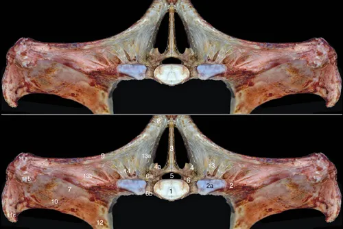
Fig. I.8 Cranial aspect of the equine pelvis: dorsal part.
1- Vertebral body of the first sacral vertebra (S1); 2- Transverse process of S1 (sacral wing), 2a- articular surface of the intertransverse lumbosacral joint; 3- Spinal process of S1; 4- Left (cranial) articular process of S1, 4a- articular surface of the right artic...