
eBook - ePub
Essentials of Clinical Anatomy of the Equine Locomotor System
Jean-Marie Denoix
This is a test
Partager le livre
- 296 pages
- English
- ePUB (adapté aux mobiles)
- Disponible sur iOS et Android
eBook - ePub
Essentials of Clinical Anatomy of the Equine Locomotor System
Jean-Marie Denoix
Détails du livre
Aperçu du livre
Table des matières
Citations
À propos de ce livre
Essentials of Clinical Anatomy of the Equine Locomotor System presents a unique photographic record of dissections showing the topographical anatomy of the locomotor system of the horse. Readers of this book will be able to see the position and relationships of the bones, joints, muscles, nerves and blood vessels that make up each region of the forelimb, vertebral column and hindlimb.
Key features:
- Important features of regional and topographical anatomy are presented using full-color photos of detailed dissections
- Anatomy is presented in a clinical context
- Preparations of cross-sectional anatomy facilitate interpretation of diagnostic imaging, such as ultrasonography, MRI images and CT scans
- All dissections are of fresh material, rather than preserved specimens, to demonstrate the appearance of tissues in the living animal, or at post mortem autopsy
This new atlas is essential for anybody involved in detailed anatomical study, complex lameness evaluation or advanced imaging techniques in horses. It will be a useful guide for veterinary students, and a reference for equine vets in practice.
Foire aux questions
Comment puis-je résilier mon abonnement ?
Il vous suffit de vous rendre dans la section compte dans paramètres et de cliquer sur « Résilier l’abonnement ». C’est aussi simple que cela ! Une fois que vous aurez résilié votre abonnement, il restera actif pour le reste de la période pour laquelle vous avez payé. Découvrez-en plus ici.
Puis-je / comment puis-je télécharger des livres ?
Pour le moment, tous nos livres en format ePub adaptés aux mobiles peuvent être téléchargés via l’application. La plupart de nos PDF sont également disponibles en téléchargement et les autres seront téléchargeables très prochainement. Découvrez-en plus ici.
Quelle est la différence entre les formules tarifaires ?
Les deux abonnements vous donnent un accès complet à la bibliothèque et à toutes les fonctionnalités de Perlego. Les seules différences sont les tarifs ainsi que la période d’abonnement : avec l’abonnement annuel, vous économiserez environ 30 % par rapport à 12 mois d’abonnement mensuel.
Qu’est-ce que Perlego ?
Nous sommes un service d’abonnement à des ouvrages universitaires en ligne, où vous pouvez accéder à toute une bibliothèque pour un prix inférieur à celui d’un seul livre par mois. Avec plus d’un million de livres sur plus de 1 000 sujets, nous avons ce qu’il vous faut ! Découvrez-en plus ici.
Prenez-vous en charge la synthèse vocale ?
Recherchez le symbole Écouter sur votre prochain livre pour voir si vous pouvez l’écouter. L’outil Écouter lit le texte à haute voix pour vous, en surlignant le passage qui est en cours de lecture. Vous pouvez le mettre sur pause, l’accélérer ou le ralentir. Découvrez-en plus ici.
Est-ce que Essentials of Clinical Anatomy of the Equine Locomotor System est un PDF/ePUB en ligne ?
Oui, vous pouvez accéder à Essentials of Clinical Anatomy of the Equine Locomotor System par Jean-Marie Denoix en format PDF et/ou ePUB ainsi qu’à d’autres livres populaires dans Medicine et Opthalmology & Optometry. Nous disposons de plus d’un million d’ouvrages à découvrir dans notre catalogue.
Informations
Chapter I
THE PELVIS
I.1 Physical aspect

Fig. I.1 Caudolateral aspect of the equine pelvis. (A) Bones; (B) superficial aspect.
1- Sacral tuber; 2- Tuber coxae; 3a- Tuber ischiadicum, 3b- point of the croup; 4a- Greater trochanter, 4b- Hip; 5- Third trochanter; 6- Gluteus medius muscle; 7- Gluteus superficialis muscle; 8- Gluteofemoralis muscle; 9- Biceps femoris muscle; 10- Semitendinosus muscle; 11- Semimembranosus muscle; 12- Tensor fascia latae muscle; 13- Sacrum (median sacral crest); 14a- Caudal vertebrae, 14b- Tail.
I.2 Bones
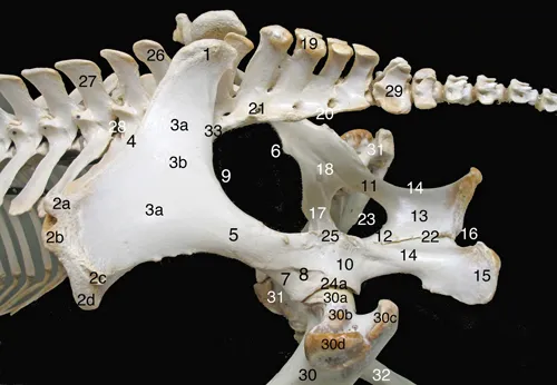
Fig. I.2 Dorsolateral aspect of the equine pelvic bones.
Ilium: 1- Sacral tuber; 2- Tuber coxae, 2a- dorsocranial cuspid, 2b- ventrocranial cuspid, 2c- dorsocaudal cuspid, 2d-ventrocaudal cuspid; 3- Ilium wing, 3a- gluteal face, 3b- accessory gluteal line; 4- Ilium crest, 5- Ilium neck; 6- Psoas minor muscle tubercle; 7- Ilium body; 8- Lateral rectus femoris muscle area; 9- Major sciatic incisura;
Ischium: 10- Ischium body; 11- Lateral (acetabular) ramus; 12- Medial ramus; 13- Ischium table; 14- Minor sciatic incisura; 15- Tuber ischiadicum (ischiatic tuberosity); 16- Ischiatic arch;
Pubis: 17- Cranial ramus; 18- Obturator sulcus;
Sacrum: 19- Median sacral crest (five spinal processes); 20- Lateral sacral crest (fused transverse processes); 21- Dorsal sacral foramen;
Pelvis and connected bones: 22- Pelvic symphysis; 23- Obturator foramen; 24- Acetabulum, 24a- Acetabular margin; 25- Ischiatic spine (sciatic crest); 26- Spinal process of the sixth lumbar vertebra; 27- Fourth lumbar vertebra; 28- Intervertebral foramen; 29- First caudal vertebra; 30- Left femur, 30a- head, 30b- neck, 30c- major trochanter (caudal part); 30d- major trochanter (cranial part); 31- Right femur; 32- Right tibia; 33- Sacroiliac joint.

Fig. I.3 Ventral aspect of the equine pelvic bones.
Ilium: 1- Tuber coxae, 1a- dorsocranial cuspid, 1b- ventrocranial cuspid, 1c- ventrocaudal cuspid; 2- Ilium wing, 2a- iliac face, 2b- sacropelvic face (insertion of the interosseous sacroiliac ligament); 3- Ilium crest; 4- Ilium neck; 5- Arch line; 6- Psoas minor muscle tubercle; 7- Ilium body; 8- Medial rectus femoris muscle area; 9- Major sciatic incisura;
Pubis: 10- Pubis body; 11- Cranial ramus; 12- Caudal ramus, 12a- symphysial face; 13- Pubis pecten; 14- Accessory ligament sulcus;
Ischium: 15- Ischium body; 16- Lateral (acetabular) ramus; 17- Ischium table; 18- Medial ramus, 18a- symphysial face; 19- Tuber ischiadicum (ischiatic tuberosity); 20- Ischiatic arch;
Pelvis: 21- Pelvic symphysis; 22- Obturator foramen; 23- Acetabulum, 23a- acetabular margin, 23b- lunar surface, 23c- acetabular fossa, 23d- acetabular notch;
Sacrum: 24- First sacral vertebra; 25- Fourth sacral vertebra; 26- Sacral wing; 27- First ventral (intervertebral) sacral foramen; 28- Third ventral (intervertebral) sacral foramen; 29- Promontory; 30- Sacroiliac joint;
Lumbar spine: 31- Sixth lumbar vertebra (L6); 32- Transverse process of the fifth lumbar vertebra (L5, fused with L6); 33- Lumbar ventral intervertebral foramen; 34- Third lumbar vertebra (L3, ventral crest); 35- Transverse process of L3; 36- Intertransverse lumbosacral joint;37- Intertransverse synostosis between L5 and L6; 38- Intervertebral symphysis between L3 and L4.

Fig. I.4 Dorsal aspect of the equine pelvic bones.
Ilium: 1- Tuber coxae, 1a- dorsocranial cuspid, 1b- dorsocaudal cuspid, 1c- ventrocaudal cuspid; 2- Sacral tuber; 3- Ilium wing; 4- Ilium crest; 5- Ilium neck; 6- Ilium body; 7- Major sciatic incisura;
Ischium: 8- Ischium body; 9- Lateral (acetabular) ramus; 10- Ischium table; 11- Medial ramus, 11a- symphysial face; 12- Tuber ischiadicum (ischiatic tuberosity); 13- Ischiatic arch;
Pubis: 14- Cranial ramus; 15- Caudal ramus, 15a- symphysial face;
Pelvis: 16- Pelvic symphysis, 16a- cranial (pubic part), 16b- caudal (ischiatic) part; 17- Obturator foramen; 18- Ischiatic spine (sciatic crest); 19- Acetabulum, 19a- acetabular margin;
Sacrum: 20- Median sacral crest; 21- Lateral sacral crest; 22- Transverse process of the first sacral vertebra (sacral wing); 23- Right lumbosacral articular process joint; 24- Sacroiliac joint; 25- Third dorsal (intervertebral) sacral foramen; 26- Sacral canal (caudal opening); 27- First caudal vertebra (vertebral body);
Lumbar spine: 28- Transverse process of the third lumbar vertebra (L3); 29- Transverse process of the fifth lumbar vertebra (L5); 30- Right articular process joint between L3 and L4.

Fig. I.5 Dorsal aspect of the equine sacrum.
1- Spinal process of the first sacral vertebra (S1); 2- Vertebral arch of S1; 3- Left cranial articular process of S1; 4- Transverse process of the first sacral vertebra (sacral wing), 4a- auricular surface (contributing to the sacroiliac joint), 4b- insertion surface of the interosseous sacroiliac ligament; 5- Articular surface of the intertransverse lumbosacral joint seen through the cranial intervertebral incisura; 6- Median sacral crest, 6a- spinal process of the second sacral vertebra, 6b- spinal process of the fifth sacral vertebra; 7- First dorsal (intervertebral) sacral foramen; 8- Second dorsal (intervertebral) sacral foramen; 9- Lateral sacral crest; 10- First caudal vertebra, 10a- vertebral arch, 10b- vertebral body.
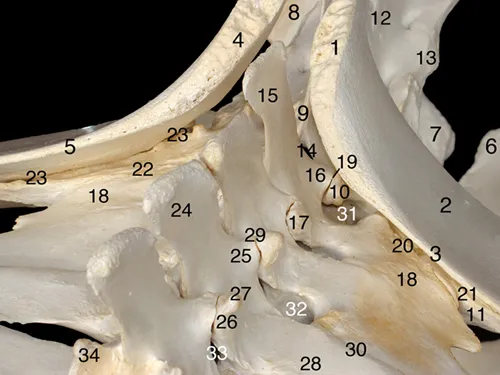
Fig. I.6 Craniolaterodorsal aspect of the lumbosacroiliac area.
Coxal bones: 1- Sacral tuber of the left ilium; 2- Ilium wing of the left ilium; 3- Ilium crest of the left ilium; 4- Sacral tuber of the right ilium; 5- Ilium crest of the right ilium; 6- Left ischium (angle between ilium arch and pelvic symphysis); 7- Right ischium (angle between ilium arch and pelvic symphysis); 8- Right tuber ischiadicum;
Sacrum: 9- Spinal process of the first sacral vertebra (S1); 10- (Cranial) articular process of S1; 11- Left transverse process of S1 (sacral wing); 12- Median sacral crest; 13- Lateral sacral crest; 14- Interarcual space between the last lumbar vertebra (L6) and S1;
Lumbar vertebrae and sacroiliac joints: 15- Spinal process of L6; 16- Caudal articular process of L6; 17- Cranial articular process of L6; 18- Transverse process of L6 (fused with the transverse process of the fifth lumbar vertebra (L5)); 19- Left lumbosacral articular process joint; 20- Left lumbosacral intertransverse joint; 21- Left sacroiliac joint; 22- Right lumbosacral intertransverse joint; 23- Right sacroiliac joint; 24- Spinal process of the fourth lumbar vertebra (L4); 25- Caudal articular process of L4; 26- Cranial articular process of L4; 27- Mammillary process; 28- Transverse process of L4; 29- Left articular process joint between L3 and L4; 30- Left intertransverse joint between L4 and L5; 31- Dorsal lumbosacral intertransverse foramen; 32- Dorsal intertransverse foramen between L4 and L5; 33- Left intervertebral foramen between L3 and L4; 34- Right cranial articular process of the third lumbar vertebra.
I.3 Dissected specimen
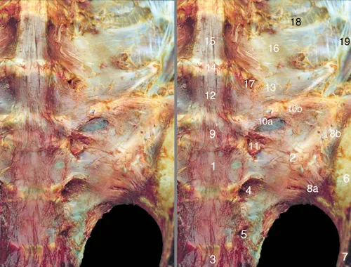
Fig. I.7 Ventral aspect of the roof of the pelvis: bones and ligaments.
1- Body of the first sacral vertebra (S1); 2- Transverse process of S1 (sacral wing); 3- Body of the third sacral vertebra (S3); 4- First ventral (intervertebral) sacral foramen; 5- Second ventral (intervertebral) sacral foramen; 6- Ilium wing; 7- Ilium neck; 8- Sacroiliac joint covered by the ventral sacroiliac ligament, 8a- caudomedial part, 8b- craniolateral part (covered by the iliopsoas muscle); 9- Lumbosacral intervertebral disc (sixth lumbar (L6) disc); 10- Lumbosacral intertransverse joint, 10a- joint space, 10b- lumbosacral intertransverse ligament; 11- Ventral ramus of the sixth lumbar nerve; 12- Body of the sixth lumbar vertebra (L6); 13- Transverse process of L6 (fused with L5 transverse process); 14- Fifth lumbar intervertebral disc; 15- Body of the fifth lumbar vertebra (L5); 16- Transverse process of L5 (fused with L6 transverse process); 17- Ventral intertransverse foramen between L5 and L6; 18- Intertransverse ligament; 19- Iliolumbar ligament.
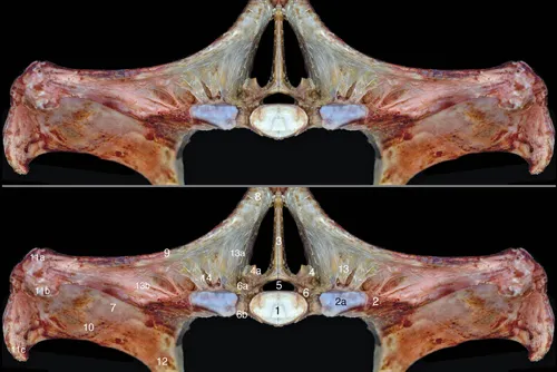
Fig. I.8 Cranial aspect of the equine pelvis: dorsal part.
1- Vertebral body of the first sacral vertebra (S1); 2- Transverse process of S1 (sacral wing), 2a- articular surface of the intertransverse lumbosacral joint; 3- Spinal process of S1; 4- Left (cranial) articular process of S1, 4a- articular surface of the right artic...