eBook - ePub
Fibroids and Reproduction
Botros R.M.B. Rizk, Yakoub Khalaf, Mostafa A. Borahay, Botros R.M.B. Rizk, Yakoub Khalaf, Mostafa A. Borahay
This is a test
- 130 pagine
- English
- ePUB (disponibile sull'app)
- Disponibile su iOS e Android
eBook - ePub
Fibroids and Reproduction
Botros R.M.B. Rizk, Yakoub Khalaf, Mostafa A. Borahay, Botros R.M.B. Rizk, Yakoub Khalaf, Mostafa A. Borahay
Dettagli del libro
Anteprima del libro
Indice dei contenuti
Citazioni
Informazioni sul libro
The most common abnormal growth of the female reproductive system, fibroids, are thought to affect the majority of women at some point during their reproductive years. This text from leading fibroid experts looks at the latest evidence on how the problem impinges on reproduction and the most up-to-date management and treatment options available to help patients with fibroids hoping to conceive.
Print versions of this book also include access to the eBook version with links to procedural videos.
Domande frequenti
Come faccio ad annullare l'abbonamento?
È semplicissimo: basta accedere alla sezione Account nelle Impostazioni e cliccare su "Annulla abbonamento". Dopo la cancellazione, l'abbonamento rimarrà attivo per il periodo rimanente già pagato. Per maggiori informazioni, clicca qui
È possibile scaricare libri? Se sì, come?
Al momento è possibile scaricare tramite l'app tutti i nostri libri ePub mobile-friendly. Anche la maggior parte dei nostri PDF è scaricabile e stiamo lavorando per rendere disponibile quanto prima il download di tutti gli altri file. Per maggiori informazioni, clicca qui
Che differenza c'è tra i piani?
Entrambi i piani ti danno accesso illimitato alla libreria e a tutte le funzionalità di Perlego. Le uniche differenze sono il prezzo e il periodo di abbonamento: con il piano annuale risparmierai circa il 30% rispetto a 12 rate con quello mensile.
Cos'è Perlego?
Perlego è un servizio di abbonamento a testi accademici, che ti permette di accedere a un'intera libreria online a un prezzo inferiore rispetto a quello che pagheresti per acquistare un singolo libro al mese. Con oltre 1 milione di testi suddivisi in più di 1.000 categorie, troverai sicuramente ciò che fa per te! Per maggiori informazioni, clicca qui.
Perlego supporta la sintesi vocale?
Cerca l'icona Sintesi vocale nel prossimo libro che leggerai per verificare se è possibile riprodurre l'audio. Questo strumento permette di leggere il testo a voce alta, evidenziandolo man mano che la lettura procede. Puoi aumentare o diminuire la velocità della sintesi vocale, oppure sospendere la riproduzione. Per maggiori informazioni, clicca qui.
Fibroids and Reproduction è disponibile online in formato PDF/ePub?
Sì, puoi accedere a Fibroids and Reproduction di Botros R.M.B. Rizk, Yakoub Khalaf, Mostafa A. Borahay, Botros R.M.B. Rizk, Yakoub Khalaf, Mostafa A. Borahay in formato PDF e/o ePub, così come ad altri libri molto apprezzati nelle sezioni relative a Medicina e Teoría, práctica y referencia médicas. Scopri oltre 1 milione di libri disponibili nel nostro catalogo.
Informazioni
1
Fibroids and Reproduction: A Bird’s-Eye View
Botros R.M.B. Rizk, Candice P. Holliday, and Yakoub Khalaf
Contents
Introduction
Classification
Diagnosis
Fertility
Conception
Miscarriage
In Vitro Fertilization Outcome
References
Introduction
A fibroid (or leiomyoma) is a benign, monoclonal, smooth muscle tumor of the uterus, which usually presents as multiple lesions (Figure 1.1) but can occur as a single lesion (Figure 1.2). Fibroids are diagnosed in 20%–40% of women, generally after age 30 years, with a stable increase in incidence with increasing age [1]. This age-related increase in incidence of fibroids should be considered when looking into the relationship between fibroids and reproductive dysfunction (subfertility or miscarriage), as both are intimately age related.
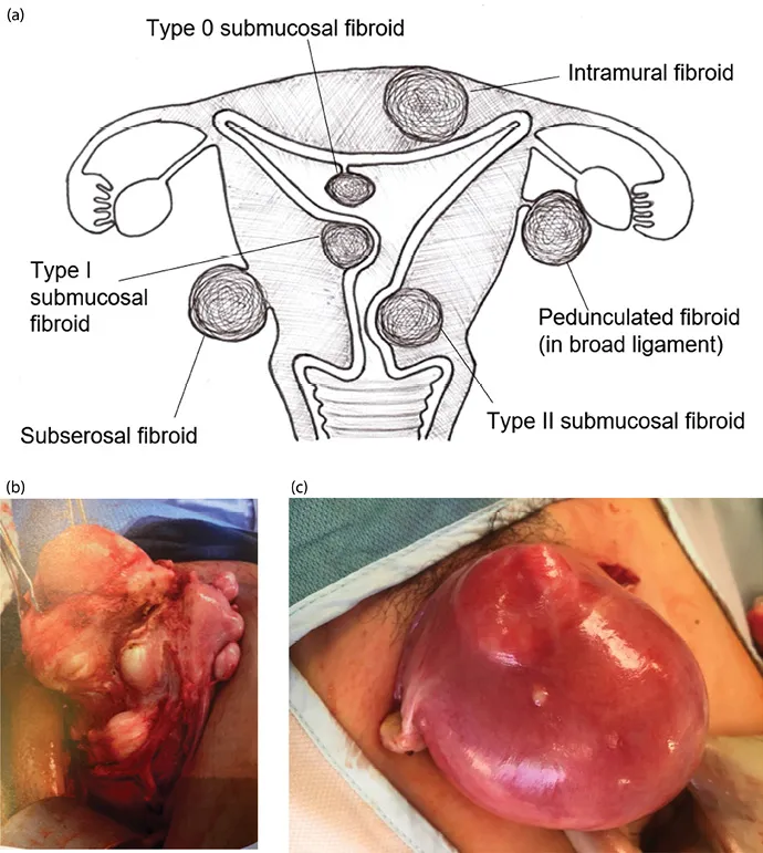
Figure 1.1 (a) Varying fibroid locations within the uterus; (b) multiple fibroids—intramural, subserosal, and submucosal; (c) uterus enlarged with intramural fibroids.
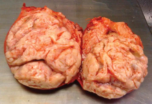
Figure 1.2 Solitary large fibroid.
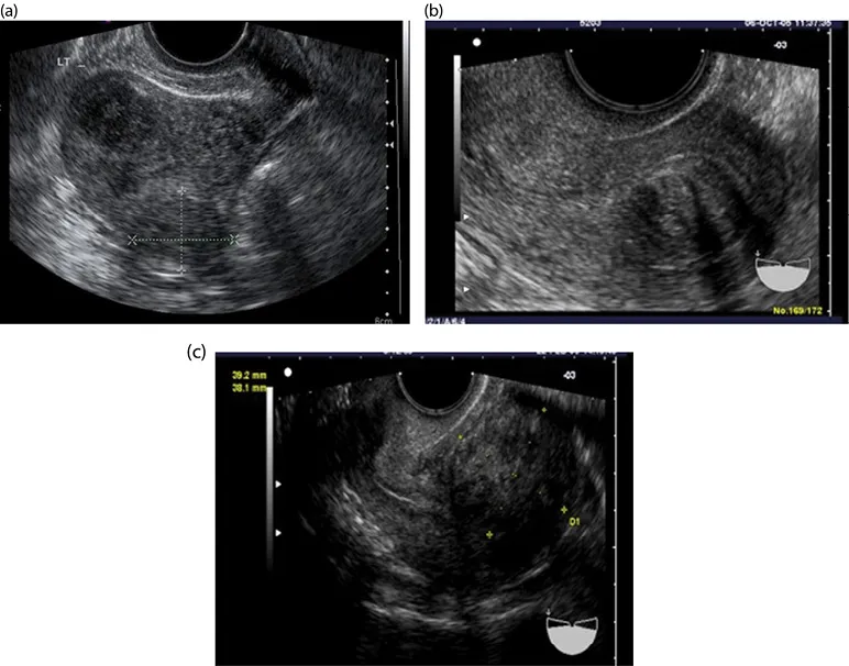
Figure 1.3 (a–c) Intramural fibroids.
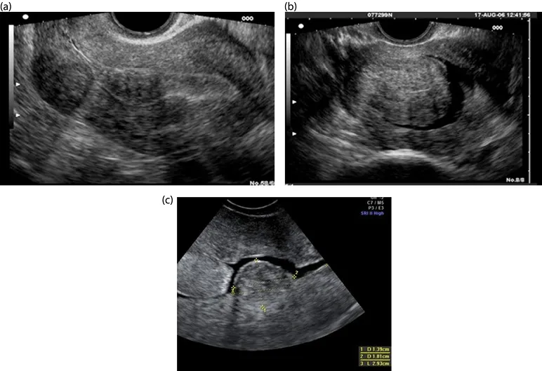
Figure 1.4 (a–c) Submucosal fibroids.
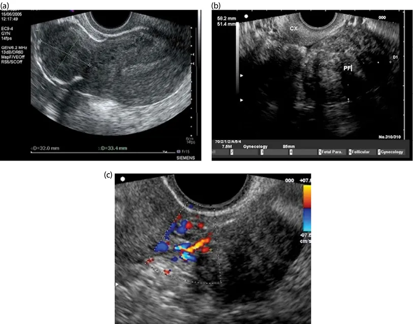
Figure 1.5 (a) Subserosal fibroid; (b) pedunculated fibroid; (c) Doppler scan showing feeder vessels of pedunculated fibroid.
Although it is biologically plausible and clinically evident that fibroids are associated with reproductive dysfunction (see Chapter 2), a cause-effect relationship has not been established.
Most symptomatic fibroids can be diagnosed clinically, but crucial clinical information can be obtained by one imaging modality or another.
Ultrasound is a noninvasive imaging modality that is well tolerated by patients, and it is a rather inexpensive way to obtain a relatively accurate assessment of fibroids within the pelvis (see Chapter 8). Assessing fibroid size and location can be beneficial for planning surgery or for monitoring changes in fibroids over time (Figures 1.3–1.5).
In some complex cases (multiple fibroids, previous surgery, and associated morbidities), magnetic resonance imaging (MRI) can provide additional valuable information that could help in planning surgery or guide the choice of alternative therapeutic approaches, such as the use of uterine artery embolization (UAE) or MRI-guided focused ultrasound (see Chapter 7).
Classification
When fibroids develop from the uterine wall but distort the uterine cavity, it is helpful to detail the degree of uterine cavity impingement (see Chapter 7) with the Wamsteker and de Blok classification [2]. Following are the types of fibroids:
•0: 100% of the fibroid is pedunculated into the uterine cavity
•I: Greater than 50% of the fibroid is within the uterine cavity
•II: Less than 50% of the fibroid is within the uterine cavity (i.e., greater than 50% of the fibroid is within the myometrium)
It can be difficult to assess on two-dimensional (2D) ultrasound scan what type of fibroid a patient has. Three-dimensional (3D) ultrasound and saline infusion sonohysterography are often used to obtain that information (Figures 1.6–1.8). A diagnosis that is accurate is critical to determine presurgical treatment, what type of surgery would be best, and what sort of prognosis a patient can expect [2]. A comparable classification system has been suggested for intramural and subserosal fibroids in order to describe what degree of myometrial involvement exists (see Chapter 2 for the detailed classification system). Three-dimensional ultrasound is becoming an increasingly valuable imaging tool to map out the relationship between the fibroid and the endometrial cavity.
Diagnosis
Two-dimensional ultrasound is the traditional manner of imaging fibroids, although other imaging modalities exist. For ideal visualization of fibroids, especially of their outline, the best technique uses a transvaginal scan (TVS) (see Figures 1.3–1.5). With a transvaginal approach, the ultrasound probe is closer to the uterus, which allows a higher frequency to be used. This higher frequency provides better definition of the tissues. A patient should empty her bladder first before a TVS is done. The TVS transducer is curvilinear, multifrequency, and endocavity, with a central frequency that is usually 6.5 MHz. The ultrasound beam can be highly attenuated by fibroids due to fibroids’ dense and mixed tissue composition. Thus, poor throug...
Indice dei contenuti
Stili delle citazioni per Fibroids and Reproduction
APA 6 Citation
[author missing]. (2020). Fibroids and Reproduction (1st ed.). CRC Press. Retrieved from https://www.perlego.com/book/1718882/fibroids-and-reproduction-pdf (Original work published 2020)
Chicago Citation
[author missing]. (2020) 2020. Fibroids and Reproduction. 1st ed. CRC Press. https://www.perlego.com/book/1718882/fibroids-and-reproduction-pdf.
Harvard Citation
[author missing] (2020) Fibroids and Reproduction. 1st edn. CRC Press. Available at: https://www.perlego.com/book/1718882/fibroids-and-reproduction-pdf (Accessed: 14 October 2022).
MLA 7 Citation
[author missing]. Fibroids and Reproduction. 1st ed. CRC Press, 2020. Web. 14 Oct. 2022.