eBook - ePub
Fibroids and Reproduction
Botros R.M.B. Rizk, Yakoub Khalaf, Mostafa A. Borahay, Botros R.M.B. Rizk, Yakoub Khalaf, Mostafa A. Borahay
This is a test
- 130 Seiten
- English
- ePUB (handyfreundlich)
- Über iOS und Android verfügbar
eBook - ePub
Fibroids and Reproduction
Botros R.M.B. Rizk, Yakoub Khalaf, Mostafa A. Borahay, Botros R.M.B. Rizk, Yakoub Khalaf, Mostafa A. Borahay
Angaben zum Buch
Buchvorschau
Inhaltsverzeichnis
Quellenangaben
Über dieses Buch
The most common abnormal growth of the female reproductive system, fibroids, are thought to affect the majority of women at some point during their reproductive years. This text from leading fibroid experts looks at the latest evidence on how the problem impinges on reproduction and the most up-to-date management and treatment options available to help patients with fibroids hoping to conceive.
Print versions of this book also include access to the eBook version with links to procedural videos.
Häufig gestellte Fragen
Wie kann ich mein Abo kündigen?
Gehe einfach zum Kontobereich in den Einstellungen und klicke auf „Abo kündigen“ – ganz einfach. Nachdem du gekündigt hast, bleibt deine Mitgliedschaft für den verbleibenden Abozeitraum, den du bereits bezahlt hast, aktiv. Mehr Informationen hier.
(Wie) Kann ich Bücher herunterladen?
Derzeit stehen all unsere auf Mobilgeräte reagierenden ePub-Bücher zum Download über die App zur Verfügung. Die meisten unserer PDFs stehen ebenfalls zum Download bereit; wir arbeiten daran, auch die übrigen PDFs zum Download anzubieten, bei denen dies aktuell noch nicht möglich ist. Weitere Informationen hier.
Welcher Unterschied besteht bei den Preisen zwischen den Aboplänen?
Mit beiden Aboplänen erhältst du vollen Zugang zur Bibliothek und allen Funktionen von Perlego. Die einzigen Unterschiede bestehen im Preis und dem Abozeitraum: Mit dem Jahresabo sparst du auf 12 Monate gerechnet im Vergleich zum Monatsabo rund 30 %.
Was ist Perlego?
Wir sind ein Online-Abodienst für Lehrbücher, bei dem du für weniger als den Preis eines einzelnen Buches pro Monat Zugang zu einer ganzen Online-Bibliothek erhältst. Mit über 1 Million Büchern zu über 1.000 verschiedenen Themen haben wir bestimmt alles, was du brauchst! Weitere Informationen hier.
Unterstützt Perlego Text-zu-Sprache?
Achte auf das Symbol zum Vorlesen in deinem nächsten Buch, um zu sehen, ob du es dir auch anhören kannst. Bei diesem Tool wird dir Text laut vorgelesen, wobei der Text beim Vorlesen auch grafisch hervorgehoben wird. Du kannst das Vorlesen jederzeit anhalten, beschleunigen und verlangsamen. Weitere Informationen hier.
Ist Fibroids and Reproduction als Online-PDF/ePub verfügbar?
Ja, du hast Zugang zu Fibroids and Reproduction von Botros R.M.B. Rizk, Yakoub Khalaf, Mostafa A. Borahay, Botros R.M.B. Rizk, Yakoub Khalaf, Mostafa A. Borahay im PDF- und/oder ePub-Format sowie zu anderen beliebten Büchern aus Medicina & Teoría, práctica y referencia médicas. Aus unserem Katalog stehen dir über 1 Million Bücher zur Verfügung.
Information
1
Fibroids and Reproduction: A Bird’s-Eye View
Botros R.M.B. Rizk, Candice P. Holliday, and Yakoub Khalaf
Contents
Introduction
Classification
Diagnosis
Fertility
Conception
Miscarriage
In Vitro Fertilization Outcome
References
Introduction
A fibroid (or leiomyoma) is a benign, monoclonal, smooth muscle tumor of the uterus, which usually presents as multiple lesions (Figure 1.1) but can occur as a single lesion (Figure 1.2). Fibroids are diagnosed in 20%–40% of women, generally after age 30 years, with a stable increase in incidence with increasing age [1]. This age-related increase in incidence of fibroids should be considered when looking into the relationship between fibroids and reproductive dysfunction (subfertility or miscarriage), as both are intimately age related.
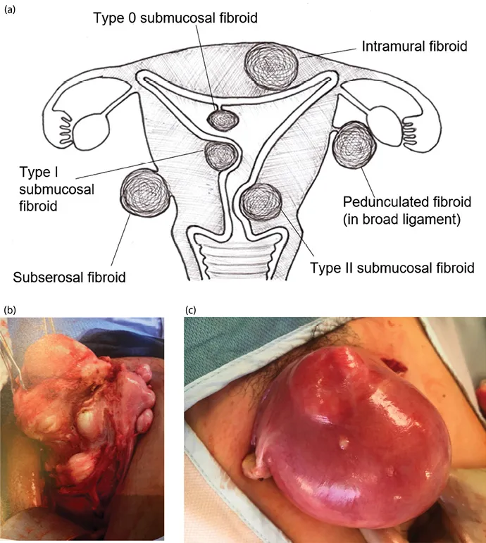
Figure 1.1 (a) Varying fibroid locations within the uterus; (b) multiple fibroids—intramural, subserosal, and submucosal; (c) uterus enlarged with intramural fibroids.
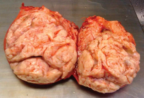
Figure 1.2 Solitary large fibroid.
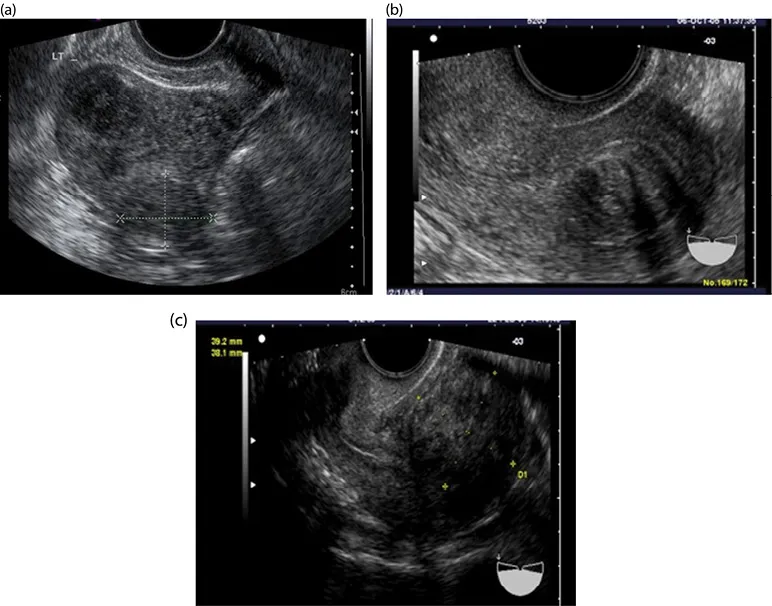
Figure 1.3 (a–c) Intramural fibroids.
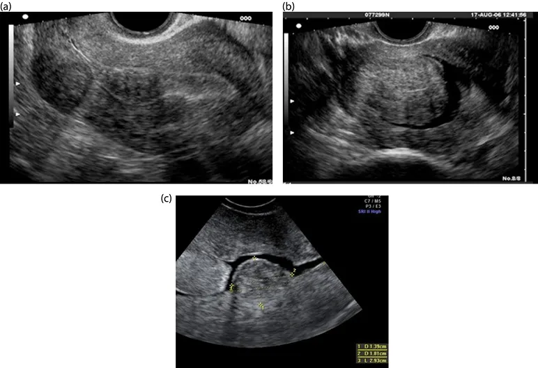
Figure 1.4 (a–c) Submucosal fibroids.
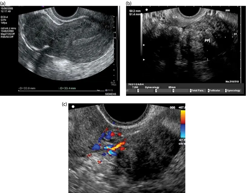
Figure 1.5 (a) Subserosal fibroid; (b) pedunculated fibroid; (c) Doppler scan showing feeder vessels of pedunculated fibroid.
Although it is biologically plausible and clinically evident that fibroids are associated with reproductive dysfunction (see Chapter 2), a cause-effect relationship has not been established.
Most symptomatic fibroids can be diagnosed clinically, but crucial clinical information can be obtained by one imaging modality or another.
Ultrasound is a noninvasive imaging modality that is well tolerated by patients, and it is a rather inexpensive way to obtain a relatively accurate assessment of fibroids within the pelvis (see Chapter 8). Assessing fibroid size and location can be beneficial for planning surgery or for monitoring changes in fibroids over time (Figures 1.3–1.5).
In some complex cases (multiple fibroids, previous surgery, and associated morbidities), magnetic resonance imaging (MRI) can provide additional valuable information that could help in planning surgery or guide the choice of alternative therapeutic approaches, such as the use of uterine artery embolization (UAE) or MRI-guided focused ultrasound (see Chapter 7).
Classification
When fibroids develop from the uterine wall but distort the uterine cavity, it is helpful to detail the degree of uterine cavity impingement (see Chapter 7) with the Wamsteker and de Blok classification [2]. Following are the types of fibroids:
•0: 100% of the fibroid is pedunculated into the uterine cavity
•I: Greater than 50% of the fibroid is within the uterine cavity
•II: Less than 50% of the fibroid is within the uterine cavity (i.e., greater than 50% of the fibroid is within the myometrium)
It can be difficult to assess on two-dimensional (2D) ultrasound scan what type of fibroid a patient has. Three-dimensional (3D) ultrasound and saline infusion sonohysterography are often used to obtain that information (Figures 1.6–1.8). A diagnosis that is accurate is critical to determine presurgical treatment, what type of surgery would be best, and what sort of prognosis a patient can expect [2]. A comparable classification system has been suggested for intramural and subserosal fibroids in order to describe what degree of myometrial involvement exists (see Chapter 2 for the detailed classification system). Three-dimensional ultrasound is becoming an increasingly valuable imaging tool to map out the relationship between the fibroid and the endometrial cavity.
Diagnosis
Two-dimensional ultrasound is the traditional manner of imaging fibroids, although other imaging modalities exist. For ideal visualization of fibroids, especially of their outline, the best technique uses a transvaginal scan (TVS) (see Figures 1.3–1.5). With a transvaginal approach, the ultrasound probe is closer to the uterus, which allows a higher frequency to be used. This higher frequency provides better definition of the tissues. A patient should empty her bladder first before a TVS is done. The TVS transducer is curvilinear, multifrequency, and endocavity, with a central frequency that is usually 6.5 MHz. The ultrasound beam can be highly attenuated by fibroids due to fibroids’ dense and mixed tissue composition. Thus, poor throug...
Inhaltsverzeichnis
Zitierstile für Fibroids and Reproduction
APA 6 Citation
[author missing]. (2020). Fibroids and Reproduction (1st ed.). CRC Press. Retrieved from https://www.perlego.com/book/1718882/fibroids-and-reproduction-pdf (Original work published 2020)
Chicago Citation
[author missing]. (2020) 2020. Fibroids and Reproduction. 1st ed. CRC Press. https://www.perlego.com/book/1718882/fibroids-and-reproduction-pdf.
Harvard Citation
[author missing] (2020) Fibroids and Reproduction. 1st edn. CRC Press. Available at: https://www.perlego.com/book/1718882/fibroids-and-reproduction-pdf (Accessed: 14 October 2022).
MLA 7 Citation
[author missing]. Fibroids and Reproduction. 1st ed. CRC Press, 2020. Web. 14 Oct. 2022.