
eBook - ePub
OCT Angiography in Retinal and Macular Diseases
F. Bandello, E. H. Souied, G. Querques
This is a test
Condividi libro
- 184 pagine
- English
- ePUB (disponibile sull'app)
- Disponibile su iOS e Android
eBook - ePub
OCT Angiography in Retinal and Macular Diseases
F. Bandello, E. H. Souied, G. Querques
Dettagli del libro
Anteprima del libro
Indice dei contenuti
Citazioni
Informazioni sul libro
In only a short period of time, the innovative procedure of OCT angiography has become an essential macula imaging technique. Now that it is routinely used in clinical practice, the investigation of retinal and choroidal circulation is non-invasive, which significantly changes the professional's approach to patients. In this volume, retina specialists and renowned experts share their experience with OCT angiography. They have included numerous color images and presented current ideas to form a base for further research and discussion. This book provides retina specialists, ophthalmologists, and researchers with a first glance at original research and clinical reports on this new methodology.
Domande frequenti
Come faccio ad annullare l'abbonamento?
È semplicissimo: basta accedere alla sezione Account nelle Impostazioni e cliccare su "Annulla abbonamento". Dopo la cancellazione, l'abbonamento rimarrà attivo per il periodo rimanente già pagato. Per maggiori informazioni, clicca qui
È possibile scaricare libri? Se sì, come?
Al momento è possibile scaricare tramite l'app tutti i nostri libri ePub mobile-friendly. Anche la maggior parte dei nostri PDF è scaricabile e stiamo lavorando per rendere disponibile quanto prima il download di tutti gli altri file. Per maggiori informazioni, clicca qui
Che differenza c'è tra i piani?
Entrambi i piani ti danno accesso illimitato alla libreria e a tutte le funzionalità di Perlego. Le uniche differenze sono il prezzo e il periodo di abbonamento: con il piano annuale risparmierai circa il 30% rispetto a 12 rate con quello mensile.
Cos'è Perlego?
Perlego è un servizio di abbonamento a testi accademici, che ti permette di accedere a un'intera libreria online a un prezzo inferiore rispetto a quello che pagheresti per acquistare un singolo libro al mese. Con oltre 1 milione di testi suddivisi in più di 1.000 categorie, troverai sicuramente ciò che fa per te! Per maggiori informazioni, clicca qui.
Perlego supporta la sintesi vocale?
Cerca l'icona Sintesi vocale nel prossimo libro che leggerai per verificare se è possibile riprodurre l'audio. Questo strumento permette di leggere il testo a voce alta, evidenziandolo man mano che la lettura procede. Puoi aumentare o diminuire la velocità della sintesi vocale, oppure sospendere la riproduzione. Per maggiori informazioni, clicca qui.
OCT Angiography in Retinal and Macular Diseases è disponibile online in formato PDF/ePub?
Sì, puoi accedere a OCT Angiography in Retinal and Macular Diseases di F. Bandello, E. H. Souied, G. Querques in formato PDF e/o ePub, così come ad altri libri molto apprezzati nelle sezioni relative a Medizin e Augenheilkunde & Optometrie. Scopri oltre 1 milione di libri disponibili nel nostro catalogo.
Informazioni
Argomento
MedizinCategoria
Augenheilkunde & OptometrieBandello F, Souied EH, Querques G (eds): OCT Angiography in Retinal and Macular Diseases.
Dev Ophthalmol. Basel, Karger, 2016, vol 56, pp 45-51 (DOI: 10.1159/000442776)
Dev Ophthalmol. Basel, Karger, 2016, vol 56, pp 45-51 (DOI: 10.1159/000442776)
______________________
Optical Coherence Tomography Angiography of Type 1 Neovascularization in Age-Related Macular Degeneration
Nicholas A. Iafea · Nopasak Phasukkijwatanaa · David Sarrafa, b
aStein Eye Institute, David Geffen School of Medicine at University of California Los Angeles, and bGreater Los Angeles VA Healthcare Center, Los Angeles, Calif., USA
______________________
Abstract
Age-related macular degeneration continues to be the leading cause of severe central vision loss in older adults of European descent. Optical coherence tomography angiography (OCT-A) enables more accurate identification of type 1 neovascularization in age-related macular degeneration than traditional fluorescein and indocyanine green angiographies. In addition, OCT-A facilitates the morphological classification of type 1 lesions, including features characteristic of early, mature, and fibrotic lesions. Vessel complex analysis, including lesion area and capillary density quantification, can also be readily measured and monitored over time. Performing this analysis following anti-vascular endothelial growth factor therapy may lead to a better understanding of the efficacies and responses to such treatments. Although some limitations currently exist, OCT-A is a promising imaging modality that could prove to have profound implications if incorporated into regular clinical practice.
© 2016 S. Karger AG, Basel
Age-related macular degeneration (AMD) continues to be the leading cause of blindness among individuals older than 50 years of age in the developed world [1]. Neovascular AMD is the etiology for severe vision loss in 90% of AMD cases. Three lesion subtypes, best classified on the basis of spectral domain optical coherence tomography (OCT), comprise the neovascular form of this disease [2]. Type 1 neovascularization originates from the choriocapillaris and is localized under the retinal pigment epithelium. Type 2 neovascularization also originates from the choriocapillaris but extends through the retinal pigment epithelium and is localized in the subretinal compartment. Type 3 neovascularization originates from the deep retinal capillary plexus [2-4] and is located in the outer retina. Type 1 neovascularization is the most common neovascular subtype of AMD [5].
Recent advancements in OCT angiography (OCT-A) have provided retinologists with a window to directly identify the morphologies of neovascular subtypes in AMD. OCT-A enables more accurate identification of type 1 lesions compared to traditional fluorescein angiography (FA). While FA can identify the superficial retinal capillary plexus, this imaging modality poorly visualizes the deep retinal capillary plexus and the choroid. Pigment epithelial detachment (PED) may demonstrate pooling or stippled fluorescence with FA, but the identification of the causative neovascular complex is very challenging and only minimally improved with indocyanine green angiography. Conversely, OCT-A utilizes amplitude or phase decorrelation technology with high-frequency and dense volumetric scanning to detect red blood cell movement and to visualize blood vessels at various depth-resolved levels of the retina and choroid [3]. As opposed to FA and indocyanine green angiography, in which the presence of an occult choroidal neovascular membrane is inferred by the presence of pooling within a PED and/or the identification of a hot spot, OCT-A reveals the vessels themselves and enables one to more accurately identify and evaluate the morphology of the neovascular complex.
OCT-A of type 1 neovascularization has lead to a detailed assessment of the microvascular morphologies of these vessel complexes, which are typically hidden under a PED. Numerous studies have identified the different morphologies of these neovascular lesions and have applied varying descriptive terms to label these structures, which are best visualized with OCT-A. These labels include ‘umbrella vessels’ , ‘seafan and medusa vessels’ , ‘tangled network pattern’ , and ‘pruned vascular and blossoming tree’ [3,6,7]. This complicated and indistinct nomenclature has caused confusion in the retina community, and a simpler classification system will certainly evolve that may reflect the chronicity of type 1 neovascularization as identified with OCT-A.
Though the precise precipitating stimulus for angiogenesis in AMD remains to be elucidated, the development of neovascular complexes has been shown to be highly dependent on the presence of vascular endothelial growth factor (VEGF) [8-10]. Hypoperfusion or alteration of the choriocapillaris is often noted in OCT-A in association with type 1 complexes [3,11-13] and is likely one cause of localized increases in VEGF production. The newly established VEGF gradient stimulates the propagation of vascular endothelial cells to form new capillaries [8]. When imaged with OCT-A in this early or acute phase, the neovascularization has the appearance of a tangled web of fine vessels (fig. 1) [8,14]. Muakkassa et al. [14] performed OCT-A on treatment-naïve eyes with type 1 neovascularization, and the lesions were typically small (less than 1 mm2) and comprised of a round tuft of small-caliber capillaries without dilated core feeder vessels.
Chronic type 1 lesions have been noted to demonstrate a distinctly different morphology. In the largest study to date using OCT-A to describe chronic type 1 lesions previously treated with multiple intravitreal anti-VEGF injections, Kuehlewein and associates [3] analyzed 33 eyes with AMD and PED associated with type 1 lesions that were large and mature; these averaged 5.79 mm2 in area. Of note, 75% of the cases showed a highly organized vascular complex with vessels branching from a core trunk (fig. 2) and multiple large, dilated feeder vessels (fig. 3). The existence of well-perfused feeder vessels in chronically treated type 1 lesions has also been identified by Coscas and associates [15]. It has been hypothesized that the feeder vessels and central trunk are more resistant to anti-VEGF therapy because their endothelial cells are protected by overlying pericytes, whereas the finer branching vessels contain unprotected endothelial cells, rendering them more responsive to continued anti-VEGF therapy [16,17]. In his seminal paper, Spaide [8] highlights the distinction between angiogenesis and arteriogenesis and proposes a theory of vascular abnormalization to describe the altered morphology of chronically treated type 1 neovascular complexes. Spaide theorizes that the process of closing smaller pericyte-poor vessels within a neovascular complex in response to anti-VEGF leads to increased vascular resistance within the lesion. The remaining pericyte-rich vessels subsequently experience higher flow and higher intraluminal pressure, thereby creating a stimulus for arteriogenesis and increased vessel size. A cycle of regrowth and pruning of the immature, pericyte-poor vessels at the leading edge of the type 1 complex takes place in response to anti-VEGF therapy, while the mature, dilated pericyte-rich core vessels progressively enlarge. This cycling has been shown to carry a risk of evolution toward subretinal fibrosis [18,19].
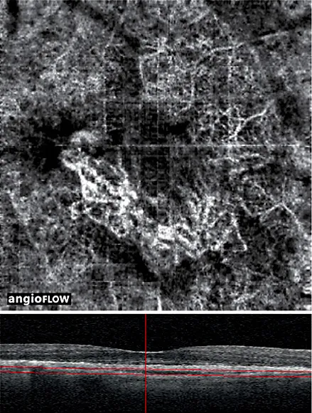
Fig. 1. An 86-year-old patient with treatment-naïve type 1 neovascularization in the right eye. (Top) A 3 mm × 3 mm motion-corrected optical coherence tomography (OCT) angiogram imaged on Avanti RTVue OCTA device showing well-circumscribed choroidal neovascularization with a tangled web of vessels. A quilting artifact due to motion is present. (Bottom) Corresponding spectral domain OCT B-scan with segmentation lines. The authors would like to acknowledge and credit Dr. Nadia Waheed and Dr. Emily Cole for providing these images.
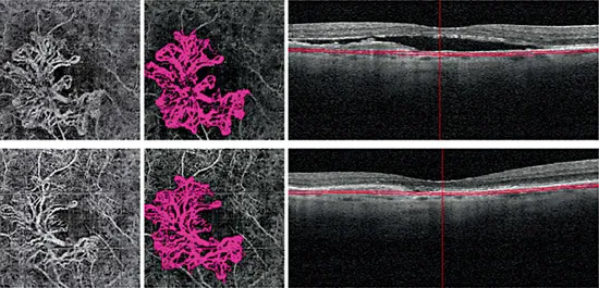
Fig. 2. A 77-year-old male patient with type 1 neovascularization in the right eye, with a visual acuity of 20/40. The patient’s status following 6 aflibercept injections and 7 ranibizumab injections is shown. (Top left) 3 mm x 3 mm OCT angiography (OCT-A) en face projection image of a mature type 1 complex with large feeder vessels and multiple dilated core vessels identified. (Top middle) Corresponding color-coded vessel complex for density analysis. (Top right) OCT B-scan image showing slab segmentation through the pigment epithelial detachment. (Bottom row) Follow-up OCT-A 8 weeks later (interim treatment: 2 aflibercept injections). (Bottom left) 3 mm x 3 mm OCT-A en face projection image of the type 1 complex. (Bottom middle) Corresponding color-coded complex for vessel density analysis. (Bottom right) OCT B-scan image showing slab segmentation. Note that the many large, dilated vessels of this mature type 1 complex are unaffected by additional anti-vascular endothelial growth factor therapy, although the finer vascular plexus may show some attenuation. The lesion area was 3.08 mm2 at baseline and 3.08 mm2 at follow-up; the vessel density was 47% at baseline and 43% at follow-up.
OCT-A has also been employed to study the late fibrotic stage of type 1 neovascularization in AMD. Miere and associates [7] analyzed 49 eyes diagnosed with subretinal fibrosis complicating neovascular AMD, 39 of which were either type 1 or combined type 1 and type 2 lesions. OCT-A demonstrated blood flow related to a persistent neovascular complex within the fibrotic scar in 46 of the 49 eyes [7]. Analysis of these complexes revealed large, dilated vessels with or without vascular loops and interlacing networks, but they typically consisted of only large, mature vessels without an associated fine dense capillary plexus. Most fibrotic lesions also had large flow void areas of the choriocapillaris or dark halos.
Additionally, OCT-A has been utilized to characterize the response of type 1 neovascularization to antiangiogenic therapy. Muakkassa and associates [14] studied six patients with treatment-naïve choroidal neovascularization (CNV), four of which had type 1 lesions. Eyes were scanned before anti-VEGF treatment and at follow-up visits in order to assess the area of each neovascular lesion and its greatest linear dimension (GLD). Follow-up images taken 2-9.5 weeks after initial injection revealed a 29.8% average decrease in CNV area and a 23.6% decrease in its GLD [14]. All patients also experienced improvement or stabilization of...